
Ultrasound for Tightening
Ultrasound for Tightening
Guilherme Bueno de Oliveira and Carlos Roberto Antonio Faculdade de Medicina Estadual de São José do Rio Preto – FAMERP, Rua Silva Jardim, 3114, Centro, São José do Rio Preto, SP, Brazil Hospital de Base de São José do Rio Preto, São Paulo, Brazil
Abstract
Ultrasound is a well-known method used for medical imaging. However, microfocused ultrasound differs by emitting a convergent beam of energy at a specific point. For this, the different types of devices that use ultrasound energy are used for a small focal point, where high temperatures are adequate to cause tissue coagulation. The technology exclusively depends on heat for its effects on the tissue. The objective is to raise the local temperature to at least 65 °C, which is a temperature suitable for collagen contraction. By directing the focused energy wave into deep areas, it causes thermal coagulation points sparing adjacent nontarget tissues. The result obtained when treating this structure is tissue contraction with effective noninvasive lifting of the skin of the neck and face, in addition to the improvement of fine lines and wrinkles. This chapter is going to describe basic concepts of this new technology and explain the procedure and its indications.
Keywords UltrasoundTighteningFlaccid skinSMASCollagen
Introduction
Dermatology participates in the revolution of development of noninvasive technologies for tissue contraction and consequent lifting effect. Among the numerous devices and variety of energy technologies that have been recently developed for this purpose, microfocused ultrasound is also included. According to Alam et al. (2010), the different types of devices that use ultrasound energy are used for a small focal point, where high temperatures are adequate to cause tissue coagulation. Similar to the traditional ultrasound used for medical imaging, the focused beam of energy can pass inoffensively through the epidermis, allowing the focal point to reach deeper tissues, such as the deep dermis, subcutaneous and superficial aponeurotic muscular system (SMAS), where the temperature reaches approximately 65 °C and causes protein denaturation within milliseconds.
According to Kim et al. (2008), one distinction must be made between the two main types of focused ultrasound used in medicine. High-intensity focused ultrasound (HIFU) uses high-energy waves and is primarily used for deep-tissue medical applications between 1.1 cm and 1.8 cm, such as for ablating adipose tissue to contour the body. In contrast, microfocused ultrasound (MFU) uses much lower energy waves to treat the more superficial layers of the skin, between 1.5 mm and 4.5 mm. Despite its lower energy, the MFU is able to stimulate tissue heating above 60 °C, producing small coagulation points (less than 1 mm3) to a depth of up to 5 mm, reaching the deep dermis and SMAS and sparing the overlying epidermis.
According to Baumann and Zelickson (2016), the MFU depends only on heat to achieve its effects on the tissue. The goal is to raise the local temperature to at least 65 °C, which is the temperature at which collagen contraction begins. By directing the focused energy wave into deep areas, the MFU causes thermal coagulation points sparing adjacent nontarget tissues. In addition to local coagulation, the application of heat causes the collagen fibers in the SMAS and subcutaneous fat layer to become denatured and contract. This occurs by breaking intramolecular hydrogen bonds, determining the fold of the collagen chains and a consequent configuration that is more stable and with thicker fibers. In addition, neo-collagen formation occurs within the areas of thermal tissue coagulation and new forms of viscoelastic collagen are produced, resulting in contraction of flaccid skin. The MFU aims to treat facial SMAS, a fan-shaped structure that covers the face and connects the facial muscles with the dermis. The result obtained when treating this structure is tissue contraction with a noninvasive lifting effect of all the skin of the neck and face, in addition to the improvement of fine lines and wrinkles.
The treatment with MFU can be customized to meet the unique physical characteristics of each patient, adjusting the energy and focal depth of the emitted ultrasound wave. These options differ in their focus and wavelength, wherein the depth and amount of energy delivered during the treatment may be varied to achieve a desired effect within the target tissue layer. Currently available transducers emit 10.0 MHz, 7.0 MHz, and 4.0 MHz frequencies with focal depths of 1.5 mm, 3.0 mm, and 4.5 mm, respectively. Two 10 MHz/1.5 mm and 7.0 MHz/3.0 mm transducers are also available to allow the release of energy into smaller anatomical regions that are harder to reach with larger transducers. Together, these transducers can be used in combination to reach the superficial dermis (1.5 mm), deep dermis (3.0 mm), or subcutaneous tissues and SMAS (4.5 mm).
According to Dayan et al. (2014), there are commercially available MFU devices that are also capable of generating high-resolution ultrasound imaging, allowing the tissue plane to be viewed at a depth of 8 mm and letting the user see where the energy will be applied. According to Hitchcock and Dobke (2014), each handpiece uses high-resolution ultrasound that is capable of clearly generating the image of the target facial anatomy, including skin, subcutaneous fat, SMAS, facial muscles, and underlying bone. This ensures treatment at the appropriate depth and allows avoiding inadvertent treatment of nontarget tissue, such as bone and larger blood vessels. The image also allows the operator to be certain of the appropriate acoustic coupling between the transducer and the skin prior to applying power from the MFU.
Patient Selection: Indications and Contraindications
According to Oni et al. (2014), there are relatively few absolute contraindications to the use of MFU. These include infections and open cutaneous lesions in the treatment area, severe or active cystic acne, and the presence of active metal implants such as pacemakers or defibrillators in the treatment area. Precautions include treatment directly over keloids and permanent dermal fillers, and the presence of factors that could alter or impair wound healing, such as smoking.
Although not all people achieve a full aesthetic benefit with MFU, patient satisfaction will be enhanced by appropriate patient selection and realistic expectations. According to MacGregor and Tanzi (2013), MFU is best suited for patients with mild to moderate muscle and skin flaccidity. An ideal patient is younger with normal healing, since the clinical response to treatment with MFU depends in part on the synthesis of new collagen and the so-called wound healing. Older patients or those heavily damaged by light, with sagging skin and marked looseness of the platysmal bands, may require a higher energy density during a single treatment or more than one treatment to achieve the maximum benefits of the technology. In this way, older patients with extensive photodamage, severe skin sagging, and very marked platysma bands are not good candidates for treatment with MFU, and surgical treatment or other adjuvant technologies should be recommended.
Application Technique
Pretreatment
In the authors' practice, patients typically receive topical application of lidocaine 12% and ingest paracetamol 500 mg plus codeine 30 mg 45 minutes before the procedure; diazepam 5–10 mg is administered only if there is significant anxiety.
Demarcation of the Area to Be Treated
MFU devices have standardized protocols of shots according to the type of transducer to be used and to the region. According to Brobst et al. (2012, 2014), these protocols have standard demarcation and a predetermined number of shots in each region. It is important to respect the demarcation to avoid side effects, as MFU should not be applied in areas where superficial nerves are present. The demarcation recommended by the industry is followed using a marking ruler that accompanies the apparatus. It has the correct size for the appropriate space between the lines of application of the transducer, providing greater security against overlapping MFU shots in the region to be treated. According to Fabi (2015), the marking should be done with a white pencil and must obey the following steps:
- Draw a line on the entire mandibular contour and mental region;
- draw a line over the arc of the zygomatic bone, bypassing the orbital region, until it reaches the nasal region;
- demarcate the regions of danger for application of MFU on the face – draw a line in the nasogenian sulcus, extending to the jaw line. At the junction of these lines, mark 2 cm laterally and 1 cm superiorly to make a square of protection for the branch of the marginal nerve of the mandible. To mark the infraorbital nerve region as a danger area, follow these steps:
- on the upper third of the face, draw a line between the lateral epicanthus and the hair implantation line and, using the ruler above this line, draw a second line, thus creating an application space; draw a line between the eyebrow tail and the hair implantation line and, with an application ruler on the medial side, draw a second line, thereby creating an application space in the frontal region;
- in the region of the neck, palpate the eminence of the thyroid cartilage, mark 1 cm above and 1 cm on each side of it and draw a safety area running down the entire region of the trachea; draw a second line over the clavicular line;
- after these defined lines, markings are made on the face and neck, all with the size of one marking ruler, vertically, totaling two to three areas of application on the face per side and two to three areas of application in the neck per side, plus one central area. Each area has a total number of shots defined by the manufacturers and varies according to the machine the user owns.
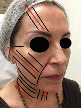
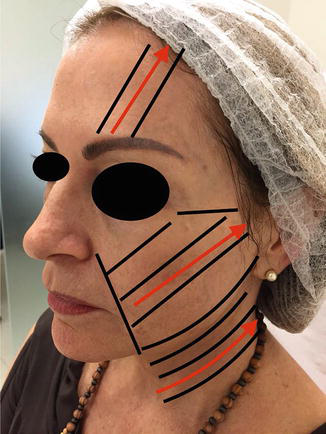
In the authors' practice, marking is performed after identification of the sagging vectors. The flaccidity vectors of the face and neck obey a diagonal fall in the median direction. This angulation of the vector is determined by palpation in clinical examination, since sagging of the SMAS is variable among patients. The evaluation technique consists of clamping the fingers on a part of the skin and applying traction to a point where a better lifting-effect response is obtained. Thus, the necessary angulation is demarcated to draw the lines of application of the MFU. All safety lines are respected, changing only the direction of the marking lines, following the order:
- Draw a line on the entire mandibular contour and mental region;
- draw a line over the arc of the zygomatic bone, bypassing the orbital region, until it reaches the nasal region;
- demarcate the regions of danger for application of MFU on the face – draw a line in the nasogenian sulcus, extending to the jaw line. At the junction of these lines, mark 2 cm laterally and 1 cm superiorly to make a square of protection for the branch of the marginal nerve of the mandible. To mark the infraorbital nerve region as a danger area, follow these steps:
- on the upper third of the face, draw a line between the lateral epicanthus and the hair implantation line and, using the ruler above this line, draw a second line, thus creating an application space; draw a line between the eyebrow tail and the hair implantation line and, with an application ruler on the medial side, draw a second line, thereby creating an application space in the frontal region;
- in the region of the neck, palpate the eminence of the thyroid cartilage, mark 1 cm above and 1 cm on each side of it and draw a safety area running down the entire region of the trachea; draw a second line over the clavicular line;
- after these defined lines, markings are made on the face and neck, all with the size of one marking ruler, diagonally opposite the sagging vectors identified by the technique described above, totaling three to five areas of application on the face per side and four to six areas of application in the neck per side, plus one central area. The total number of shots per area is defined by the immediate clinical response at the time of the procedure. The total number of shots in an area never exceeds the total released by the company, but the total per application space is not equal between them; they depend on the immediate response of the patient's skin (Figs. 1 and 2).
Application
The number of shots with each transducer has been previously determined between minimum and maximum numbers of shots by the company for each region. Preference is given to the maximum energy of the device, which is decreased if the patient's pain sensitivity is low. Treatment should always be started with transducers that reach a greater depth and then come more superficially with the other transducers.
In the authors' technique, the number of shots is kept between the minimum and maximum predetermined by the company. However, the final number is determined clinically by the immediate treatment outcome.
Results
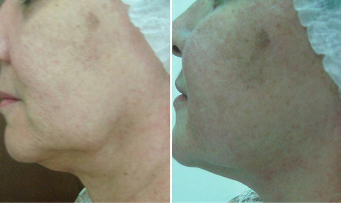
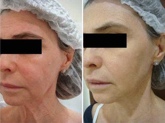
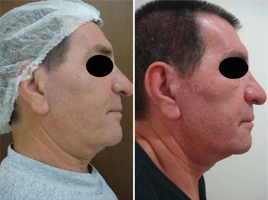
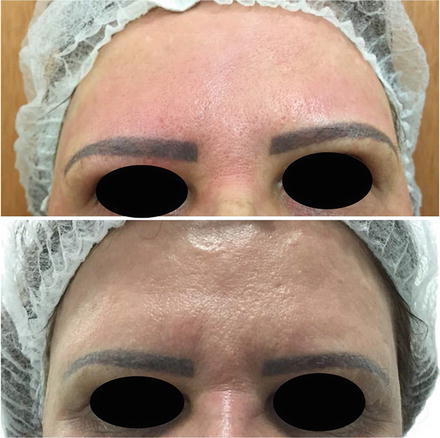
The effectiveness of MFU treatment is superior when multiple transducers are used. The beneficial effects of double-depth treatment are evaluated by the lifting effect of the middle third of the face, mandibular definition, and reduction of sagging of the submental skin. The final result occurs at 90 days post treatment. A new session can only be held after this time period. Maintenance is usually carried out between 10 and 18 months.
See results with Ulthera® (Ultherapy®; Ulthera Inc.) in Figs. 3, 4, and 5 and with AccuTyte® (Vydence®) in Fig. 6.
Side Effects and Their Management
Pain
According to Pak et al. (2014), the most commonly reported side effect associated with MFU is painful discomfort during the treatment session. In the literature, studies do not report significant pain. According to Kakar et al. (2014), suggestions for minimizing discomfort during treatment include pretreatment with oral paracetamol, a nonsteroidal anti-inflammatory drug or a narcotic analgesic, topical anesthetics with lidocaine when using the 1.5 mm transducer, and applying as much energy as possible according to tolerance. In the authors' practice, patients typically receive topical application of lidocaine 12% and ingest paracetamol 500 mg plus codeine 30 mg 45 minutes before the procedure. Diazepam 5–10 mg is administered only in cases of great anxiety.
Transient Erythema
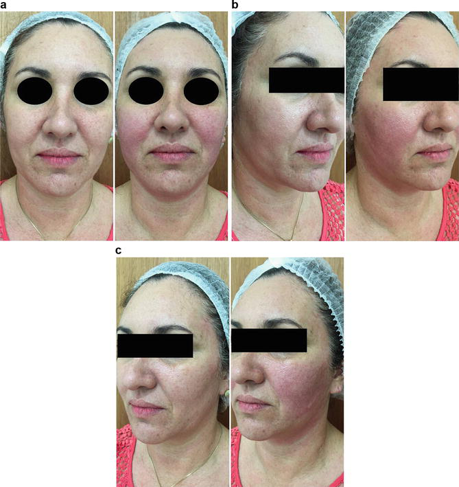
The second most commonly reported side effect is the combination of transient erythema and edema (Fig. 7a–c). This is usually transient, resolving in most cases within 3 hours after the session. It is due to regional warming with stimulation of the inflammatory reaction and vasodilation. Patients do not usually complain about it; however, cold compresses can be applied at most to speed up improvement of these symptoms.
Less Common Side Effects
Occasional side effects include postinflammatory hyperpigmentation 1 month after treatment, muscle weakness and transient local numbness, striated linear skin excoriations, or papule formation. The papules appear to be due to nonideal technique and are more likely to be associated with the use of 3 mm and 1.5 mm transducers.
The most serious effects reported in the literature are facial paralysis. The reported cases had a modification of the facial anatomy from a previous surgical facelift or application in areas known not to be safe.
Take Home Messages
- Microfocused ultrasound (MFU) aims to treat facial SMAS, a fan-like structure that covers the face and connects the facial muscles to the dermis.
- MFU uses special acoustic transducers that direct the energy of the ultrasound to a small focal point, where the high temperatures are able to cause tissue coagulation.
- Treatment with MFU should be customized to meet the unique physical characteristics of each patient, adjusting the energy and focal depth of the emitted ultrasound wave. These options differ in their focus and wavelength, varying the depth and amount of energy supplied during the treatment.
- MFU is best suited for patients with mild to moderate muscle and skin flaccidity. Patient satisfaction will be enhanced by appropriate patient selection and realistic expectations.
- Older patients with extensive photodamage, severe sagging skin, and very marked platysma bands are not good candidates for treatment with MFU, and surgical treatment or other adjuvant technologies should be recommended.
References
Alam M, White LE, Martin N, Witherspoon J, Yoo S, West DP. Ultrasound tightening of facial and neck skin: a rater-blinded prospective cohort study. J Am Acad Dermatol. 2010;62:262–9.
Baumann L, Zelickson B. Evaluation of micro-focused ultrasound for lifting and tightening neck laxity. J Drugs Dermatol. 2016;15(5):607–14.
Brobst RW, Ferguson M, Perkins SW. Ulthera: initial and six month results. Facial Plast Surg Clin North Am. 2012;20:163–76.
Brobst RW, Ferguson M, Perkins SW. Noninvasive treatment of the neck. Facial Plast Surg Clin North Am. 2014;22:191–202.
Dayan SH, Fabi SG, Goldman MP, Kilmer SL, Gold MH. Prospective, multi-center, pivotal trial evaluating the safety and effectiveness of micro-focused ultrasound with visualization (MFU-V) for improvement in lines and wrinkles of the décolletage. Plast Reconstr Surg. 2014;134(4 suppl 1):123–4.
Fabi SG. Noninvasive skin tightening: focus on new ultrasound techniques. Clin Cosmet Investig Dermatol. 2015;8:47–52.
Hitchcock TM, Dobke MK. Review of the safety profile for microfocused ultrasound with visualization. J Cosmet Dermatol. 2014;13(4):329–35.
Kakar R, Ibrahim O, Disphanurat W, et al. Pain in naïve and non-naïve subjects undergoing nonablative skin tightening dermatologic procedures: a nested randomized control trial. Dermatol Surg. 2014;40(4):398–404.
Kim YS, Rhim H, Choi MJ, Lim HK, Choi D. High-intensity focused ultrasound therapy: an overview for radiologists. Korean J Radiol. 2008;9:291–302.
MacGregor JL, Tanzi EL. Microfocused ultrasound for skin tightening. Semin Cutan Med Surg. 2013;32:18–25.
Oni G, Hoxworth R, Teotia S, Brown S, Kenkel JM. Evaluation of a microfocused ultrasound system for improving skin laxity and tightening in the lower face. Aesthet Surg J. 2014;34(7):1099–110.
Pak CS, Lee YK, Jeong JH, Kim JH, Seo JD, Heo CY. Safety and efficacy of ulthera in the rejuvenation of aging lower eyelids: a pivotal clinical trial. Aesthetic Plast Surg. 2014;38(5):861–8.







