
Intense Pulsed Light for Photorejuvenation
Intense Pulsed Light for Photorejuvenation
Sílvia Karina Kaminsky Jedwab and Caio Roberto Shwafaty de Siqueira Escola Paulista de Medicina, Universidade Federal de São Paulo, and Skinlaser Brasil, São Paulo, Brazil (Sílvia Karina Kaminsky Jedwab) Faculdade de Medicina de Botucatu, Universidade Estadual Paulista, São Paulo, Brazil (Caio Roberto Shwafaty de Siqueira)
Abstract
Intense pulsed light (IPL) is a non-laser, versatile light technology that is a key tool in dermatology. It is widely used for the treatment of photoaged skin. It is also used for hair removal, acne treatment, and treatment of vascular lesions. IPL works through selective photothermolysis to reduce dark spots, telangiectasia, wrinkles, and skin laxity (photorejuvenation) caused by chronic sun exposure. This chapter discusses all aspects of the physics behind IPL technology, clinical indications, treatment, and possible complications.
Keywords IPLPhotorejuvenationPhotoageDark spotsTelangiectasiaWrinklesSkin laxity
Introduction
The appearance of the skin has been a constantly growing reason to visit dermatologists’ clinics, not only for aesthetic reasons aiming at youth but also for medical reasons, such as treating and preventing skin diseases. Therefore, the number of procedures to improve the quality of the skin with laser and non-laser light devices grows each year (El-Domyati et al. 2015).
The main goal of this chapter is to discuss all the issues regarding the use of intense pulsed light (IPL) for photorejuvenation of the skin. This treatment modality is popular because it does not disrupt the epidermis, limiting adverse effects and minimizing downtime (El-Domyati et al. 2015).
Basic Concepts
The Biology of Photoaging
The aging process of the skin reflects both extrinsic and intrinsic factors (Table 1) (Balibas et al. 2010; Friedmann et al. 2014).
| Intrinsic factors | Extrinsic factors |
|---|---|
| Chronological senescence of dermal fibroblasts | Chronic exposure to UV light |
| Lower production of collagen and hyaluronic acid | Pollution |
| Poor extracellular dermal matrix | Tobacco smoke |
| Production of reactive oxygen species and activator protein-1 | |
| Higher expression of metalloproteinases | |
| Higher fragmentation of collagen fibers | |
| Decreased TGF-β1 levels | |
| Chronic inflammatory response | |
| Defects in the DNA repair system |
The intrinsic factors are related to chronological senescence of dermal fibroblasts, leading to lower collagen and hyaluronic acid production that results in a poor extracellular dermal matrix (Goldberg 2012; Friedmann et al. 2014).
The extrinsic factors are related to long-term, chronic ultraviolet (UV) light exposure (usually from the sun), pollution, and tobacco smoke. All of them enhance the breakdown and fragmentation of collagen fibers through the production of reactive oxygen species and activator protein-1, which upregulates abnormal expression of matrix metalloproteinases, the main agents that destroy collagen fibers (El-Domyati et al. 2015; Friedmann et al. 2014).
In normal skin, transforming growth factor-β (TGF-β) upregulates collagen production (Ali et al. 2013). In photo-damaged skin, TGF-β1 levels are decreased, causing reduced collagen production. This results in poor levels of collagen I and III in the dermal matrix (Ali et al. 2013; El-Domyati et al. 2015).
Also, it is well known that the chronic inflammatory response to long-term UV light exposure leads to permanent DNA damage. Locally in the skin, UV light may trigger DNA photoproducts, isomerization of the trans- to the cis-isoform of urocanic acid in the stratum corneum, and UV-induced alteration of the membrane redox potential. All these events lead to a UV-induced immune response that causes immunosuppressive effects, UV mutagenesis, and UV carcinogenesis. The DNA repair pathways are no longer capable of detecting and repairing the DNA defects effectively, leading to replication and transcription of damaged genes. Besides the obvious carcinogenic and mutagenic consequences, these events also change the normal half-life of epidermal cells, leading to excessive (or even abnormal) proliferation of melanocytes and keratinocytes in the basal and Malpighi’s layers, respectively (Bolognia et al. 2008).
Clinically, all these events result in laxity, chronically tanned skin, wrinkles, telangiectasia, rough and sagging skin, and melanocytic lesions (like lentigos and ephelides), all together named photoaging (Gold and Biron 2013; Sasaya et al. 2011).
The Biophysics Behind IPL
IPL devices are non-laser, non-ablative sources of high-intensity light that use a high-output xenon flash pump light source to produce a polychromatic, noncoherent, non-collimated diffused light with a broad wavelength range (500–1,300 nm). There are simultaneous emissions of green, yellow, red, and infrared wavelengths (Mattos et al. 2009; Balibas et al. 2010; Goldberg 2012; Ali et al. 2013; El-Domyati et al. 2011; Friedmann et al. 2014).
IPL was first approved by the Food and Drug Administration (FDA, USA) in 1998 for the treatment of photoaged skin.
Nowadays, there are more than 300 different IPL devices available on the market. The latest generation of devices is much safer because all of them have protective features to prevent epidermal damage from excessive heat. Most use sapphire or quartz interface cooling systems to provide this safety at the epidermal layer (Goldberg 2012). The light pulse is usually single, but it can be double or triple depending on the device, with smooth energy delivery and peak flow occurring at the early shot, square shaped. It is not possible to deliver energy in nanoseconds as with Q-switched lasers. This system prevents prolonged heating of the epidermis, treating the majority of the chromophores at the same time with one single shot. The pulse duration should be equal to or below the thermal relaxation time (TRT) of the target structures to prevent unselected damage to surrounding tissue, but should be at least 50% of the targets’ TRT to generate their cell death (Mattos et al. 2009; Balibas et al. 2010; Goldberg 2012). Normal skin has an average TRT of 10 milliseconds (ms). Vessels with a diameter of 0.1 mm have a TRT of 10 ms; larger vessels (0.3 mm) have a TRT of 100 ms (Goldberg 2012).
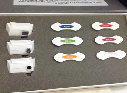
The handpiece allows the physician to choose which wavelength will be applied to the patient’s skin. These cut-off filters block the lower wavelengths that are not desired for treatment but do not filter the higher wavelengths. The usual filters available on the market are 400, 515, 540, 550, 560, 570, 590, 595, 615, 650, 695, and 750 nm (Fig. 1). Energy fluence varies from 8 to 100 joules (J), and pulse time varies from 5 to 100 ms, depending on each device.
IPL works at skin sites based on the principle of selective photothermolysis, first described by Anderson and Parrish with pulsed dye lasers (Railan and Kilmer 2000). Normal skin contains substances that act as chromophores (i.e., substances that absorb energy depending on their intrinsic color, transforming it into heat), such as hemoglobin, carotene, melanin, and water (Fig. 2) (Goldberg 2012). Each substance has its own curve of maximum and minimum light absorption (medium wavelengths of light absorption for deoxyhemoglobin are 550–560 nm, for oxyhemoglobin are 540 nm and 575–580 nm, and for vascular lesions and melanin are 400–755 nm) (Friedmann et al. 2014).
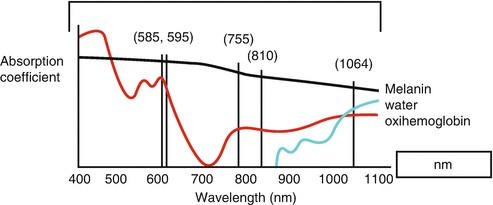
Photo-damaged skin contains higher amounts of these chromophores, distributed in different forms (localized spots or in a diffuse, multiple-layer manner). These substances have the capacity to absorb the photons, transforming them into heat, which destroys the colored target and dissipates the heat into the surrounding area, promoting activation of collagen production by the fibroblasts (collagen I and III) through cytokine activation and upregulation of TGF-β1 (Balibas et al. 2010; Ali et al. 2013).
IPL light targets all the chromophores at the same time, especially melanin and hemoglobin. The overall immediate endpoint after application of IPL to the skin is darkening of melanocytic lesions, blurred telangiectasia, and light redness over the entire treated skin surface.
The goal of treating vascular lesions is to raise the blood vessel temperature high enough to cause its coagulation, leading to its destruction and replacement by fibrous granulation tissue. Oxyhemoglobin (red lesions), deoxygenated hemoglobin (blue lesions), and methemoglobin can be targeted by IPL (peak absorption of 418 nm, 542 nm, and 577 nm, respectively). Therefore, the best IPL filters to treat vascular lesions lie between 400 and 600 nm. Some devices allow a vascular tip to be fitted to the handpiece, to increase the delivered energy only at the desired target (Fig. 1).
The goal of treating pigmentary lesions is to elevate the rapid differentiation of keratinocytes induced by thermal heating, using melanin as the target. This results in an upward transfer of destroyed and non-destroyed melanosomes along the necrotic keratinocytes, resulting in their elimination as crusts that can be seen in the days after the treatment session. Lower cut-off filters are the usual best choice for treating melanocytic lesions, usually around 400–540 nm (Goldberg 2012).
The goal of treating skin laxity and wrinkles is to heat dermal water to stimulate dermal fibroblasts, which increases the synthesis of extracellular matrix proteins, such as collagen I and III and elastin. Higher cut-off filters are the best choice to achieve these goals, usually around 600–1,200 nm (Goldberg 2012; El-Domyati et al. 2011).
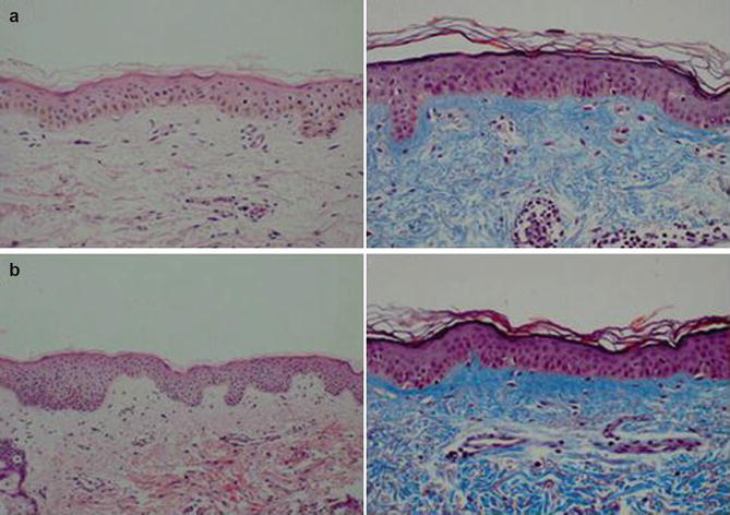
The histopathology findings that confirm all these changes are seen as an increase in the number of fibroblasts associated with an increase in collagen compaction, thickening, and density. This also indicates collagen remodeling. Also, the dermal papillae and the interpapillary crests are more pronounced, showing the higher deposition of collagen after IPL treatments. The melanin pigment has a more homogeneous distribution at the basal layer. The number and diameter of blood vessels from the superficial plexus were reduced as well, in one-third of treated patients (Fig. 3) (Scattone et al. 2012; Friedmann et al. 2014).
The Clinical Aspects of IPL on the Daily Practice
IPL is a key tool in the treatment of photoaged skin.
In the first place, the physician should establish the skin color type of the photoaged skin according to Fitzpatrick’s classification for skin types (Table 2). The use of IPL on higher skin color types (≥ IV) and on highly photo-damaged skin (too many targets) demands lower fluences, higher milliseconds, cooling systems on, and multiple treatment sessions.
| Skin type | Skin color | Characteristics |
|---|---|---|
| I | White; very fair, red or blonde hair; blue eyes; freckles | Always burns, never tans |
| II | White, fair, red or blond hair; blue, hazel or green eyes | Usually burns, tans with difficulty |
| III | Cream white, fair with any eye or hair color (common) | Sometimes mild burn, gradually tans |
| IV | Brown; typical Mediterranean Caucasian skin | Rarely burns, tans with ease |
| V | Dark brown; Middle Eastern skin types | Very rarely burns, tans easily |
| VI | Black | Never burns, tans very easily |
Pregnancy, lactation, infected skin, use of photosensitizing medications or oral retinoids, photosensitivity from any disease, surreal expectancies, keloids, hypertrophic scars, suntan, and photo-induced skin disorders are exclusion criteria for treatment (Table 3) (Balibas et al. 2010).
| Exclusion criteria |
|---|
| Pregnancy |
| Lactation |
| Infected skin |
| Photosensitizing medications/oral retinoid |
| Photosensitivity |
| Surreal expectancies |
| Scars: keloids, hypertrophic |
| Suntan |
| Photo-induced skin disorders |
The number of sessions varies depending on each patient, i.e., it depends on the skin color type and the degree of photoaging. Usually four to six sessions are required to achieve the overall improvement of the skin (Fig. 4).
Before the treatment starts at the office, the patient should lie down in a comfortable office chair; the skin area to be treated should be cleaned with lotions without high concentrations of alcohol, removing makeup and sunscreen. Photos should be taken, and a consent form should be signed explaining the possible outcomes and results from the treatment, as well as possible complications. The use of topical anesthetic creams is not necessary. In fact, they can cause vasoconstriction, decreasing the quantity of hemoglobin at the target site and reducing overall results (Mattos et al. 2009).
The patient and the staff must use eye protection at all times during application. The skin should be covered with a thin layer of transparent gel (like those used for ultrasound exams), cooled or not.
Before initiating the treatment, the doctor must tell the patient that the light will be triggered, to reduce patient anxiety (Fig. 5). Usually application starts with the chosen parameters at the border of the area to be treated, especially covered areas, to prevent any complications on exposed sites (Balibas et al. 2010). The handpiece should be applied perpendicularly to the skin surface, gently touching it. After three to four shots, treatment should stop and the skin inspected.
The immediate endpoint should be slight redness of the skin, darkening of brown spots, and blurriness of telangiectasias (Fig. 6).
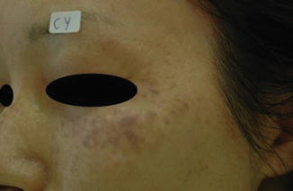
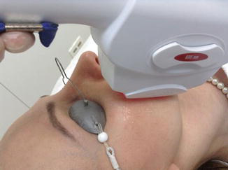
Each spot should overlap the previous one by 10%. When treating isolated and resilient lesions, the physician may use a perforated plastic shield with varying aperture sizes (Fig. 7) or small special tips applied to the handpiece (Fig. 1), to deliver higher energy only to that target (Balibas et al. 2010).
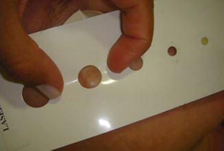
If urticaria, blistering (Fig. 4), or gray-toned skin is seen, treatment should stop immediately, the parameters reviewed, and ice or cooling substances and topical corticosteroid creams should be applied immediately, because these are signs of excessive heating of the skin, with possible burning.
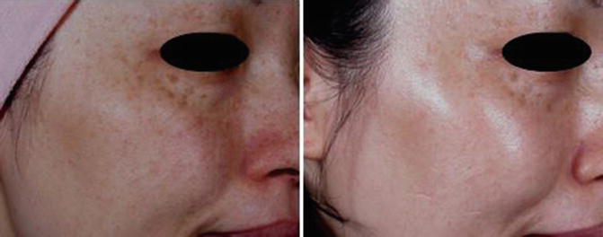
In areas where more results are needed, shots may be applied twice, but the second application should be perpendicular to the first one, avoiding “zebra marking.” Usually patients feel little discomfort, little heat, and little pain during the session. Anything different from that should alert the doctor to review the parameters that have been used, because it can mean excessive heating of the skin.
At the end of treatment, the staff should apply thermal spring water spray and soothing creams to reduce skin redness and discomfort, and also broadband sunscreen creams.
Overall, the physician should keep in mind that the targets should be treated in layers, because this is actually how the excessive pigments are displayed on the skin. First, darker brown spots and superficial telangiectasia should be reduced, and then collagen at the deeper layers can be stimulated. Most complications occur when trying to destroy all the targets, at all layers, at the same time, with the same high energy. Another reminder is that parameters should be reduced (lower energies, higher pulse time duration) when the patient has too many targets at the skin area to be treated, to prevent excessive heating of the skin.
For skin color types ≤ III, treatment can start by choosing the 515–540 nm filters, 10–20 ms, 10–15 J/cm2. If there are too many targets, use 15–20 ms, 8–13 J/cm2 (Table 4). With the progression of the treatment, targets at the middle layers will reduce, so the physician can elevate the energy delivered and reduce the pulse duration to target the remaining lighter chromophores (localized vessels, lighter dark spots) at the superficial layers, using lower filters, usually applied specifically at the target. The physician can also elevate the energy delivered to the deeper layers, using higher filters and higher milliseconds to stimulate overall skin tightening.
For skin color types ≥ IV, treatment can start using 570–695 nm filters, trying to avoid the naturally higher concentrations of melanin present at the basal layer of the epidermis in these skin types: 20–100 ms, 6–10 J/cm2 (Table 4). With the progression of the treatment, targets will reduce, so the physician can elevate the energy (while maintaining the long milliseconds) and deepen light penetration using higher filters.
| Targets | Skin color type ≤ III | Skin color type ≥ IV |
|---|---|---|
| Little to moderate quantity | 515–540 nm 10–20 ms 10–15 J/cm2 |
570–695 nm 20–100 ms 6–10 J/cm2 |
| Moderate to high quantity | 515–540 nm 15–20 ms 8–13 J/cm2 |
570–695 nm 100 ms 6–8 J/cm2 |
The healing process of facial skin differs from the skin of other areas, because the higher density of sebaceous glands in facial skin provides quicker healing through prompt cell restoration. So, application of IPL outside facial skin demands higher caution, lower fluences, higher pulse time durations, and overlapping the application of light so it does not cause a “zebra effect.” Usually it takes 7–10 days to complete healing of facial skin; outside the facial area healing time may double.
The authors mostly use the 540 nm and 695 nm filters in their routine dermatologic practice, because these two filters target both superficial and deep chromophores in a global manner.
IPL can be associated with other treatments, with or without devices, at the same session. We must advise that the parameters chosen for these multiple simultaneous treatments should be more conservative, since different ways of treating the whole skin are being combined in the same session.
The authors have experience combining IPL with lasers, chemical peelings, and laser hair removal in the same session:
- Alexandrite laser 755 nm may be used when dark spots are resilient to IPL, to epilate a few facial hairs before any other laser is applied, for angiomas, or for larger telangiectasias.
- Ruby laser 694 nm Q-switched (QS) and KTP (potassium titanyl phosphate) laser 532 nm QS may be used to treat lighter brown spots resilient to IPL and to the Alexandrite laser.
- Nd:YAG (neodymium-doped yttrium aluminum garnet) laser 1,064 nm long-pulsed may be used for telangiectasias larger than 0.1 mm and for epilation of facial hair in dark-skinned patients.
- Nd:YAG QS laser may be used for skin tightening, to treat lighter freckles and dark spots, and for reduction of sebaceous skin pores.
- Fractionated ablative lasers (CO2 10,600 nm and erbium 2,940 nm) may be used for skin tightening, wrinkle reduction, comedone reduction, and improvement of rough aged skin.
Also, IPL can be associated with all types of chemical peelings, such as retinoid acid, glycolic acid, Jessner solution (a combination of resorcinol, salicylic acid, and lactic acid), salicylic acid, trichloroacetic acid (TCA), phenol, mandelic acid, and so on. It must be pointed out that the association between IPL and chemical peelings should not occur when the endpoint has already been achieved by the IPL itself and/or the patient reports that the skin is burning too much after IPL treatment. This prevents complications that may arise from overtreatment of the skin.
Applications with botulinum toxin and hyaluronic acid fillers may be associated with IPL sessions, usually right after the IPL session ends.
After treatment, at home the patient must use products to minimize excessive inflammation. In all situations, the patient should use broadband sunscreen, cleansing gel to wash the treated skin, thermal spring water spray for burning relief, topical low-potency corticosteroid cream if the patient presents with a stronger burning or itching sensation, and herpes simplex prophylaxis if there is a personal history of this disease (Balibas et al. 2010). If the patient was treated only with IPL and/or non-ablative lasers, simple creams or gel creams should be used, usually containing alpha-bisabolol, vitamin C, and essential oils (grape seed, sunflower seed). If the patient was treated with IPL associated with ablative lasers, creams or ointments containing healing substances as well as antimicrobial agents (copper, zinc, triclosan, petrolatum) should be used.
During the 7–15 days after the session, the treated skin will peel off gently. The dark spots will become darker with small crusts, and the vanished telangiectasias may reappear blurred or bluish. After a minimum of 7 days after the initial treatment, the patient can be submitted to other healing/tightening procedures, like radiofrequency and micro-focused ultrasound.
Possible side effects are (Table 5) blistering, purpura, excessive crusting, persistent erythema, dyschromias, irritant contact dermatitis, atrophy, scarring, hypertrophic scarring, keloid formation, and infection (bacterial and/or viral) (Balibas et al. 2010; Friedmann et al. 2014). All of these can be prevented through correct indication and individualization of parameters and respect for the skin color type of the patient. The complications can be divided into two groups: immediate complications and late complications.
| Side effects |
|---|
| Blister |
| Purpura |
| Excessive crusting |
| Persistent erythema |
| Dyschromias |
| Irritant contact dermatitis |
| Scars: atrophic, hypertrophic, keloid |
| Infections |
Immediate complications are those related to excessive energy delivered into the skin. Possible causes are the following wrong parameters: excessive joules delivered or too little time for thermal relaxation, tanned skin or too many targets at the skin location, and misclassification of the patient’s true skin color type.
The patient may present with acute burning pain, urticaria, purpura, gray skin, and blisters right after treatment (Fig. 4).
When these occur, the physician must immediately stop the procedure, apply cooling substances, and apply high-potency corticosteroid cream to the skin site. Low-intensity laser (LIL) can be applied to the burned areas on a daily basis until total healing, because this treatment blocks excessive inflammation. Patients should maintain use of medium- to high-potency corticosteroid creams at home, twice a day, until total healing (usually 7–10 days), and avoid the sun by physical blocking and use of broadband sunscreens.
Late complications are scars (atrophy, keloids, hypertrophic scars), infections, allergy and dyschromias, and both postinflammatory hyperchromia (PIHE) and hypochromia (PIHO). These are usually transient complications, resolving spontaneously within 2–3 months after they start.
The scars (Fig. 8) usually occur after episodes with burned skin, especially after blistering. Overall, the scars may become white and atrophic. Depending on the personal background of the patient, keloids and hypertrophic scars can develop in specific locations (the jaw line, earlobes, chest, back, neck, upper arms, and abdomen).
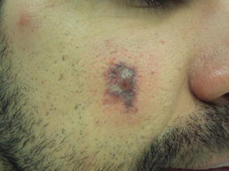
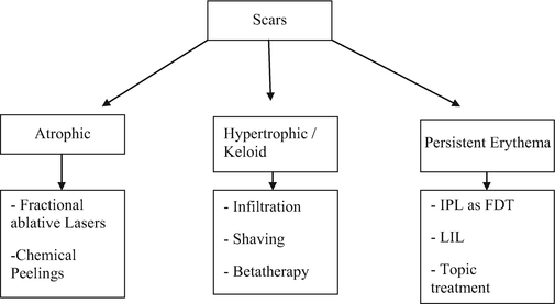
Just after the healing process has ended after the immediate complications, the skin may show persistent erythema. This may signal that excessive inflammation is ongoing, which may trigger formation of the scars mentioned. To prevent them, besides treating the immediate complications, the physician should apply IPL as photodynamic therapy to the persistent erythematous area every week (very low fluence, between 6 and 10 J, filters 540–695 nm, 100 ms pulse time), LIL on a daily basis, and anti-inflammatory creams (medium-potency corticosteroid creams, rose hip oil, silicone oil). If keloid scars or hypertrophic scars occur even after the preventive treatment, the scars may be treated with local infiltration of injectable corticosteroids or bleomycin, once a month until total regression. For cases of keloid scars resistant to infiltration therapy, surgical options may be indicated, like shaving of the lesion associated with beta therapy. If atrophic scars occur, they may be treated with fractional ablative lasers and chemical peelings (like the chemical reconstruction of skin scars [CROSS] chemical peeling), in multiple sessions, once a month (Fig. 9).
Infections are rare complications, but they can be devastating since they may arise suddenly without warning. The most common is activation of herpes simplex virus, which may lead to local or widespread infection at the treated area (simulating the Kaposi varicelliform eruption seen on atopic skin). Bacterial infections are rare, taking place most often around contaminated skin sites (perioral, paranasal, perineum, hands, feet, axillae, skin sites with bacterial folliculitis, or any other bacterial skin infection), usually caused by Gram-positive bacteria (Streptococcus sp., Staphylococcus sp.). Risk factors for them are high-energy ablative fractionated lasers, high-energy IPL, burning complications, medium-potency chemical peelings (Jessner solution associated with TCA), and large areas treated at the same time. The most important measure to prevent them is to anticipate them, by prescribing herpes simplex prophylaxis for patients with this background when they are submitted to the treatments listed above (to be initiated 1 day before the treatment), as well as topical and/or oral antibiotics or antimicrobial topical creams to use after the procedure, until total healing of the treated skin site. We do not recommend treating any skin site with IPL with evident infection of any kind.
Allergies are rare; they may occur when epidermal disruption is seen, leading to possible higher absorption of allergens and/or irritating substances. In all cases, known allergenic substances reported by the patient should not be used; after treatment, the use of creams for better healing of the skin minimizes the risks of allergic contact dermatitis and/or irritant contact dermatitis. If it happens, low- to moderate-potency corticosteroid creams should be applied to the skin twice a day for 7–10 days.
Dyschromias are the most common late complication seen after IPL treatments. Persistent PIHE can be treated with superficial chemical peelings (like retinoid acid and Jessner solution), associated or not with the Nd:YAG QS 1,064 nm laser. At home, patients should use Kligman’s formula or a similar formula.

Persistent PIHE will heal within 3–12 months after the complication.
When persistent PIHO occurs, the physician must examine the site through a Wood’s lamp (WL). If the area is highlighted by the WL, it indicates that the hypochromia may be truly persistent, so it should be treated with narrowband UVB phototherapy weekly until total healing.
If the area is not highlighted by the WL, it indicates that it is transient, so the physician may observe the natural healing or try to make the surrounding skin lighter with IPL and/or chemical peelings.
For all cases of PIHO, patients should also avoid direct sun exposure, use broadband sunscreens, and use Kligman’s formula at home. PIHO usually will heal within 3–15 months.
IPL treatment sessions can be repeated at 30–45 day intervals. Normally, patients require three to six sessions to achieve global improvement, but it must be pointed out that this depends on individual characteristics. Overall, treatment results in a better clinical appearance in skin texture, reduction in mottled appearance, and clearance of pigmented and vascular lesions (Scattone et al. 2012; Gold and Biron 2013).
Take Home Messages
- IPL is a key treatment for patients with photoaged skin.
- IPL devices are versatile, but it should always be kept in mind that individualization of treatment should play the leading role in successfully achieving the goals of youthful and healthy skin.
- Defining the patient’s true skin color type is the main measure to avoid complications and to achieve the main goals of the treatment.
- As with lasers, IPL works through selective photothermolysis, in which the skin chromophores absorb photons from the IPL light, causing heat damage.
- Supportive care after sessions should be emphasized, because this measure helps quick healing of the skin, reducing the chances of all complications.
References
Ali MM, Porter RM, Gonzalez ML. Intense pulsed light enhances transforming growth factor beta1/Smad3 signaling in acne-prone skin. J Cosmet Dermatol. 2013;12:195–203.
Balibas P, Schreml S, Szeimies RM, Landthaler M. Intense pulsed light (IPL): a review. Lasers Surg Med. 2010;42:93–104.
Bolognia JL, Jorizzo JL, Rapini RP. Dermatology. 2nd ed. London: Mosby; 2008. p. 1321–31.
El-Domyati M, El-Ammawi TS, Moawad O, Medhat W, Mahoney MG, Uitto J. Intense pulsed light photorejuvenation: a histological and immunohistochemical evaluation. J Drugs Dermatol. 2011;10(11):1246–52.
El-Domyati M, El-Ammawi TS, Moawad O, Medhat W, Mahoney MG, Uitto J. Expression of transforming growth factor-β after different non-invasive facial rejuvenation modalities. Int J Dermatol. 2015;54:396–404.
Friedmann DP, Fabi SG, Goldman MP. Combination of intense pulsed light, Sculptra, and Ultherapy for treatment of the aging face. J Cosmet Dermatol. 2014;13:109–18.
Gold MH, Biron JA. Safety and cosmetic effects of photodynamic therapy using hexyl aminolevulinate and intense pulsed light: a pilot study conducted in subjects with mild-to-moderate facial photodamage. J Clin Aesthet Dermatol. 2013;6(10):27–31.
Goldberg DJ. Current trends in intense pulsed light. J Clin Aesthet Dermatol. 2012;5(6):45–53.
Mattos R, Filippo A, Torezan L, Campos V. Non-laser energy sources on rejuvenescence: part II. Surg Cosmet Dermatol. 2009;1(2):80–6.
Railan D, Kilmer S. Treatment of benign pigmented cutaneous lesions. In: Goldman’s Cutaneous and Cosmetic Laser Surgery. Elsevier; 2000. p. 93–108.
Sasaya H, Kawada A, Wada T, Hirao A, Oiso N. Clinical effectiveness of intense pulsed light therapy for solar lentigines of the hands. Dermatol Ther. 2011;24:584–6.
Scattone L, Alchorne MMA, Michalany N, Miot HA, Higashi VS. Histopathologic changes induced by intense pulsed light in the treatment of poikiloderma of Civatte. Dermatol Surg. 2012;38:1010–6.







