
Ultrasound for Lipolysis
Ultrasound for Lipolysis
Shirlei Schnaider Borelli, Maria Fernanda Longo Borsato and Bruna Backsmann Braga Brazilian Society of Dermatology, Private Office in São Paulo, São Paulo, Brazil
Abstract
Body sculpting refers to the use of either surgical or noninvasive techniques to modify the appearance of the body. Throughout the years noninvasive procedures for body sculpting gained interest of patients because of the reduced risks and complications. Technologies aiming aesthetic body sculpting include cryolipolysis, radiofrequency, light energy, low-intensity nonthermal focused ultrasound, or the combination of low-energy sources with mechanical manipulation. In this following chapter, we are going to discuss a new noninvasive technology using high-intensity focused ultrasound (HIFU) for ablation of the unwanted adipose tissue, evidencing its mechanisms and possible results for body sculpting.
Keywords Body sculptingAdipose tissueUltrasoundHigh-intensity focused ultrasoundNoninvasive techniques
Introduction
Body sculpting procedures are becoming increasingly popular all over the world. Body sculpting refers to the use of either surgical or noninvasive techniques to modify the appearance of the body (Jewell et al. 2011a). Body sculpting is indicated for focal adiposity (abdomen, thighs, or hips); skin laxity (neck, arms); or both. Patients who have both focal adiposity and skin laxity usually require combined treatment (Jewell et al. 2011a; Kennedy et al. 2015).
For many years a wide range of invasive procedures including liposuction were available for body sculpting (Jewell et al. 2011a; Mordon and Plot 2009). Although liposuction is extremely effective at removing large amounts of excess fat, it is accompanied by significant risk of complications and severe adverse effects. These may include postprocedural pain, infection, prolonged recovery, scarring, hematoma, ecchymosis, or edema (Kennedy et al. 2015; Mordon and Plot 2009).
Recently there is a tendency to achieve body fat reduction without substantial risks and financial costs, without long recovery time and with less postprocedure complications. The number of patients seeking cosmetic minimally invasive procedures had increased 110% from 2000 to 2010. Technologies aiming aesthetic body sculpting include cryolipolysis, radiofrequency, light energy, low-intensity nonthermal focused ultrasound, or the combination of low-energy sources with mechanical manipulation. A new noninvasive technology using high-intensity focused ultrasound (HIFU) for ablation of unwanted adipose tissue has shown positive results (Jewell et al. 2011a, b).
Basic Concepts
Adipocytes, commonly known as fat cells, are connective-tissue cells specialized to synthesize and contain large globules of fat. Ultrasound can lyse adipocytes through mechanical and thermal mechanisms (Jewell et al. 2011a). Ultrasound is an oscillating pressure wave that propagates at the speed of sound. Ultrasonic waves are characterized by intensity, expressed in W/cm2, and frequency, expressed as kilohertz (kHz) or megahertz (MHz). When ultrasonic waves penetrate and travel through tissue, they lose energy as they are reflected, scattered, or absorbed by the tissues. Sufficient quantities of absorbed energy create molecular vibrations in body tissues, generating heat (Haar and Coussios 2007).
There are two categories of ultrasound used for body sculpting: a low-intensity/low-frequency nonthermal ultrasound and HIFU. The nonthermal ultrasound disrupts adipose tissue through mechanical stress generated from inertial cavitation. In general, cavitation is less predictable than HIFU thermal effects (Jewell et al. 2011a).
HIFU Device
The ultrasonic energy used in HIFU device is generated by an internally focused transducer, which focuses the waves so that they converge at a specified depth and location. LipoSonix system (Medicis Technologies Corporation, Scottsdale, AZ, USA) uses this technology.
Mechanism of Action
Sound is a mechanical compression wave that travels through a medium. They are not electromagnetic waves like RF, Laser or IPL. High-intensity focused ultrasound (HIFU) is a noninvasive technology which mechanisms involve thermal ablation of the adipose tissue. It has intensity 1000 times greater than diagnostic ultrasound. HIFU crosses skin and superficial tissue without causing injury. This energy quickly deposited in a small area, creating heat. High intensities in the HIFU focal zone result in nonlinear effects in the ultrasonic beam that multiply the heating rate severalfold (Jewell et al. 2011a). By raising local temperature above 56 °C at a focal point within subcutaneous tissue, HIFU results in a coagulative necrosis and subsequently rapid cell death within the targeted area, but it doesn’t affect the tissue in the ultrasound propagation path (Fatemi and Kane 2010). Irreversible cell death of fat cells occurs after 1 s at temperature >56 °C.
For body sculpting, thermal HIFU focuses energy adequate for ablation of targeted adipose tissue using ultrasonic waves at a frequency of 2 MHz and an intensity exceeding 1000 W/cm2. At 2 MHz, a tightly focused HIFU beam creates a lesion in adipose tissue about 1 mm in diameter and 10 mm in length (Haar and Coussios 2007). This frequency has been proven capable of disrupting adipocytes and contracting collagen fibers present in the subcutaneous layer to tighten skin (Ferraro et al. 2008). Lesions do not affect the overlying dermis or underlying fascia. There are no injuries to major vascular and nervous tissue outside the targeted treatment areas (Gadsden et al. 2010).
The disrupted adipocytes and the released triglyceride content are subsequently resorbed through a normal physiological pathway via inflammatory cells. The adipose tissue removal does not induce any clinically significant acute or chronic changes in lipid metabolism, free fatty acids, glucose metabolism, and liver function (Murray et al. 2005a).
Pathophysiological changes, induced by HIFU, occur from several hours until 8 weeks. Histological findings during the peri-acute (hours) and acute (7 days) phase revealed a well-demarcated zone of adipocyte disruption with minimal inflammatory response consisting predominantly of macrophages. Scavenger macrophages with abundant foamy cytoplasm are found within the treatment zones at the fourth week. After 8 weeks, 75% of resolution of the treated adipose tissue occurs with collapse of the surrounding fibrovascular stroma (Murray et al. 2005b).
Indications and Contraindications
The indication of the treatment areas may vary depending on the locality:
- In USA: indicated for noninvasive circumference reduction (i.e., abdomen and flanks)
- In Europe: indicated for trunk and lower extremities, excluding inner leg (i.e., abdomen, flanks, thighs, buttocks)
- In Canada: indicated for abdomen, flanks, thighs, buttocks.
The procedure is indicated for patients with body mass index lower than 30, an overall healthy life style, an average skin tightness, and lack of skin folds in the treated area. There needs to be a minimal quantity (1.0 cm) of subcutaneous tissue below the focal area, which can be detected by an impingement test. Impinge test must measure at least 2.5 cm of fat to indicate the treatment.
Patients with body mass index higher than 30, scars and/or wounds in the focal areas, severe skin flaccidity, or redundant folds in the skin are considered inappropriate candidates for the procedure.
Contraindications include the presence of hernia in the treated area, pregnancy or the suspicion of pregnancy. Additional contraindications include implants or foreign bodies of any kind in the treatment area; the use of medicaments to prevent blood coagulation or platelets aggregation (low daily dose aspirin are allowed); the use of steroid or chronic immunosuppressive therapy; systemic or localized skin disease; past of liposuction, lipolysis injection, abdominoplasty; surgeries, laser, RF, cryolipolysis within the last 90 days.
Efficacy and Fat Reduction
In literature, studies about HIFU efficacy for abdomen area or waist treatment report circumferential reduction greater than 2 cm (Jewell et al. 2011a, b). Three HIFU studies reported reductions of 4.1–4.7 cm (Fatemi and Kane 2010; Fatemi 2009; Teitelbaum et al. 2007) and four showed reductions of 2.1–2.5 cm (Shek et al. 2014; Solish et al. 2012; Hotta 2010; Gadsden et al. 2011). One HIFU study showed a 1.6 cm reduction in the thighs (Teitelbaum et al. 2007). In our experience, it is possible to achieve reductions of approximately 10% of the initial measurement of the area treated after one single session.
Side Effects and Their Managements
Patients should be advised about all the stages of HIFU treatment. Possible sensations during treatment include discomfort, cold, heat, tingling, and stinging. HIFU studies reported procedural pain (90.2%), postprocedural pain (56.6%), ecchymosis (66.4%), edema (9–72%), dysesthesia (59%), and erythema on treatment sites (45%) (Solish et al. 2012; Hotta 2010). Most of these adverse events resolved spontaneously within 4 weeks, but they can last 12 weeks. Hard lumps, prolonged tenderness, discomfort, burning sensation, mild blisters, and purpura had also been reported (Fatemi 2009; Teitelbaum et al. 2007; Hotta 2010).
The pain is handled with oral analgesic, which can be offered just after the procedure and sustained until necessary.
Ecchymosis can last 2–3 weeks and it is solved by itself, but if patients ask for treatment some creams containing cumarin and heparin or vitamin K can be applied until complete resolution.
If nodules occur, therapeutic massages are indicated after 3 weeks.
Procedure
- Evaluate, measure, and photograph the patient.
- Shave (if the area is very hairy) and clean the treatment area.
- Mark the circle of treatment(s) while the patient is standing. Reassess the area of treatment, with patient in dorsal decubitus.
- Mark the treatment area with the numbered grid.
- Remove excess paint.
- Select the treatment model and fluence.
- Align the treatment head on the treatment site and activate the device.
- Execute three or more shots at the same location (site) until reaching the appropriate total fluence 140–180 J/cm2.
- Clear the area after completion of all treatment points.
After Treatment
The way to handle the expectations of the patient is very important. The patient should be advised that the reduction of the measure is gradual. The maximum effect of treatment with HIFU will usually be seen approximately 8–12 weeks after treatment (Figs. 1, 2, and 3).
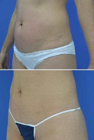
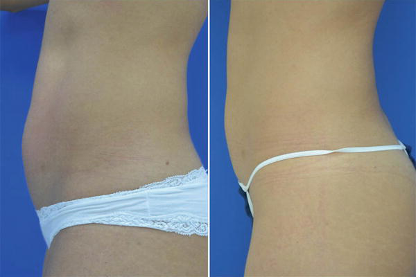
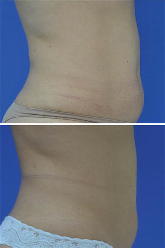
Some studies have reported satisfaction rates ranging from 47.5% to 85% (Fatemi and Kane 2010; Fatemi 2009; Hotta 2010). However, as other aesthetic procedures, up to 20% of patients may not have satisfactory results.
Measures and good photography are essential for patient satisfaction. Follow-up visits should be scheduled with the patient for measurements and photos. The same person should perform the patient’s measurements at each visit to decrease variability.
Take Home Messages
- With the aim of reaching best results the appropriate patient selection is essential.
- Adjusting realistic expectation is always a concern when approaching body-sculpting techniques.
- Results are usually seen between 8 and 12 weeks after the treatment.
- Individual results may vary. It is possible to achieve reductions of approximately 10% of the initial measurement.
Cross-References
- Cryolipolysis for Body Sculpting
References
Fatemi A. High-intensity focused ultrasound effectively reduces adipose tissue. Semin Cutan Med Surg. 2009;28(4):257–62.
Fatemi A, Kane MA. High-intensity focused ultrasound effectively reduces waist circumference by ablating adipose tissue from the abdomen and flanks: a retrospective case series. Aesthet Plast Surg. 2010;34(5):577–82.
Ferraro GA, De Francesco F, Nicoletti G, Rossano F, D’Andrea F. Histologic effects of external ultrasound-assisted lipectomy on adipose tissue. Aesthet Plast Surg. 2008;32(1):111–5.
Gadsden E, Smoller B, Rock L, Aguilar MT, Jewell M, Glogau R. The clinical safety and histologic changes associated with the use of a novel high-intensity focused ultrasound device for noninvasive body sculpting. J Am Acad Dermatol. 2010;62(3):AB142.
Gadsden E, Aguilar MT, Smoller BR, Jewell ML. Evaluation of a novel high-intensity focused ultrasound device for ablating subcutaneous adipose tissue for noninvasive body contouring: safety studies in human volunteers. Aesthet Surg J. 2011;31(4):401–10.
Haar GT, Coussios C. High intensity focused ultrasound: physical principles and devices. Int J Hyperth. 2007;23(2):89–104.
Hotta TA. Nonsurgical body contouring with focused ultrasound. Plast Surg Nurs. 2010;30(2):77–82; quiz 83–4.
Jewell ML, Solish NJ, Desilets CS. Noninvasive body sculpting technologies with an emphasis on high-intensity focused ultrasound. Aesthet Plast Surg. 2011a;35(5):901–12.
Jewell ML, Baxter RA, Cox SE, Donofrio LM, Dover JS, Glogau RG, et al. Randomized sham-controlled trial to evaluate the safety and effectiveness of a high-intensity focused ultrasound device for noninvasive body sculpting. Plast Reconstr Surg. 2011b;128(1):253–62.
Kennedy J, Verne S, Griffith R, Falto-Aizpurua L, Nouri K. Non-invasive subcutaneous fat reduction: a review. J Eur Acad Dermatol Venereol. 2015;29(9):1679–88.
Mordon S, Plot E. Laser lipolysis versus traditional liposuction for fat removal. Expert Rev Med Devices. 2009;6(6):677–88.
Murray EG, Rivas OEA, Stecco KA, Desilets CS, Kunz L. Evaluation of the acute and chronic systemic and metabolic effects from the use of high intensity focused ultrasound for adipose tissue removal and non-invasive body sculpting: P27. Plast Reconstr Surg. 2005a;116(3):151–2.
Murray EG, Rivas OEA, Stecco KA, Desilets CS, Kunz L. The use and mechanism of action of high intensity focused ultrasound for adipose tissue removal and non-invasive body sculpting: P80. Plast Reconstr Surg. 2005b;116(3):222–3.
Shek SY, Yeung CK, Chan JC, Chan HH. Efficacy of high-intensity focused ultrasonography for noninvasive body sculpting in Chinese patients. Lasers Surg Med. 2014;46(4):263–9.
Solish N, Lin X, Axford-Gatley RA, Strangman NM, Kane M. A randomized, single-blind, postmarketing study of multiple energy levels of high-intensity focused ultrasound for noninvasive body sculpting. Dermatol Surg. 2012;38(1):58–67.
Teitelbaum SA, Burns JL, Kubota J, Matsuda H, Otto MJ, Shirakabe Y, et al. Noninvasive body contouring by focused ultrasound: safety and efficacy of the Contour I device in a multicenter, controlled, clinical study. Plast Reconstr Surg. 2007;120(3):779–89.







