
Ultrasound-Assisted Drug Delivery in Fractional Cutaneous Applications
Ultrasound-Assisted Drug Delivery in Fractional Cutaneous Applications
Joseph Lepselter, Alex Britva, Ziv Karni and Maria Claudia Almeida Issa Laser and Ultrasound Laboratory, Alma Lasers Ltd., Caesarea, Israel Department of Clinical Medicine – Dermatology, Fluminense Federal University, Praia de Icarai 139, 702 Niterói, RJ, Brazil
Abstract
Cutaneous biodistribution and bioavailability of most topically applied drugs are quite low. For a topical agent to be active, it must first traverse the rate-limiting barrier of the stratum corneum. Many medications are too large (size >500 Da in molecular weight) to penetrate this barrier, which requires either an injectable or systemic delivery. Fractional ablative skin therapy became a common modality in procedural dermatology, with the ability to create ablative microscopic channels of varying depths in the stratum corneum and other epidermal layers in a predictable manner, thus creating new opportunities in drug delivery. Pairing fractional ablation skin perforation with a physical-assisted modality for the enhancement of drug delivery would seem an attractive option for various skin indications. That said, the extent of collateral damage created by the ablative fractional technology on the microchannels and the unavoidable exudates created by the natural wound-healing process would prove problematic for passive drug delivery. Recently, a novel ultrasound-assisted drug delivery device (IMPACT™) was introduced and clinically used, in pairing with fractional ablation technology, for pushing and enhancing the delivery of substances immediately after the skin perforation, for the purpose of increasing biodistribution and bioavailability through the microchannels for a variety of cutaneous indications, such as scars, striae distensae, actinic keratosis, or dysplastic skin lesions in photodynamic therapy. This ultrasound-assisted drug delivery device should be used by a qualified practitioner who must possess an understanding of cutaneous anatomy in relation to the local target (indication), appropriate training, and a comfort level with and familiarity with the properties and appropriate selection of the topical agent.
Keywords Transepidermal drug deliveryTransdermal drug deliveryTranscutaneous administrationTransdermal administrationLaserUltrasound
Introduction
The skin is one of the most readily accessible organs of the human body, covering a surface area of approximately 2 m2 and receiving about one third of the body’s blood circulation. The skin is a complex system consisting of the epidermis, the dermis, and skin appendages, such as hairs, interwoven within the two layers. The outermost layer of the skin, the epidermis, is avascular, receiving nutrients from the underlying dermal capillaries by diffusion through a basement membrane. The outermost layer of the epidermis is called the stratum corneum, the protective covering that serves as a barrier to prevent desiccation of the underlying tissues and to exclude the entry of noxious substances from the environment, including agents applied to the skin. This layer consists of corneocytes embedded in lipid regions (Michael et al. 2002).
Transdermal (or transcutaneous) drug delivery offers advantages over traditional drug delivery methods, such as injections and oral delivery. In particular, transdermal drug delivery avoids gastrointestinal drug metabolism, reduces side effects, and can provide sustained release of therapeutic compounds. The term “transdermal” is used generically because, in reality, transport of compounds by passive diffusion occurs only across the epidermis, where absorption into the blood via capillaries occurs (Elias and Menon 1991).
However, the diffusion rate of topically applied compounds will vary because of both internal (physiological) and external (environmental) factors. The diffusion rate is also dependent upon physical and chemical properties of the compounds being delivered. The patient’s skin needs to be carefully evaluated to minimize the natural, internal barriers to transdermal drug delivery (e.g., dry skin, thick skin, dehydration, poor circulation, poor metabolism) and to maximize the natural enhancers (e.g., ensuring the patient is well hydrated and selecting an area of skin that is thin, warm, moist, and well perfused). The stratum corneum is considered to be the rate-limiting barrier for transdermal delivery, and so diffusion is often enhanced. Various methods include preheating the skin to increase kinetic energy and dilate hair follicles and covering the area with an occlusive dressing after the drug application to maintain moisture and activate the reservoir capacity of the skin (Scheuplein and Blank 1971; Prausnitz et al. 2012; Kurihara-Bergstrom and Good 1987).
Enhancers of transcutaneous drugs are usually used to alter the nature of the stratum corneum to ease diffusion. This alteration may result from denaturing the structural keratin proteins in the stratum corneum, stripping or delaminating the cornified layers of the stratum corneum, changing cell permeability, or altering the lipid-enriched intercellular structure between corneocytes. Enhancers are incorporated into transdermal controlled drug delivery systems, or they are used prior to, during, or after the topical application of a drug. Preferred enhancers allow drugs to diffuse actively and quickly, but do not inactivate the drug molecules, damage healthy epidermis, cause pain, or have toxicological side effects (Haftek et al. 1998; Menon and Elias 1997; Akomeah 2010).
Even though ultrasound has been used extensively for medical diagnostics and physical therapy, it has only recently become popular as an enhancer of drug delivery. Numerous studies have demonstrated that ultrasound is generally safe, with no negative long-term or short-term side effects, but the mechanisms by which ultrasound works for the purpose of transepidermal drug delivery as an enhancer are less clearly understood (Nino et al. 2010; Prausnitz and Langer 2008; Smith 2007).
Ultrasound-Skin Interaction in Cutaneous Application
Ultrasound is defined as an acoustic wave at a frequency of between 20 kHz and 10 MHz. The properties defining the ultrasound are the amplitude and the frequency of the acoustic waves. Similar to audible sound, ultrasound waves undergo reflection and refraction when they encounter another medium with dissimilar properties. If the properties of the encountered medium are different from those of the transmitting medium, the acoustic energy of the transmitted ultrasound beam is attenuated by being reflected from this medium. Attenuation of ultrasound by its absorption and scattering in tissue also limits its depth of penetration (Anselmo and Mitragotri 2014; Byl 1995; Mitragotri 2004a).
Ultrasound may bring about various reactions when propagated in biological tissue. The resulting effects include thermal, mechanical, chemical, and optical reactions. Mechanical effects, more specifically, may consist of acoustic cavitation, radiation force, shear stress, and acoustic streaming/microstreaming (David and Gary 2010).
The use of ultrasound to enhance the transport of a substance through a liquid medium is referred to as sonophoresis or phonophoresis. It may be used alone or in combination with other enhancers, such as chemical enhancers, iontophoresis, electroporation, magnetic force fields, electromagnetic forces, mechanical pressure fields, or electrical fields (Mitragotri 2004b).
Both the thermal and non-thermal characteristics of high-frequency sound waves can enhance the diffusion of topically applied drugs. Heating from ultrasound increases the kinetic energy of the molecules (mobility) in the drug and in the cell membrane, dilates points of entry such as the hair follicles and the sweat glands, and increases the circulation to the treated area. These physiological changes enhance the opportunity for drug molecules to diffuse through the stratum corneum and be collected by the capillary network in the dermis. Both the thermal and non-thermal effects of ultrasound increase cell permeability. The mechanical characteristics of the sound wave also enhance drug diffusion by oscillating the cells at high speed, changing the resting potential of the cell membrane and potentially disrupting the cell membrane of some of the cells in the area (David and Gary 2010; Mitragotri 2004b). One of the theories of ultrasound phoresis postulates that the main factor is increasing the permeability of skin by creating lipid bridges between keratin layers in the stratum corneum (Mason 2011; Guy 2010; Pitt et al. 2004).
Another important factor that may affect drug diffusion is related to the shear forces (or shock waves) that occur when adjacent portions of the same membranous structures vibrate with different displacement amplitudes. The acoustic waves cause streaming and/or cavitation in the drug medium and the skin layers, which helps the drug molecules to diffuse into and through the skin. “Streaming” is essentially oscillation in a liquid that forces the liquid away from the source of the energy, while “cavitation” is the formation of bubbles in a liquid that is subjected to intense vibration. Cavitation is the result of rarefaction areas during propagation of longitudinal acoustic waves in the liquid when the waves have an amplitude above a certain threshold (Mitragotri 2005; Pua and Pei 2009).
When these bubbles occur in specific cells of the skin, fatigue or rupture of the cells can occur as the bubbles reach an unstable size. Destruction of cells in the transmission path of the ultrasound may facilitate intercellular diffusion of drug molecules. Cavitation may also disrupt the organization of lipids in the stratum corneum, resulting in an increase in the distance between the lipid layers. As a result, the amount of water phase in the stratum corneum increases, thereby enhancing the diffusion of water-soluble components through the intercellular space (Sivakumari et al. 2005). As the permeation pathway for topically applied products is mainly along the tortuous intercellular route, the lipids in the stratum corneum play a crucial role in proper skin barrier function.
Ultrasound (sonophoresis, phonophoresis, and ultraphonophoresis) is a technique for increasing the skin permeation of drugs using ultrasound (20 kHz–16 MHz) as a physical force. It is a combination of ultrasound therapy with topical drug therapy to achieve therapeutic drug concentrations at selected sites in the skin. In this technique, the drug is mixed with a coupling agent, usually a gel but sometimes a cream or ointment, which transfers ultrasonic energy from the device to the skin through this coupling agent (Lee and Zhou 2015). Application of low-frequency ultrasound (20–100 kHz) enhances skin permeability more effectively than high-frequency ultrasound (1–16 MHz). The mechanism of transdermal skin permeation involves disruption of the stratum corneum lipids, thus allowing the drug to pass through the skin. A corresponding reduction in skin resistance was observed due to cavitation, microstreaming, and heat generation (Park et al. 2014; Mormito et al. 2005; Mitragotri and Kost 2001).
With this in mind, burning and abrasion of the epidermis are a serious consideration when using ultrasound. Ultrasound should act as a mechanical acoustic pressure agent without damaging the epidermis of the skin. However, cavitation in the liquid coupling the sonotrode to the skin may produce cavitation erosion of the epidermis and the dermis. While this cavitation assists with active penetration of the substance being delivered, it may also damage the epidermis. A further challenge when using ultrasound is that it is low in efficiency, due to cavitation in the buffer suspension and the low acoustic pressure and ultrasonic energy that is required to prevent burning and other injury (Tang et al. 2002; Terahara et al. 2002; Mitragotri and Kost 2000; Mitragotri et al. 1995).
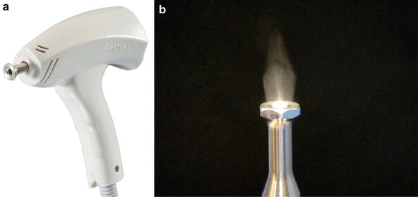
There is an ongoing need for improved devices and methods to enhance transdermal medical and cosmetic compound delivery. In particular, there is an ongoing need for ultrasound-based devices and techniques for transdermal drug or cosmetic delivery. A low-frequency (kHz) ultrasound-assisted drug delivery technology was developed for the purpose of transepidermal delivery of topical ingredients, i.e., drugs or cosmeceuticals, used in pairing with fractional laser- or radiofrequency-assisted technology (Fig. 1a, b).
Laser-Assisted Skin Permeation in Transepidermal Drug Delivery
Fractional ablation of the skin is a special laser–skin interaction event of photothermolysis first described in 2004. Fractional ablative lasers (erbium:YAG and CO2) or other fractional ablative modalities, such as radiofrequency, ablate the skin in fractions, splitting the laser beam into microbeams. These microbeams create microscopic vertical channels of ablation in the skin. The creation of these channels may provide access pathways for topically applied drug molecules that would otherwise be too large to traverse the epidermal layer. The location, diameter, depth, and other characteristics of these channels can be controlled or manipulated by the settings of the laser or radiofrequency technology (Manstein et al. 2004; Alexiades-Armenakas et al. 2008; Haak et al. 2011; Lin et al. 2014; Carniol et al. 2015; Forster et al. 2010).
Investigation into the in vitro/in vivo effectiveness of fractional laser devices for facilitating drug permeation in animals is abundant. However, few studies have examined clinical laser-assisted drug delivery in humans. Oni et al. (2012) demonstrated in vivo lidocaine absorption using the fractional Er:YAG laser, with the pig as the animal model. The drug level in the serum was detected after topical application of 4% lidocaine. The serum concentration was undetectable in the nontreated group. The serum level was detectable following laser treatment. Peak levels of lidocaine were significantly greater at 250-μm pore depth (0.62 mg/l), compared to 500 μm (0.45 mg/l), 50 μm (0.48 mg/l), and 25 μm (0.3 mg/l). The greater depth of the microchannels did not guarantee greater enhancement.
Haedersdal et al. (2010) evaluated skin delivery assisted by the fractional CO2 laser by using methyl aminolevulinate (MAL) as the model permeant. MAL is an ester prodrug of aminolevulinic acid (ALA). Yorkshire swine were treated with the fractional CO2 laser and were subsequently treated with MAL. Fluorescence derived from protoporphyrin IX (PpIX) was measured by fluorescence microscopy at a skin depth of 1,800 μm. The fractional laser created cone-shaped channels of 300 μm in diameter and 1,850 μm in depth. This ablation enhanced drug delivery, with higher fluorescence in the hair follicles and dermis as compared to intact skin. Fractional laser irradiation facilitates topical delivery of the porphyrin precursor deep into the skin. Haedersdal et al. (2011) also investigated MAL permeation enhanced by the fractional CO2 laser, quantifying PpIX skin distribution and photodynamic therapy (PDT)-induced photobleaching from the skin surface to the depth of 1,800 μm in Yorkshire swine. The red light for creating photobleaching was delivered by light-emitting diode arrays at fluences of 37 and 200 J/cm2. The fraction of porphyrin fluorescence lost by photobleaching was less after 37 J/cm2 than after 200 J/cm2. The fractional laser greatly facilitated skin PpIX, and the fraction of photobleached porphyrins was similar for the superficial and deep skin layers.
Distribution of PpIX into laser-treated skin may depend on the microchannel depth and the drug incubation period. Haak et al. (2012a) further evaluated whether the depth of the fractional laser and the incubation time affect methyl ALA permeation. Yorkshire swine were treated with the fractional CO2 laser at 37, 190, and 380 mJ to create microchannel depths of 300 (superficial), 1,400 (mid), and 2,100 μm (deep dermis/subcutaneous), respectively. The incubation times for methyl ALA cream were 30, 60, 120, and 180 min. Similar fluorescence of PpIX was induced throughout the skin layers independent of the laser channel depth by a 180-min incubation. Laser irradiation and the following methyl ALA incubation for 60 min increased fluorescence at the skin surface compared to intact skin. Laser exposure and the subsequent methyl ALA incubation for 120 min increased fluorescence in the follicles and the dermis compared to intact skin.
Treatment of scars, including hypertrophic scars and keloids, intrinsically poses a delivery challenge given their variable and fibrotic nature, especially when considering penetration into the mid-to-deep dermis. Corticosteroids, 5-fluorouracil (5-FU), imiquimod, methotrexate, and other immunomodulators have been used as adjuvant scar therapies for years. However, when used topically, these agents demonstrate only mild efficacy and may carry risks such as corticosteroid-related epidermal atrophy. Intralesional use may prove more efficacious but may also be associated with significant pain, atrophy, pigmentary changes, and a high recurrence rate (Haak et al. 2012a; Brauer et al. 2014).
Furthermore, the extent of collateral damage created by fractional ablative lasers or non-laser fractional technology and the post-trauma exudate (interstitial fluid or fibrin) within the microchannels would prove problematic for drug delivery. This is because the hydrostatic forces pushing the exudative fluid out of the tissue into the channels would directly compete with the diffusion of the substance applied topically. This, therefore, reduces any potential for passive transdermal diffusion and emphasizes the need for pressure-assisted technology that will immediately push the substance into the viable microchannel space (Haedersdal et al. 2011; Haak et al. 2012a, b; Erlendsson et al. 2014a).
Ultrasound-Assisted Drug Delivery in Fractional Cutaneous Applications
In the past two decades, numerous studies have described the usefulness of low-frequency ultrasound to improve skin permeability. Only recently has application of fractional ablative methods using lasers and radiofrequency been described for potential therapeutic purposes in investigational and procedural dermatology, aiming at creating microchannels in the epidermis to increase permeability of topically applied drugs in indications such as hypertrophic and atrophic scars and other skin imperfections (Bloom et al. 2013; Sklar et al. 2014; Gauglitz 2013; Arno et al. 2014; Waibel and Rudnick 2015; Rkein et al. 2014; Waibel et al. 2013; Togsverd-Bo et al. 2012, 2015; Taudorf et al. 2014; Erlendsson et al. 2014b). Table 1 depicts assisted-drug delivery compounds currently tested and used with fractional ablative applications.
| Compounds delivered |
|---|
| ALA and MAL (PDT) |
| Topical anesthetics |
| NSAIDs |
| Opioids |
| Chemotherapeutic drugs |
| Corticosteroids |
| Vaccinations |
| Topical ascorbic acid (vitamin C) |
| Imiquimod |
| Allogenic mesenchymal stem cells |
| Botulinum toxin |
| Antioxidants |
| Beta-blockers |
| Antifungals |
| Bone marrow transplantation |
| 5-Fluorouracil (5-FU) |
| Interferons |
| Filler agents |
A novel ultrasound-assisted drug delivery device (IMPACT™) is displayed in Fig. 1. The ultrasound applicator is an acoustic-wave proprietary technology (Britva et al.) that operates at low frequency (kHz). The ultrasound applicator has a sonotrode that emits acoustic waves and creates mechanical air pressure which helps to advance topical cosmetic products/substances into the top layers of the skin, creating cycles of negative/positive pressure – a “push and pull” effect (Fig. 2a, b) – within the preliminary organized channels to release the buildup of intracellular fluid and help the cosmetic product more rapidly advance into the skin to the targeted tissue depth.
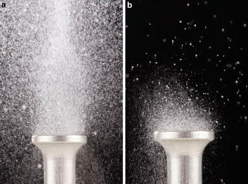
The sonotrode serves various functions, such as conversion of the acoustic waves by increasing the amplitude of the oscillations, modifying the distribution, and matching acoustic impedance to that of the substrate. Resonance of the sonotrode, which increases the amplitude of the acoustic wave, occurs at a frequency determined by the characteristics of modulus elasticity and density of the material from which the sonotrode is made, the speed of sound through the material, and the ultrasonic frequency. The size and shape (round, square, profiled) of a sonotrode will depend on the quantity of vibratory energy and the physical requirements for each specific application. Sonotrodes connected to ultrasound transducers, especially those employed for transdermal delivery of drugs, typically have a length L, expressed by the equation L = n(λ/2), where λ is the wavelength of the ultrasound in the sonotrode and n is a positive integer. In this case, the maximum amplitudes of the acoustic wave are found at the proximal end of the sonotrode and at the λ/2 length beyond the foot of the sonotrode. The vibrating column of air within the bore in the distal end of the sonotrode, together with the vibration of the annular end surface of the foot portion, has the effect of cyclically reducing and increasing the pressure at the skin interface, and this sucking and blowing action facilitates the transport of the compound to be delivered through the microchannel perforations created by ablative fractional laser or radiofrequency across the stratum corneum (Britva et al.).
The mode of operation is based on mechanical (acoustic) pressure and torques by propagation of the US wave via the sonotrode to the distal horn and the creation of a hammering-like effect (“push and pull”) in the thin layer between the cosmetic products and the distal surface of the sonotrode. The sonotrode is applied normally to the surface of the skin, contacting continuously with the skin surface. The vibrational cycles (push–pull) enhance its delivery via the skin microchannels. Thus, this ultrasound-assisted technology emits acoustic waves producing ultrasonic pressure and creating a “push and pull” effect within the channels to release the buildup of intracellular fluid and enhances the cosmetic ingredients or the drugs to the target tissue depth.
The ultrasonic acoustic waves emitted by the flared foot (Figs. 1 and 2) of the sonotrode are believed to assist in spreading through the dermis the compound that has been transported through the perforated stratum corneum, and the flaring of the foot serves to spread the ultrasonic waves over a greater area to assist in diffusion of the compound.
The shape of the sonotrode is believed to achieve two effects. First, the hollow neck creates a vibrating air column that blows and sucks alternately, serving to transfer the medicament through the stratum corneum. Second, the setting of the length of the sonotrode to λ/6 allows the acoustic energy to be focused to a point that is between approximately 0.3 and 2 mm beneath the surface of the skin. This positioning of the maximum acoustic wave amplitude at a point deeper in the tissue magnifies the sonophoresis effect of cavitation, lipid destruction, etc. and increases absorption of the drug or cosmetic.
The pairing of fractional ablative technology and ultrasound-assisted technology in procedural dermatology has gained attention for the purpose of skin permeation to facilitate transepidermal drug delivery. In the past 5 years, clinical efficacy and safety of the IMPACT technology has been tested and validated in various preclinical and clinical ex vivo and in vivo models.
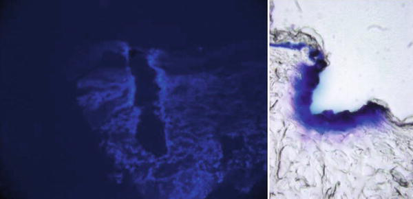
Lepselter et al. (2013) analyzed transepidermal enhancement following fractional ablative skin permeation in a rat model. Rats (Sprague Dawley, either sex) were anesthetized with a mixture of ketamine (60 mg/kg) and xylazine (10 mg/kg) injected i.p. or i.m. Rats were shaved and prepped on their dorsal and lateral sides. Skin perforation (SP) followed by the low-frequency ultrasound-assisted device (IMPACT) was studied in five different conditions: normal skin (control), topical Evans blue (EB), topical EB + US, topical SP + EB, and topical SP + EB + US. Frozen sections from skin biopsies were taken at 0 and 15 min of incubation time for histological examination using a high-resolution digital microscope. Penetration area (penetration depth + width) and coloration between distances of penetration were analyzed by advanced imaging software. Reflectance intensity by spectroscopy was measured under two wavelength conditions: 665 nm (EB sensitive) and 772 nm (EB insensitive). The 665/772-nm ratio was used as a penetration indicator – a low ratio denoted higher EB penetration and a high ratio low EB penetration. Biopsies were taken and frozen for histological examination using a digital microscope (Olympus, Japan), with data analysis using the image-processing software ImageJ (Burger and Burg, Hagenberg, Austria). Penetration area was defined as the colored area divided by the crater area and expressed in %. Penetration width was defined as the width line divided by crater width, expressed in %. Penetration depth was defined as the depth line divided by crater depth, expressed in %. To visualize and analyze the ultrasound-assisted device effect, a polarization imaging system was used. A CCD camera (Olympus, Japan) was located above the prepared skin sample slide for image acquisition.
EB color intensity versus distance (depth and width) was significantly higher for the SP + EB + US (99.66 ± 23.67 pixels) versus SP + EB (52.33 ± 25.34 pixels), and for the SP + EB + US (80.83 ± 15.41 pixels) versus SP + EB (66.83 ± 28.56 pixels), respectively (p < 0.05–0.01). Similarly, the spectrometry reflectance intensity ratios for topical EB (2.1 ± 0.4) and topical EB + US (1.8 ± 0.3) were high, indicating low EB penetration. In contrast, SP + EB + US (0.4 ± 0.02) versus SP + EB (1.4 ± 0.08) ratios were low, indicating significantly higher EB penetration for the former (p < 0.01). Histology frozen sections from the high-resolution digital photographs were in agreement with the objective measurements. At 0-min incubation time, EB penetration was significantly higher at 70 versus 40 W (p < 0.01). At 15 min, EB penetration was significantly higher when compared to 0 min for 70 versus 40 W, respectively. It was concluded that the ultrasound-assisted device following ablative RF permeation significantly enhances the amount of EB penetration, as evidenced by depth, width, and color intensity (Fig. 3a–d).
The assisted-ultrasound drug delivery device’s first clinical experience was reported in 2011 by Kassuga and colleagues (2011). This case study described two female patients with multiple actinic keratoses (AK). The study evaluated the effectiveness of the transepidermal (TED) application of methyl aminolevulinate (MAL) prodrug in PDT combined with the ultrasound-assisted drug delivery device. Clinical efficacy evaluation was based on reducing the AK number and improving skin texture and color. The two patients were simultaneously treated with the standard MAL-PDT (left forearm) and a modified protocol – fractional radiofrequency (RF) combined with MAL and the ultrasound device on the opposite forearm (MAL-GT). Improvements in the texture and pigmentation of the skin were observed after a single treatment on both sides and were more evident on the side treated with the MAL-GT modality (at the 6-month follow-up visit). Patient 1 had 34 lesions on the right forearm (MAL-GT protocol) and 54 lesions on the contralateral forearm (standard MAL-PDT protocol). After 6 months of protocol implementation, there were 8 AK lesions on the MAL-GT treated side and 34 on the MAL-PDT treated side. These results demonstrated a 76.4% and a 37% decrease in the right and left treated forearms, respectively. Patient 2, initially with 24 and 21 lesions on the right and left forearms, respectively, presented after PDT with two and six lesions, respectively, which represents a 91.6% and a 71.4% decrease, respectively. Overall, it was concluded that for these cases, PDT associated with TED of the MAL with an incubation time of 1 h not only is effective in AK treatment but also shows better results than the standard protocol.
Issa et al. (2013a) evaluated efficacy, safety, and patient satisfaction in using ablative fractional RF technology (Pixel RF) associated with retinoic acid 0.05% cream and an acoustic pressure wave ultrasound (US) in patients with alba-type striae distensae (SD) on the breast. Eight patients were treated with a three-step procedure: fractional ablative RF for skin perforation, topical application of retinoic acid 0.05% on the perforated skin, and US applied to enhance penetration. An additional eight patients with abdominal alba-type SD were submitted to RF treatment alone, without retinoic acid or US. Three patients with SD on the breast improved from “severe” to “moderate,” two patients from “severe” to “mild,” two patients from “moderate” to “mild,” and one patient from “marked” to “mild.” Clinical assessment demonstrated significant improvement in the appearance of SD in all these patients (P = 0.008), with a low incidence of side effects and a high level of patient satisfaction. Among the patients treated only with RF, two patients improved from “severe” to “marked,” one patient from “marked” to “moderate,” and one patient from “marked” to “mild.” Four patients did not show any sort of improvement. Clinical assessment demonstrated no significant improvement in the appearance of SD treated with RF alone, with a low incidence of side effects but a low level of patient satisfaction. It was concluded that ablative fractional RF and acoustic pressure US associated with retinoic acid 0.05% cream is safe and effective for alba-type SD treatment.
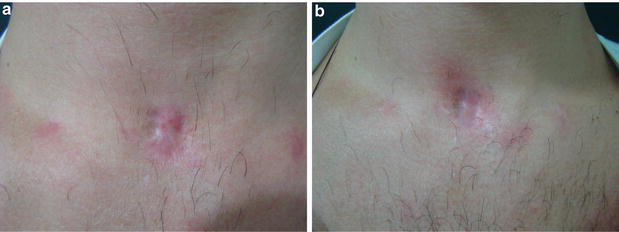
The aim of the study conducted by Issa et al. (2013b) was to evaluate clinical response and side effects of transepidermal drug delivery (TED) technology in hypertrophic scars on the body and face using ablative fractional radiofrequency (RF) associated with low-frequency acoustic pressure ultrasound (US). Four patients with hypertrophic scars were treated with triamcinolone using fractional ablative RF and US. The treatment procedure comprised three steps: (i) ablative fractional RF for skin perforation, (ii) topical application of triamcinolone acetonide 20 mg/ml on the perforated skin, and (iii) acoustic pressure wave US applied to enhance penetration. The study resulted in complete resolution after one session in patients with scars on the nose and mandibular area. The scar on the neck (tracheostomy) showed complete resolution after four sessions (Fig. 4a, b). The scar on the knee showed marked improvement after four sessions. Mild and homogeneous atrophy was observed in the hypertrophic scar on the neck.
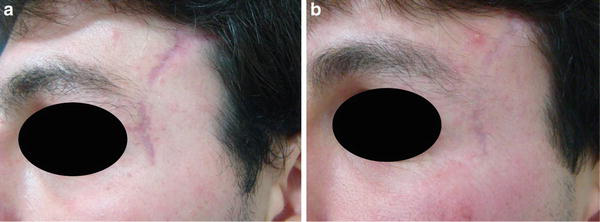
The method used in this study was shown to improve the efficacy of steroids in hypertrophic scar treatment, minimizing risks of localized atrophy and irregular appearance of the treated lesion. The same method, with the three-step procedure (Fig. 5), can be used to improve the quality of non-hypertrophic scars, in which retinoic acid and vitamin C, instead of triamcinolone, are better indicated (Fig. 6a, b).
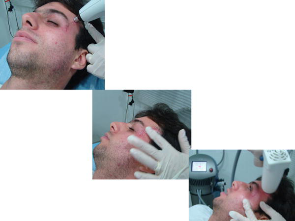
In a recent study, Trelles et al. (Trelles and Martínez-Carpio 2014) aimed to evaluate the efficacy and safety of the transepidermal delivery (TED) method for treating acne scarring. A total of 19 patients with moderate to severe scarring were treated using unipolar fractional ablative RF technology (Pixel RF) to create dermal microchannels, followed by acoustic pressure ultrasound. All patients underwent four treatment sessions at 3-week intervals. The study demonstrated significant improvement in scarring on the face, back, and shoulders (P < 0.0001). In a 2-month follow-up, fading of total scarring was 57% on the face and 49% on the back and shoulders. After 6 months, the percentage increased to 62% and 58% on the face and on the back/shoulders, respectively. Patients reported being somewhat satisfied (16%), satisfied (53%), and very satisfied (31%). No unexpected side effects to the ablation and no hypersensitivity were observed. It was concluded that the bimodal procedure is safe and effective in reducing acne scarring.
Earlier, Trelles et al. (2013) aimed to determine the efficacy and safety of facial rejuvenation using the fractional carbon dioxide (CO2) laser (iPixel CO2), an ultrasound emitter (IMPACT), and a cosmeceutical preparation to be applied intraoperatively. Fourteen patients were enrolled in this split-face, double-blind, randomized prospective study; for each patient, one half of the face was treated with a fractional CO2 laser alone while the other half received the same laser followed by acoustic pressure ultrasound of cosmeceuticals. Both treatments achieved significant improvements in all parameters evaluated, yet the combined US and cosmeceutical treatment had better scores for reduced fine lines and wrinkles as well as an 80% overall improvement of facial aging at the 6-month follow-up. Treatment was well tolerated and no adverse effects were observed. Eighty-six percent of patients stated that they were satisfied or very satisfied with their results. The conclusion was that one session of fractional ablative CO2 laser and acoustic pressure ultrasound technology for TED of cosmeceuticals is an effective method for facial rejuvenation.
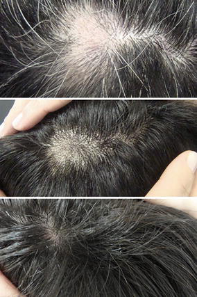
Recently, Issa and her colleagues (2015) studied the clinical efficacy and side effects of TED in areata alopecia (AA) using ablative fractional methods and acoustic pressure ultrasound (US) to deliver triamcinolone solution into the skin. Treatment comprised three steps: ablative fractioned RF or CO2 laser followed by topical application of triamcinolone and inserting it using acoustic pressure wave US. The number of sessions varied according to clinical response (range 1–6). Treatment resulted in complete recovery of the area treated in all patients; four of them maintained the result at the 12-month follow-up. Two of the patients were treated with ablative fractional RF + triamcinolone + US and had a complete response after three and six sessions. The other two, treated with ablative fractional CO2 + triamcinolone + US, had a complete response after one session (Fig. 7). Researchers concluded that fractioned ablative resurfacing associated with acoustic pressure wave US is a new option for AA treatment, with good clinical results and a low incidence of side effects.
In a recent study Waibel et al. (2015) evaluated in vivo whether there is increased efficacy of fractional ablative laser with immediate transdermal acoustic waves (IMPACT) to enhance drug delivery, via histologic immunofluorescent evaluation. Aminolevulinic acid (ALA) was chosen to evaluate whether the combination of the fractional ablative laser with immediate transdermal ultrasound enhances drug delivery. Six patients were treated with four treatment sites – one area treated with topically applied ALA, one area with fractional ablative laser (iPixel CO2) and ALA topically applied, one area with fractional ablative laser and the transdermal delivery system, and a final area of ALA topically applied with the transdermal delivery system. Comparison of the magnitude of diffusion, both lateral spread of ALA and depth diffusion of ALA, was measured by fluorescence microscopy. With laser + ALA + the acoustic device, the protoporphyrin IX lateral fluorescence was 0.024 mm on average versus 0.0084 mm with fractional laser and ALA. Diffusion with the acoustic air device was an order of magnitude greater. The authors concluded that the combined approach of the fractional CO2 laser and the IMPACT device demonstrated the best results in increasing the depth of penetration of the ALA (Fig. 3).
Suh et al. (2012) conducted a study to evaluate the effectiveness and safety of enhanced penetration of platelet-rich plasma (PRP) with ultrasound after plasma fractional radiofrequency for the treatment of striae distensae. Participants were treated with the ultrasound-assisted drug delivery device in pairing with fractional radiofrequency technology (Pixel RF) for three sessions at 3-week intervals; ultrasound was applied to enhance platelet-rich plasma penetration. Two months after the last treatment, the average width of the widest striae had decreased from 0.75 to 0.27 mm; from a total of 18 participants, 13 were evaluated by two blinded reviewers as showing “excellent” or “very good” overall improvement. In total, 72.2% of the participants reported being “very satisfied” or “extremely satisfied” with overall improvement. The only side effect reported was postinflammatory hyperpigmentation (11.1%). The researchers concluded that fractional radiofrequency and transepidermal delivery of PRP using ultrasound are effective and safe in the treatment of striae distensae.
In a recent prospective, controlled study, Trelles et al. (2015) used a florescent technique (fluorescein) to qualitatively and quantitatively determine the transepidermal penetration of a cosmeceutical after permeabilizing the skin using the ultrasound-assisted drug delivery device and fractional ablative radiofrequency technology (Pixel RF). The treatments were performed in the retroauricular area in 16 patients, and biopsies were taken at 10 min and at 15 h after the procedure. The intensity of dermal fluorescence in the treated samples was compared to that of autofluorescence controls (AC) and technical controls (TC). The results demonstrated that the samples treated with Pixel RF + US displayed a greater intensity of fluorescence than the AC and TC, both at 10 min and 15 h after the treatment. The increases in fluorescence were graded as moderate or intense, but in no case as nil or slight. The results at 10 min were Pixel RF + US (55.4 ± 10.1), AC (8.6 ± 2.8), and TC (8.2 ± 3.6). At 15 h, the results were Pixel RF + US (54.2 ± 7.2), AC (8.9 ± 1.7), and TC (8.3 ± 2.4). The differences between the samples and the controls were significant, both at 10 min and at 15 h (p < 0.0008). The authors concluded that the transepidermal delivery procedure carried out facilitated a prolonged and effective dermal penetration of the topically applied products.
Summary
This reviewed, novel ultrasound-assisted drug delivery device creates mechanical air pressure, which helps to advance topical cosmetic products/substances into the top layers of the skin and, more importantly, creates cycles of negative/positive pressure (“push and pull” effect) within the perforated microchannels that may release the buildup of intracellular fluid and help the cosmetic product, thus enhancing the entry of the substance into the skin to the targeted tissue depth.
The idea of ablative fractionation treatment prior to application of topical agents in an attempt to enhance drug delivery is one that continues to garner increasing attention and interest in procedural dermatology (Brauer et al. 2014; Loesch et al. 2014).
With a view to mitigating the drawbacks and limitations of topically applied compounds, this novel ultrasound-assisted drug delivery device provides new possibilities for assisted drug delivery. The use of this ultrasound-assisted drug delivery device in pairing with fractional energy delivery technology has been tested and validated clinically in the past 5 years and is a promising method that can pave the way for future opportunities in transepidermal delivery in challenging cutaneous indications, such as photodynamic therapy or skin cancer, or aesthetic indications such as scars, acne, or alopecia, in procedural dermatology.
Take Home Messages
- Many medications are too large (size >500 Da in molecular weight) to penetrate the barrier of the stratum corneum and require either an injectable or systemic delivery.
- Fractional ablative skin therapy became a common modality in procedural dermatology, with the ability to create ablative microscopic channels of varying depths in the stratum corneum and other epidermal layers in a predictable manner, thus creating new opportunities in drug delivery.
- The novel ultrasound-assisted drug delivery device creates mechanical air pressure, creating cycles of negative/positive pressure within the perforated microchannels pre-formed by ablative methods.
- This push-and-pull effect may release the buildup of intracellular fluid inside the microchannels, increasing cosmetic/drug penetration into the skin.
- The use of this ultrasound-assisted drug delivery device in pairing with fractional energy delivery technology has been reported as an effective method to improve drug permeation into the skin in many different dermatoses (Bloom et al. 2013).
References
Akomeah FK. Topical dermatological drug delivery: Quo Vadis? Curr Drug Deliv. 2010;7:283–96.
Alexiades-Armenakas MR, Dover JS, Arndt KA. The spectrum of laser skin resurfacing: nonablative, fractional, and ablative laser resurfacing. J Am Acad Dermatol. 2008;58(5):719–37.
Anselmo AC, Mitragotri S. An overview of clinical and commercial impact of drug delivery systems. J Control Release. 2014;190:15–28.
Arno AI, Gauglitz GG, Barret JP, Jeschke MG. Up-to-date approach to manage keloids and hypertrophic scars: a useful guide. Burns. 2014;40(7):1255–66.
Bloom BS, Brauer JA, Geronemus RG. Ablative fractional resurfacing in topical drug delivery: an update and outlook. Dermatol Surg. 2013;39(6):839–48.
Brauer JA, Krakowski AC, Bloom BS, Nguyen TA, Geronemus RG. Convergence of anatomy, technology, and therapeutics: a review of laser-assisted drug delivery. Semin Cutan Med Surg. 2014;33:176–81.
Britva A, et al. European patent EP2459268B1 “A sonotrode”.
Byl NN. The use of ultrasound as an enhancer for transcutaneous drug delivery: phonophoresis. Phys Ther. 1995;75:539–53.
Carniol PJ, Hamilton MM, Carniol ET. Current status of fractional laser resurfacing. JAMA Facial Plast Surg. 2015;17:360–6.
David SJ, Gary PM. Themed issue: recent advances in transdermal drug delivery. J Pharm Pharmacol. 2010;62:669–70.
Elias PM, Menon GK. Structural and lipid biochemical correlates of the epidermal permeability barrier. Adv Lipid Res. 1991;24:1–26.
Erlendsson AM, Anderson RR, Manstein D, Waibel JS. Developing technology: ablative fractional lasers enhance topical drug delivery. Dermatol Surg. 2014a;40 Suppl 1:S142–6.
Erlendsson AM, Philipsen PA, Anderson RR, Paasch U, Haedersdal M. Fractional ablative erbium YAG laser: histological characterization of relationships between laser settings and micropore dimensions. Lasers Surg Med. 2014b;46(4):281–9.
Forster B, Klein A, Szeimies RM, Maisch T. Penetration enhancement of two topical 5-aminolaevulinic acid formulations for photodynamic therapy by erbium:YAG laser ablation of the stratum corneum: continuous versus fractional ablation. Exp Dermatol. 2010;19(9):806–12.
Gauglitz GG. Management of keloids and hypertrophic scars: current and emerging options. Clin Cosmet Investig Dermatol. 2013;6:103–14.
Guy RH. Transdermal drug delivery. Handb Exp Pharmacol. 2010;197:399–410.
Haak CS, Illes M, Paasch U, Haedersdal M. Histological evaluation of vertical laser channels from ablative fractional resurfacing: an ex vivo pig skin model. Lasers Med Sci. 2011;26(4):465–71.
Haak CS, Bhayana B, Farinelli WA, Anderson RR, Haedersdal M. The impact of treatment density and molecular weight for fractional laser-assisted drug delivery. J Control Release. 2012a;163:335–41.
Haak CS, Farinelli WA, Tam J, et al. Fractional laser-assisted delivery of methyl aminolevulinate: impact of laser channel depth and incubation time. Lasers Surg Med. 2012b;44:787–95.
Haedersdal M, Sakamoto FH, Fairinelli WA, et al. Fractional CO2 laser-assisted drug delivery. Lasers Surg Med. 2010;42:113–22.
Haedersdal M, Katsnelson J, Sakamoto FH, et al. Enhanced uptake and photoactivation of topical methyl aminolevulinate after fractional CO2 pretreatment. Lasers Surg Med. 2011;43:804–13.
Haftek M, Teillon MH, Schmitt D. Stratum corneum, corneodesmosomes and ex vivo percutaneous penetration. Microsc Res Tech. 1998;43(3):242–9.
Issa MC, de Britto Pereira Kassuga LE, Chevrand NS, do Nascimento Barbosa L, Luiz RR, Pantaleão L, Vilar EG, Rochael MC. Transepidermal retinoic acid delivery using ablative fractional radiofrequency associated with acoustic pressure ultrasound for stretch marks treatment. Lasers Surg Med. 2013a;45(2):81–8.
Issa MC, Kassuga LE, Chevrand NS, Pires MT. Topical delivery of triamcinolone via skin pretreated with ablative radiofrequency: a new method in hypertrophic scar treatment. Int J Dermatol. 2013b;52(3):367–70.
Issa MC, Pires M, Silveira P, Xavier de Brito E, Sasajima C. Transepidermal drug delivery: a new treatment option for areata alopecia? J Cosmet Laser Ther. 2015;17(1):37–40.
Kassuga PEL, Issa MC, Chevrand NS. Transepidermal medication application associated with photodynamic therapy in actinic keratosis treatment. Surg Cosmet Dermatol. 2011;3(4):89–92.
Kurihara-Bergstrom T, Good WR. Skin development and permeability. J Control Release. 1987;6:51–8.
Lee KL, Zhou Y. Quantitative evaluation of sonophoresis efficiency and its dependence on sonication parameters and particle size. J Ultrasound Med. 2015;34(3):519–26.
Lepselter J, Ben-Yosef T, Kostenich G, Orenstein A. Enhanced trans-epidermal delivery using fractional radiofrequency ablation and ultrasound pressure: in-vivo rat model pilot study. ASLMS Annual Conference, Boston; 2013.
Lin CH, Aljuffali IA, Fang JY. Lasers as an approach for promoting drug delivery via skin. Expert Opin Drug Deliv. 2014;11(4):599–614.
Loesch MM, Somani AK, Kingsley MM, Travers JB, Spandau DF. Skin resurfacing procedures: new and emerging options. Clin Cosmet Investig Dermatol. 2014;7:231–41.
Manstein D, Herron GS, Sink RK, Tanner H, Anderson RR. Fractional photothermolysis: a new concept for cutaneous remodeling using microscopic patterns of thermal injury. Lasers Surg Med. 2004;34:426–38.
Mason TJ. Therapeutic ultrasound: an overview. Ultrason Sonochem. 2011;18:847–52.
Menon GK, Elias PM. Morphologic basis for a pore-pathway in mammalian stratum corneum. Skin Pharmacol. 1997;10(5–6):235–46.
Michael R, Mark P, Sheree Elizabeth C. Skin transport. In: Walters K, editor. Dermatological and transdermal formulations. New York: Informa Healthcare; 2002. p. 89–195.
Mitragotri S. Breaking the skin barrier. Adv Drug Deliv Rev. 2004a;56:555–6.
Mitragotri S. Breaking the skin barrier. Adv Drug Deliv Rev. 2004b;56:555–64.
Mitragotri S. Healing sound: the use of ultrasound in drug delivery and other therapeutic applications. Nat Rev Drug Discov. 2005;4:255–60.
Mitragotri S, Kost J. Low-frequency sonophoresis: a noninvasive method of drug delivery and diagnostics. Biotechnol Prog. 2000;16:488–92.
Mitragotri S, Kost J. Transdermal delivery of heparin and low-molecular-weight heparin using low-frequency ultrasound. Pharm Res. 2001;18:1151–6.
Mitragotri S, Edwards D, Blankschtein D, Langer R. A mechanistic study of ultrasonically enhanced transdermal drug delivery. J Pharm Sci. 1995;84:697–706.
Mormito Y, Mutoh M, Ueda H, Fang L, Hirayama K, Atobe M, Kobayashi D. Elucidation of the transport pathway in hairless rat skin enhanced by low-frequency sonophoresis based on the solute–water transport relationship and confocal microscopy. J Control Release. 2005;103:587–97.
Nino M, Calabro G, Santoianni P. Topical delivery of active principles: the field of dermatological research. Dermatol Online J. 2010;16(1):4.
Oni G, Brown SA, Kenkel JM. Can fractional lasers enhance transdermal absorption of topical lidocaine in an in vivo animal model? Lasers Surg Med. 2012;44:168–74.
Park D, Park H, Seo J, Lee S. Sonophoresis in transdermal drug delivery. Ultrasonics. 2014;54(1):56–65.
Pitt WG, Husseini GA, Staples BJ. Ultrasonic drug delivery – a general review. Expert Opin Drug Deliv. 2004;1:37–56.
Prausnitz MR, Langer R. Transdermal drug delivery. Nat Biotechnol. 2008;26:1261–8.
Prausnitz MR, Elias PM, Franz TJ, et al. Skin barrier and transdermal drug delivery. In: Bolognia JL, Jorizzo JL, Schaffer JV, editors. Dermatology. 3rd ed. St. Louis: Elsevier Health Sciences; 2012.
Pua EC, Pei Z. Ultrasound-mediated drug delivery. IEEE Eng Med Biol Mag. 2009;28:64–75.
Rkein A, Ozog D, Waibel JS. Treatment of atrophic scars with fractionated CO2 laser facilitating delivery of topically applied poly-L-lactic acid. Dermatol Surg. 2014;40(6):624–31.
Scheuplein RJ, Blank IH. Permeability of the skin. Physiol Rev. 1971;51(4):702–47.
Sivakumari M, Tachibana K, Pandit AB, Yasui K, Tuziuti T, Towatai A, Iidaa Y. Transdermal drug delivery using ultrasound: theory, understanding and critical analysis. Cell Mol Biol. 2005;51:OL767–84.
Sklar LR, Burnett CT, Waibel JS, Moy RL, Ozog DM. Laser assisted drug delivery: a review of an evolving technology. Lasers Surg Med. 2014;262:249–62.
Smith NB. Perspectives on transdermal ultrasound mediated drug delivery. Int J Nanomedicine. 2007;2(4):585–94.
Suh DH, Lee SJ, Lee JH, Kim HJ, Shin MK, et al. Treatment of striae distensae combined with enhanced penetration of platelet-rich plasma and ultrasound after plasma fractional radiofrequency. J Cosmet Laser Ther. 2012;14(6):272–6.
Tang H, Wang CCJ, Blankschtein D, Langer R. An investigation of the role of cavitation in low-frequency ultrasound-mediated transdermal drug transport. Pharm Res. 2002;19:1160–9.
Taudorf EH, Haak CS, Erlendsson AM, Philipsen PA, Anderson RR, Paasch U, Haedersdal M. Fractional ablative erbium YAG laser: histological characterization of relationships between laser settings and micropore dimensions. Lasers Surg Med. 2014;46(4):281–9.
Terahara T, Mitragotri S, Langer R. Porous resins as a cavitation enhancer for low-frequency sonophoresis. J Pharm Sci. 2002;91:753–9.
Togsverd-Bo K, Haak CS, Thaysen-Petersen D, Wulf HC, Anderson RR, Haedersdal M. Intensified photodynamic therapy of actinic keratoses with fractional CO2 laser: a randomized clinical trial. Br J Dermatol. 2012;166(6):1262–9.
Togsverd-Bo K, Lei U, Erlendsson AM, Taudorf EH, Philipsen PA, Wulf HC, Skov L, Haedersdal M. Combination of ablative fractional laser and daylight-mediated photodynamic therapy for actinic keratosis in organ transplant recipients – a randomized controlled trial. Br J Dermatol. 2015;172(2):467–74.
Trelles MA, Martínez-Carpio PA. Attenuation of acne scars using high-power fractional ablative unipolar radiofrequency and ultrasound for transepidermal delivery of bioactive compounds through microchannels. Lasers Surg Med. 2014;46(2):152–9.
Trelles MA, Leclère FM, Martínez-Carpio PA. Fractional carbon dioxide laser and acoustic pressure ultrasound for transepidermal delivery of cosmeceuticals: a novel method of facial rejuvenation. Aesth Plast Surg. 2013;37:965–72.
Trelles MA, Alcolea JM, Martínez-Carpio PA. Transepidermal delivery of cosmeceuticals using radiofrequency and ultrasound: study of the penetration of a cosmetic gel in vivo by fluorescence microscopy. Glob Dermatol. 2015;2(3):143–6.
Waibel JS, Rudnick A. Current trends and future considerations in scar treatment. Semin Cutan Med Surg. 2015;34:13–6.
Waibel JS, Wulkan AJ, Shumaker PR. Treatment of hypertrophic scars using laser and laser-assisted corticosteroid delivery. Lasers Surg Med. 2013;45(3):135–40.
Waibel J, Rudnick A, Nousari C. Fractional ablative laser followed by transdermal acoustic pressure wave device to enhance the drug delivery of aminolevulinic acid – in-vivo fluorescence microscopy study. Abstract AAD San Francisco; 2015.







