
Transepidermal Drug Delivery: Overview, Concept, and Applications
Transepidermal Drug Delivery: Overview, Concept, and Applications
Andrés Már Erlendsson, Emily Wenande and Merete Haedersdal Department of Dermatology, Bispebjerg Hospital, University of Copenhagen, Copenhagen, Denmark
Abstract
Laser-assisted drug delivery (LADD) is currently being implemented in the dermatological clinic as a new method to enhance skin uptake of topical therapeutics. Compared to conventional topical use, clinical evidence shows benefit for neoplastic lesions, photodamaged skin, scars, onychomycosis, and topical anesthetic procedures. Particularly compelling evidence is available for photodynamic therapy (PDT), where improved and longer-lasting remission using laser-assisted methyl aminolevulinate (MAL) treatment for actinic keratosis is established compared to conventional PDT. Still, safety concerns related to increased risks of local and systemic side effects remain, especially when performing LADD over large skin areas. Provided responsible development, however, LADD holds promise as a new delivery modality with the potential to improve treatment of numerous dermatological conditions.
Keywords Ablative fractional laserNon-ablative fractional laserCutaneousDrug deliveryLaserLaser-assisted drug deliveryTopical administrationTransepidermalTransepidermal drug delivery
Introduction
The Skin Barrier
Topical drug therapy is a basic principle in dermatology. The therapeutic efficacy of topical drugs relates to both their inherent potency and ability to penetrate different skin layers. The major rate-limiting step of drug permeation is passage through stratum corneum (SC). Composed of densely packed corneocytes embedded in a hydrophobic nonpolar extracellular lipid matrix organized in a “brick and mortar” architecture, this outermost epidermal skin layer is an efficient barrier to cutaneous drug delivery (Elias and Menon 1991). Transport across the SC is primarily by passive diffusion in accordance with Fick’s law. Thus, topically applied drugs pass along concentration gradients and penetrate into skin via intercellular and follicular diffusion pathways before reaching target cells in specific skin compartments. In general, topical therapeutics demonstrate poor total absorption and cutaneous bioavailability, with only 1–5% being absorbed into the skin (Surber and Davis 2002).
Overcoming the Skin Barrier
Intact SC is permeable for small, lipophilic, uncharged molecules up to approximately 500 Daltons (Da). In contrast, hydrophilic, charged, and lipophilic compounds with molecular weights over 500 Da do not readily penetrate the skin barrier (Morrow et al. 2007; Govil 1988). Accordingly, many topical drugs are limited in their ability to reach target cells at deeper skin layers. There has therefore been considerable interest in developing novel drug delivery methods.
Currently available drug delivery strategies include chemical biomodulation as well as physical energy-based techniques to disrupt the skin barrier (Benson 2005). Chemical biomodulation of topical medications increases skin permeability and drug diffusion due to optimized drug-vehicle composition, achieved through penetration enhancers, supersaturated systems, prodrugs, liposomes, nanoparticles, and other carrier systems (Benson 2005; Pathan and Setty 2009; Brown et al. 2006). However, limitations to overall drug delivery persist, as the cutaneous barrier is not fundamentally changed. Chemical biomodulation has therefore traditionally been used to deliver small compounds and compared to physical enhancement techniques shows only limited success in enhancing cutaneous penetration of macromolecules (Paudel et al. 2010).
Physical Enhancement Techniques
Physical enhancement techniques involve the use of external energy to disrupt the skin barrier, aiding uptake of topically applied drugs. Several concepts have been developed, involving the use of electroporation, iontophoresis, lasers, microdermabrasion, microneedles, pressure, radiofrequency, and sonophoresis (Table 1). This armamentarium of techniques has demonstrated improved cutaneous and transcutaneous delivery of various therapeutics, ranging from small topical and systemic drugs (e.g., aminolevulinic acid (ALA) (Fang et al. 2004; Mikolajewska et al. 2010; Krishnan et al. 2013) and methotrexate (MTX) (Lee et al. 2008; Alvarez-Figueroa and Blanco-Méndez 2001)) to larger macromolecules exceeding 20,000 Da [e.g., human growth hormone (Fukushima et al. 2011; Ameri et al. 2014) and erythropoietin (Mitragotri et al. 1995)]. The literature however largely relies on in vitro experiments, and the majority of techniques have yet to gain substantial clinical impact due to various practical impediments. Still, many strategies including microneedling, radio frequency, and fractional lasers show promise and are increasingly gaining precedence in the dermatological clinic. In the following chapter, we focus on fractional laser-assisted drug delivery.
| Type | Technique | External driving force | Proposed MoA | Examples of delivered compound |
|---|---|---|---|---|
| Electroporation | High-voltage (≥100 V) electric pulses | — | Formation of transient transmembrane pores and disruption of cell membranes | ALA (177 Da) (Fang et al. 2004); Methotrexate (455 Da) (Lee et al. 2008); Bleomycin (1500 Da) (Gothelf et al. 2003); Vaccines (Sardesai and Weiner 2011) |
| Iontophoresis | Low-level electric current (max 0.5 mA cm−2) | — | Active ion flow driven by an applied electric field | ALA (177 Da) (Fang et al. 2004); Lidocaine (234 Da) (Marro et al. 2001); Methotrexate (455 Da) (Alvarez-Figueroa and Blanco-Méndez 2001); Botulinum toxin (150 kDa) (Pacini et al. 2007) |
| Laser techniques | (1) Tissue ablation; (2) Photomechanical waves; (3) Fractional tissue ablation | — | (1) Thermal removal of stratum corneum and cutis; (2) Light energy converted to mechanical energy; (3) Fractional ablative and non-ablative resurfacing | 5-Fluorouracil (130 Da) (Lee et al. 2002; Wenande et al. 2016); ALA/MAL (177–182 Da) (Fang et al. 2004; Doukas and Kollias 2004); Lidocaine (234 Da) (Baron et al. 2014); Methotrexate (455 Da) (Lee et al. 2008) |
| Microdermabrasion | Mechanical abrasion | Exfoliative crystals or sandpaper, mechanically removing stratum corneum | — | 5-Fluorouracil (130 Da) (Lee et al. 2006); Ascorbic acid (176 Da) (Lee et al. 2003); ALA (177 Da) (Fang et al. 2004); Insulin (5.8 kDa) (Andrews et al. 2011) |
| Microneedles | Mechanical introduction of an array of needles | — | Physical disruption of skin barrier with vertical microchannels through the skin | Ascorbic acid (176 Da) (You et al. 2010); ALA/MAL (177–182 Da) (Mikolajewska et al. 2010); Tretinoin (300 Da) (Kim et al. 2013); hGH (22.1 kDa) (Fukushima et al. 2011) |
| Pressure | Mechanical pressure force | External pressure | — | Caffeine (194 Da) (Treffel et al. 1993); Polyethylene glycol (400 Da) |
| Radiofrequency | High-frequency alternating current (~100 kHz) | — | Ionic vibrations within cells, causing localized heating and ablation | ALA (177 Da) (Park et al. 2016b); hGH (22.1 kDa) (Levin et al. 2005) |
| Sonophoresis | Ultrasound. Most often low-frequency waves are used in the range 20–100 kHz. High-frequency sonophoresis may also be used (>3 MHz) | — | Primary mechanism is considered transient cavitation in intercellular lipids. Also thermal effects, induction of convective transport, and mechanical effects due to pressure variation | ALA (177 Da) (Krishnan et al. 2013); Diclofenac (296 Da) (Rosim et al. 2005); Hydrocortisone (363 Da) (Griffin et al. 1967); EPO (48.0 kDa) (Mitragotri et al. 1995) |
ALA aminolevulinic acid, EPO erythropoietin, hGH human growth hormone, MAL methyl aminolevulinate.
Fractional Laser-Assisted Drug Delivery
History
Laser-assisted drug delivery (LADD) was first described in 1988, initially practiced with fully ablative lasers that removed the stratum corneum in its entirety (Jacques et al. 1987). The concept of fractional photothermolysis was developed in 2004, using focused laser beams to create arrays of microscopic injuries in the skin while leaving intermediate skin intact (Manstein et al. 2004). The first devices operated at non-ablative wavelengths, generating localized tissue coagulation while preserving the SC layer (Laubach et al. 2006). In 2007, ablative fractional lasers (AFXL) were introduced. By generating small ablation channels in the skin, AFXL provided a seemingly straightforward and useful means of drug delivery to, and through, the skin (Hantash et al. 2007a; Haedersdal et al. 2010).
Fractional Laser Systems
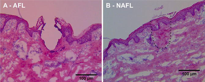
Fractional laser systems comprise both non-ablative (NAFL) and ablative devices. The systems in most widespread use include erbium-doped yttrium aluminum garnet (Er:YAG: AFXL; λ = 2940 nm), carbon dioxide (CO2; AFXL; λ = 10,600 nm), and erbium-doped glass (NAFL; λ = 1530–1560 nm) lasers, all operating in the absorption spectra of water (>1000 nm). Wavelength-dependent variations in water absorption result in vastly different tissue responses between devices. By operating in the near-infrared spectra where water absorption is fair, NAFLs result in localized tissue denaturation with heat deposition. Emitting in the mid-infrared spectra where water absorption is enhanced, AFXLs on the other hand generate greater energy depositions and localized tissue evaporation. The histological response to NAFL and AFXL treatments are depicted in Fig. 1.
AFXL
AFXL is an advantageous drug delivery technique as it provides predictable, controlled tissue responses and enables fast, sterile, and concurrent treatment of large skin areas (Hantash et al. 2007b; Taudorf et al. 2014). In AFXL-assisted drug delivery, there are two main parameters that can be adjusted for a given laser: laser channel density and depth. Density represents the ablated skin surface area, which is regulated by spot size and number of applied channels per unit skin area. Channel depth represents how deep laser channels extend into the skin and is mainly controlled by pulse energy. By calibrating laser density and depth, it is possible to (1) increase the accumulated drug amount in the skin to improve clinical efficacy and (2) adjust drug delivery rate, which can be used to reduce incubation time. Depending on the applied pulse energy and wavelength, residual thermal damage may vary; CO2 lasers induce greater coagulation zones than Er:YAG lasers, due to lower water absorbance at 10,600 nm (800 cm−1) (Marini and Krunic 2015) compared to 2940 nm (12,800 cm−1) (Walsh & Deutsch 1989). Although the importance of the residual thermal damage is currently unknown, bleeding is less common after treatment with CO2 lasers, a factor that may prove advantageous for AFXL-assisted drug delivery.
AFXL: Theoretical Concepts
The basic concept of AFXL-assisted cutaneous drug delivery can be illustrated by Fick’s law of diffusion, which describes passive diffusion through a medium (flux) as

The simplest way to describe how AFXL impacts drug delivery is to assume steady-state conditions with a constant drug concentration in the vehicle and a negligible drug concentration at the bottom of the dermal layer. Flux is then constant and can be described as

Under these assumptions, delivery of a particular drug depends on four conditions: (1) concentration gradient (ΔC), (2) partition coefficient between vehicle and skin (K), (3) diffusivity in skin (D), and (4) diffusion distance (L) (Franz 1983). By removing fractions of the SC, drug diffusion is facilitated by creating direct access to cellular epidermis and dermis. Over the laser channels, partition (K) thus occurs between vehicle and aqueous viable skin, enabling improved delivery of hydrophilic compounds. Upon leaving the vehicle, the drug enters directly into cellular skin where diffusivity (D) is higher than in SC, which results in a wide and rapid distribution of both small and large molecules around the channel. For therapeutic targets located in deeper dermis, the depth of the laser channels can be increased to minimize diffusion distance (L), in theory aiding delivery to deeper skin layers.
AFXL: Drug Delivery
AFXL has successfully enhanced delivery of the vast majority of drugs investigated thus far, including both lipophilic and hydrophilic molecules with molecular weights ranging from 177 to 13,300 Da. In preclinical trials, examined compounds include ALA, MAL (Haedersdal et al. 2010, 2011, 2014; Haak et al. 2012a, 2016; Forster et al. 2010; Huth et al. 2016), imiquimod (Lee et al. 2011a), ingenol mebutate (Erlendsson et al. 2015), diclofenac (Bachhav et al. 2011), methotrexate (Taudorf et al. 2015, 2016), 5-fluorouracil (Wenande et al. 2016), prednisolone (Yu et al. 2010), tranexamic acid (Hsiao et al. 2015), tretinoin (Chen et al. 2013), tetracycline (Chen et al. 2013), ascorbic acid (Hsiao et al. 2012), lidocaine (Bachhav et al. 2010a; Oni and Brown 2012), minoxidil (Lee et al. 2014a), diphencyprone (Lee et al. 2014a), small interfering RNA (siRNA) (Lee et al. 2014b), and polymeric microparticles containing triamcinolone acetonide (Singhal et al. 2016).
Drug delivery can be adjusted with laser density, and cutaneous drug accumulation increases with density to a point of saturation after which enhancement ceases. The specific relationship between laser density and skin deposition is best established for MAL, where laser density up to 5% coverage results in increased uptake, while no further enhancement is obtained from use of higher laser densities (Haak et al. 2016).
MAL distributes horizontally up to 1.5 mm away from single laser holes, providing the rationale for why low laser densities may suffice (Haedersdal et al. 2010). Analogous results have been confirmed for other small-size drugs such as ingenol mebutate (431 Da) (Erlendsson et al. 2015), diclofenac (296 Da) (Bachhav et al. 2011), and tretinoin (300 Da) (Chen et al. 2012). At present, densities beyond 5% seem unwarranted for use in AFXL-assisted drug delivery, although more information concerning the parameter’s effect on cutaneous diffusion pattern and biodistribution is currently needed (Haak et al. 2012b; Bachhav et al. 2010b).
Laser channel depth could in theory be regulated to target delivery to a specific, predetermined skin layer. However, studies have failed to agree on the relation between channel depth and drug deposition. While depth-dependent uptake has been described for hydrophilic compounds, e.g., methotrexate, (logP −1.85), and slightly lipophilic compounds, e.g., prednisone (logP 1.46) and diclofenac (logP 1.90), more hydrophobic drugs, such as lidocaine (logP 2.44), ingenol mebutate (logP 2.51), and imiquimod (logP 2.7), demonstrate no such relationship. Interstitial fluid and fibrin plugs have been reported to occupy the channels shortly after laser treatment, possibly inhibiting drugs and vehicles from filling deeper channel portions. Taking advantage of channel depth may thus depend on the individual drug’s ability to partition and diffuse in the medium filling channels, and various methods to actively fill the channels are currently under investigation (Erlendsson et al. 2016; Waibel et al. 2016; Alexiades 2015). At present, however, the specific relationship between laser channel depth and drug accumulation remains to be clarified for individual drugs.
Clinical Applications of AFXL Drug Delivery
AFXL-assisted delivery has been shown to enhance topical treatment efficacy for different dermatological conditions, including actinic keratoses (AKs), non-melanoma skin cancer (NMSC), actinic cheilitis, topical anesthetic treatment, rhytids, scars, wound healing, hemangiomas, vitiligo, and cutaneous infections such as onychomycosis, warts, and leishmaniasis (Vachiramon et al. 2016; Haedersdal et al. 2016; Park et al. 2016a; Gupta and Studholme 2016; Basnett et al. 2015; Ma et al. n.d.; Waibel et al. 2015). Principal findings for the most common of these indications are summarized below.
Neoplastic Lesions
The bulk of evidence on dysplastic lesions centers on PDT with methyl aminolevulinate (MAL) for AKs, demonstrating superior efficacy with AFXL compared to PDT alone. Thus, randomized controlled clinical trials report AK clearance rates of 87–92% for AFXL-assisted PDT versus 61–67% PDT alone 3 months posttreatment (Choi et al. 2015a; Ko et al. 2014a; Togsverd-Bo et al. 2012).
The long-term benefit of AFXL-assisted PDT versus conventional PDT is similarly supported for actinic cheilitis (85% vs. 29%) (Choi et al. 2015b) and Bowen’s disease (79% vs. 45%) (Ko et al. 2014b). Prolonged remission after AFXL-assisted PDT has also been described with recurrence rates of 8–10% at 12 months follow-up compared to 22–27% with conventional PDT (Haedersdal et al. 2016). In addition to improved efficacy, AFXL may reduce PDT incubation time, and AK clearance rates after AFXL-assisted PDT with 2-h (77%) (Choi et al. 2015a) and 1.5 h (71.4%) (Song et al. 2015) incubation are similar to conventional 3-h PDT (64.7–66%). Although side effects occur more frequently following AFXL-assisted versus PDT alone, treatments appear safe and side effects tolerable (Choi et al. 2015a, b; Ko et al. 2014a, b; Togsverd-Bo et al. 2012, 2015; Haak et al. 2015). For individuals taking immunosuppressants or with fields of severe actinic damage, AFXL-assisted PDT may further provide a more potent therapy requiring fewer treatment courses than conventional PDT (Togsverd-Bo et al. 2015; Helsing et al. 2013).
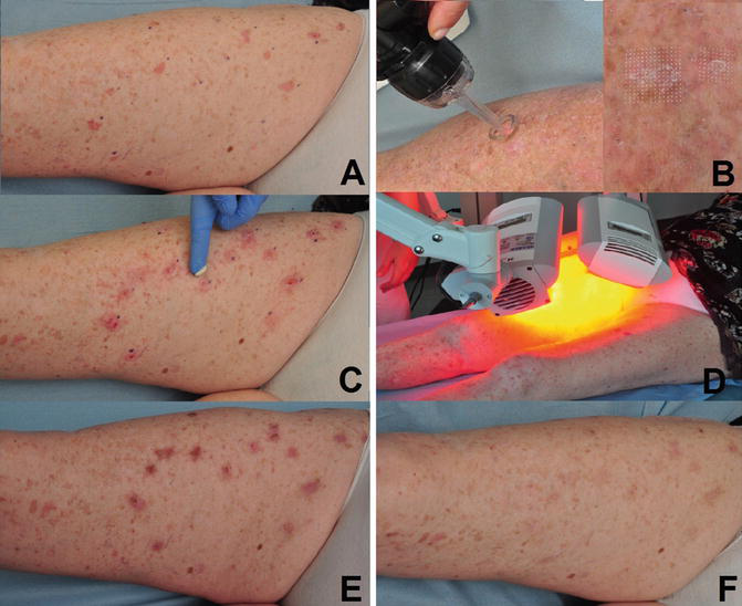
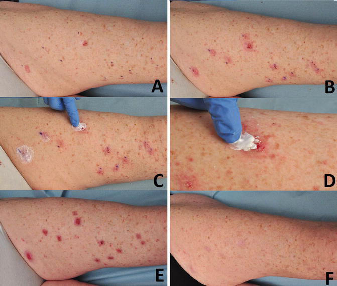
In contrast to AK treatment, there is not sufficient evidence supporting a beneficial effect of AFXL-assisted PDT for nodular basal cell carcinoma (BCC), although AFXL-assisted topical 5% 5-fluorouracil (5-FU) for Bowen’s disease and superficial BCC has shown initial promise (Hsu et al. 2016). Still, both AFXL-assisted 5-FU and PDT require further improvement to warrant recommendation for NMSC (Haak et al. 2015; Lippert et al. 2013). Figures 2 and 3 offer a practical, stepwise illustration of AFXL-assisted PDT using MAL (Fig. 2) and 5-FU (Fig. 3) for the treatment of multiple AKs.
Anesthetics
AFXL prior to application of topical anesthetics has been shown to offer significant, subject-reported pain reduction compared to sham laser pretreatment in few clinical trials (Meesters et al. 2016; Tian et al. 2016). As an indication of the importance of vehicle formulation, greater benefit of AFXL has also been noted using articaine hydrochloride + epinephrine liquid solution (AHES) compared to topical lidocaine + prilocaine in cream (Meesters et al. 2016).
Aesthetics
In addition to providing a new administration strategy, fractional laser-assisted delivery offers the potential for improved treatment outcomes with aesthetic and antiaging agents (Alexiades 2015; Mahmoud et al. 2015; Shin et al. 2012a). Of note, AFXL-assisted topical botulinum toxin A delivery has offered superior clinical efficacy for rhytides compared to AFXL-delivery of normal saline (Mahmoud et al. 2015). Superior outcomes following AFXL-delivery of cosmeceuticals are further reported for treatment of photoaging, dyschromia, and acne scarring (Alexiades 2015). AFXL-delivery of aesthetic agents remains new, however, and future studies are needed to reveal both its full potential and safety for this indication.
Scars
Initial evidence on topical AFXL-assisted drug delivery in the treatment of scars appears promising (Ali and Al-Niaimi 2016). AFXL-assisted betamethasone is reported to offer a 50% clinical improvement average for treatment-resistant keloids (Cavalié et al. 2015), and enhanced appearance consisting of improved scar texture, reduced hypertrophy, and dyschromia are noted for hypertrophic scars following AFXL-delivery of triamcinolone acetonide (average improvement 2.73 on a 0–3 scale) (Waibel et al. 2013). For atrophic scars, combined AFXL + poly-L-lactic acid (PLLA) treatment has offered a reported average clinical enhancement of 2.18 (scale 0–3) (Rkein et al. 2014), and improvement following AFXL + autologous platelet-rich plasma is shown to be comparable to intradermal injection (Gawdat et al. 2014). Though a multitude of scar types may benefit from AFXL-assisted treatments, randomized controlled clinical trials are nonetheless needed in the future before recommendations can be made.
Onychomycosis
Indicating increased efficacy with LADD, AFXL-assisted topical amorolfine treatment for Trichophyton (T) rubrum-, T. mentagrophytes-, and Epidermophyton floccosum-infected nail plates has demonstrated a 50% clinical and mycological cure rate 12 weeks after three treatment sessions (Lim et al. 2014a). More recently, fractional CO2 laser-assisted delivery of terbinafine resulted in a 92% negative culture rate at 3 months and 80% rate 6 months after three treatment sessions (Bhatta et al. 2016). As with scar treatment, future randomized controlled trials are needed to substantiate the benefits of AFXL-delivery of antimycotic drugs.
NAFL
Due to the weaker absorption by water, NAFLs do not establish direct access by microporation, but rather induce cylindrical zones of thermal damage, also known as microthermal zones (MTZs; Fig. 1). MTZs extend into underlying epidermis and dermis, leaving the SC with its low water content relatively intact (Laubach et al. 2006; Alexiades-Armenakas et al. 2008; Ganti and Banga 2016). Similar to AFXL, depth and density of MTZs represent adjustable parameters during NAFL treatment (Kim et al. 2016) and may prove valuable to regulate LADD. However, investigation of the relation between laser settings and drug uptake remains in its initial phase for NAFL.
Increased TEWL values after NAFL give indication of temporary disruption of the skin’s barrier function (Ganti and Banga 2016; Lim et al. 2014b; Kim et al. 2016). However, the mechanisms by which NAFLs increase topical drug delivery remain unclear. Beyond generation of MTZs via NAFL’s direct photothermal effect, possible theories include temporary expansion of cutaneous intercellular spaces by photomechanical wave (PW) with formation of epidermal vacuoles and dermal-epidermal junction disruption (Laubach et al. 2006; Ganti and Banga 2016; Lim et al. 2014b; Lee et al. 2002; Ruiz-Rodriguez et al. 2007). At this time, however, additional studies are needed before a mechanism of action for NAFL-assisted drug delivery can be definitively established.
NAFL Drug Delivery
Compared to AFXL, fewer studies to date examine the applicability of NAFL as a drug delivery strategy. In preclinical settings, NAFLs have been shown to enhance topical drug uptake of diclofenac, sumatriptan succinate, ALA, imiquimod, tretinoin, and peptides (Ganti and Banga 2016; Lee et al. 2016). In addition, clinical trials have indicated benefit of NAFL in combination with the following drugs: topical tretinoin (Prens et al. 2013), bimatoprost (Massaki et al. 2012), MAL (Ruiz-Rodriguez et al. 2007), ALA (Lim et al. 2014b), platelet-rich plasma (Shin et al. 2012b), tacrolimus (Chitvanich et al. 2016; Wolfshohl et al. 2016), and botulinum toxin A (Fan et al. 2016). Going forward, it is conceivable that NAFL’s combined drug delivery capabilities and favorable safety profile will provide new avenues for fractional LADD. However, whether NAFL is as effective as AFXL in enhancing cutaneous topical drug penetration has yet to be seen.
Safety Aspects
AFXL-assisted delivery breaks the natural skin barrier and provides access to the viable skin and papillary plexus (Oni and Brown 2012). Local skin responses are often aggravated by the combined effects of laser and topical drugs, and proximate access to the vascular system may result in systemic toxicity and introduction of virulent pathogens from the cutaneous flora or from nonsterile formulations (Oni and Brown 2012; Togsverd-Bo et al. 2012). Topical preparations designed for intact skin often include ingredients not intended for intradermal or systemic entry. Though these risks are reported to be significantly lower compared to fully ablative procedures (Oni et al. 2013a; Zaleski-Larsen and Fabi 2016), introduction of such agents may cause unwanted toxicity and potential immunological sensitization, resulting in hypersensitivity or anaphylaxis.
The reduced impact on the SC by NAFL significantly decreases the severity and duration of treatment-related side effects (Hantash and Mahmood 2007). While NAFL- and AFXL-assisted drug delivery safety profiles have yet to be adequately examined in direct comparison, potential advantages of NAFL include lower downtime and reduced post-inflammatory hyperpigmentation, erythema, crusting, and pain (Fan et al. 2016; Yang and Lee 2011). NAFL further carries a lower risk of infection, and in contrast to AFXL, skin permeation of bacteria following NAFL is reported comparable to that of intact skin (Lim et al. 2014b). When determining which laser technique to apply during LADD, the improved safety profile of NAFL should however be balanced against the prospect of reduced efficacy as compared to AFXL (Laubach et al. 2006; Prens et al. 2013).
In sum, LADD should be exercised with caution, and it seems appropriate to consider the technique only in well-controlled settings, using formulations and doses suitable for local injection. When performing fractional LADD, providers must be observant of known, laser-related side effects, signs of infection, hypersensitivity reactions, the risk of systemic uptake and potential for adverse events not previously described. Growth factors and platelet-rich plasma are increasingly applied to reduce downtime after AFXL treatments, and whether they promote proliferation of aberrant cells has yet to be investigated; the long-term consequences of AFXL-assisted delivery are thus currently unknown, and clinical studies are needed to fully evaluate the safety profile of combination with individual topical drugs (Lee et al. 2011b; Ai et al. 2013).
Perspectives
The full potential of fractional LADD has yet to be realized. To date, a number of emerging studies demonstrate that AFXL not only enhances cutaneous and transdermal delivery of topical drugs but also therapeutic antibodies, macromolecules, nucleic acids, allergens, scaffold materials, cells, as well as assist in delivery of light (Lee et al. 2013, 2014b; Yu et al. 2011; Oni et al. 2013b; Bachhav et al. 2013; Bach et al. 2012). Successful immunization with AFXL-assisted delivery of ovalbumin vaccines has been conducted, and the combination of AFXL with other drug delivery techniques such as iontophoresis, electroporation, and acoustic pressure wave is forthcoming. Topical application of systemic drugs not previously delivered through the skin is further made possible by AFXL and NAFL, providing new pharmaceutical treatment options and administration routes in the management of a multitude of diseases. Though still in its infant phase, fractional LADD thus holds promise as a useful, minimally invasive drug delivery system – both in dermatology and beyond.
Take Home Messages
- Fractional ablative and non-ablative laser-assisted drug delivery (LADD) is increasingly used to enhance cutaneous uptake and intensify clinical efficacy of topical drugs.
- In particular, current preclinical and clinical evidence substantiates the use of ablative fractional laser (AFXL) in photodynamic therapy for actinic keratosis.
- Fractional LADD potentially offers increased potency of currently approved, topical treatment regimes.
- When performing fractional LADD, providers must be observant of enhanced local skin reactions, potential for systemic uptake, signs of infection, hypersensitivity to drug formulation ingredients, as well as new adverse events not previously described.
Cross-References
- Biophotonics
- Laser Safety
- Transepidermal Drug Delivery with Ablative Methods (Lasers and Radiofrequency)
- Transepidermal Drug Delivery and Photodynamic Therapy
References
Ai J-J, Zha W-F, Guo B, Song W-M. A randomized guinea pig study on external cell growth factors after fractional ultrapulsed CO2 laser therapy. J Cosmet Laser Ther. 2013;15(4):219–24.
Alexiades M. Randomized, double-blind, split-face study evaluating fractional ablative erbium:YAG laser-mediated trans-epidermal delivery of cosmetic actives and a novel acoustic pressure wave ultrasound technology for the treatment of skin aging, melasma, and acne scars. J Drugs Dermatol. 2015;14(11):1191–8.
Alexiades-Armenakas MR, Dover JS, Arndt KA. The spectrum of laser skin resurfacing: nonablative, fractional, and ablative laser resurfacing. J Am Acad Dermatol. 2008;58(5):719–37.
Ali FR, Al-Niaimi F. Laser-assisted drug delivery in dermatology: from animal models to clinical practice. Lasers Med Sci. 2016;31(2):373–81.
Alvarez-Figueroa MJ, Blanco-Méndez J. Transdermal delivery of methotrexate: iontophoretic delivery from hydrogels and passive delivery from microemulsions. Int J Pharm. 2001;215(1–2):57–65.
Ameri M, Kadkhodayan M, Nguyen J, et al. Human growth hormone delivery with a microneedle transdermal system: preclinical formulation, stability, delivery and PK of therapeutically relevant doses. Pharmaceutics. 2014;6(2):220–34.
Andrews S, Lee JW, Choi S-O, Prausnitz MR. Transdermal insulin delivery using microdermabrasion. Pharm Res. 2011;28(9):2110–8.
Bach D, Weiss R, Hessenberger M, et al. Transcutaneous immunotherapy via laser-generated micropores efficiently alleviates allergic asthma in Phl p 5-sensitized mice. Allergy. 2012;67(11):1365–74.
Bachhav Y, Summer S, Heinrich A, Bragagna T, Böhler C, Kalia Y. Effect of controlled laser microporation on drug transport kinetics into and across the skin. J Control Release. 2010a;146(1):31–6.
Bachhav YG, Summer S, Heinrich A, Bragagna T, Böhler C, Kalia YN. Effect of controlled laser microporation on drug transport kinetics into and across the skin. J Control Release. 2010b;146:31–6.
Bachhav YG, Heinrich A, Kalia YN. Using laser microporation to improve transdermal delivery of diclofenac: increasing bioavailability and the range of therapeutic applications. Eur J Pharm Biopharm. 2011;78(3):408–14.
Bachhav YG, Heinrich A, Kalia YN. Controlled intra- and transdermal protein delivery using a minimally invasive Erbium:YAG fractional laser ablation technology. Eur J Pharm Biopharm. 2013;84(2):355–64.
Baron ED, Harris L, Redpath WS, Shapiro H. Laser-assisted penetration of topical anesthetic in adults. 2014;139(4):1288–90.
Basnett A, Nguyen TA, Cannavino C, Krakowski AC. Ablative fractional laser resurfacing with topical paromomycin as adjunctive treatment for a recalcitrant cutaneous leishmaniasis wound. Lasers Surg Med. 2015;47(10):788–91.
Benson HAE. Transdermal drug delivery: penetration enhancement techniques. Curr Drug Deliv. 2005;2(1):23–33.
Bhatta AK, Keyal U, Huang X, Zhao JJ. Fractional carbon-dioxide (CO2) laser-assisted topical therapy for the treatment of onychomycosis. J Am Acad Dermatol. 2016;74(5):916–23.
Brown MB, Martin GP, Jones SA, Akomeah FK. Dermal and transdermal drug delivery systems: current and future prospects. Drug Deliv. 2006;13(3):175–87.
Cavalié M, Sillard L, Montaudié H, Bahadoran P, Lacour J-P, Passeron T. Treatment of keloids with laser-assisted topical steroid delivery: a retrospective study of 23 cases. Dermatol Ther. 2015;28(2):74–8.
Chen X, Shah D, Kositratna G, Manstein D, Anderson RR, Wu MX. Facilitation of transcutaneous drug delivery and vaccine immunization by a safe laser technology. J Control Release. 2012;159(1):43–51.
Chen W-Y, Fang C-L, Al-Suwayeh SA, Yang H-H, Li Y-C, Fang J-Y. Risk assessment of excess drug and sunscreen absorption via skin with ablative fractional laser resurfacing: optimization of the applied dose for postoperative care. Lasers Med Sci. 2013;28(5):1363–74.
Chitvanich S, Rerknimitr P, Panchaprateep R, Pongprutthipan M, Asawanonda P. Combination of non-ablative fractional photothermolysis and 0.1% tacrolimus ointment is efficacious for treating idiopathic guttate hypomelanosis. J Dermatolog Treat. 2016;27(5):456–60.
Choi SH, Kim KH, Song KH. Efficacy of ablative fractional laser-assisted photodynamic therapy with short-incubation time for the treatment of facial and scalp actinic keratosis: 12-month follow-up results of a randomized, prospective, comparative trial. J Eur Acad Dermatology Venereol. 2015a;29(8):1598–605.
Choi SH, Kim KH, Song K-H. Efficacy of ablative fractional laser-assisted photodynamic therapy for the treatment of actinic cheilitis: 12-month follow-up results of a prospective, randomized, comparative trial. Br J Dermatol. 2015b;173(1):184–91.
Doukas AG, Kollias N. Transdermal drug delivery with a pressure wave. Adv Drug Deliv Rev. 2004;56(5):559–79.
Elias PM, Menon GK. Structural and lipid biochemical correlates of the epidermal permeability barrier. Adv Lipid Res. 1991;24:1–26.
Erlendsson AM, Taudorf EH, Eriksson AH, et al. Ablative fractional laser alters biodistribution of ingenol mebutate in the skin. Arch Dermatol Res. 2015;307(6):512–22.
Erlendsson AM, Doukas AG, Farinelli WA, Bhayana B, Anderson RR, Haedersdal M. Fractional laser-assisted drug delivery: active filling of laser channels with pressure and vacuum alteration. Lasers Surg Med. 2016;48(2):116–24.
Fan X, Yin Y, Wang S, et al. Clinical assessment of the safety and effectiveness of nonablative fractional laser combined with transdermal delivery of botulinum toxin A in treating periocular wrinkles. Plast Reconstr Surg Glob Open. 2016;4(8):e1004.
Fang J-Y, Lee W-R, Shen S-C, Fang Y-P, Hu C-H. Enhancement of topical 5-aminolaevulinic acid delivery by erbium:YAG laser and microdermabrasion: a comparison with iontophoresis and electroporation. Br J Dermatol. 2004;151(1):132–40.
Forster B, Klein A, Szeimies R-M, Maisch T. Penetration enhancement of two topical 5-aminolaevulinic acid formulations for photodynamic therapy by erbium:YAG laser ablation of the stratum corneum: continuous versus fractional ablation. Exp Dermatol. 2010;19(9):806–12.
Franz TJ. Kinetics of cutaneous drug penetration. Int J Dermatol. 1983;22(9):499–505.
Fukushima K, Ise A, Morita H, et al. Two-layered dissolving microneedles for percutaneous delivery of peptide/protein drugs in rats. Pharm Res. 2011;28(1):7–21.
Ganti SS, Banga AK. Non-ablative fractional laser to facilitate transdermal delivery. J Pharm Sci. 2016;105(11):3324–32.
Gawdat HI, Hegazy RA, Fawzy MM, Fathy M. Autologous platelet rich plasma: topical versus intradermal after fractional ablative carbon dioxide laser treatment of atrophic acne scars. Dermatol Surg. 2014;40(2):152–61.
Gothelf A, Mir LM, Gehl J. Electrochemotherapy: results of cancer treatment using enhanced delivery of bleomycin by electroporation. Cancer Treat Rev. 2003;29(5):371–87.
Govil S. Transdermal drug delivery systems. In: Tyle P, editor. Drug delivery devices: fundamentals and applications. 1st ed. New York: Marcel Dekker; 1988. p. 385–420.
Griffin JE, Echternach JL, Price RE, Touchstone JC. Patients treated with ultrasonic driven hydrocortisone and with ultrasound alone. Phys Ther. 1967;47(7):594–601.
Gupta AK, Studholme C. Novel investigational therapies for onychomycosis: an update. Expert Opin Investig Drugs. 2016;25(3):297–305.
Haak CS, Farinelli WA, Tam J, Doukas AG, Anderson RR, Haedersdal M. Fractional laser-assisted delivery of methyl aminolevulinate: impact of laser channel depth and incubation time. Lasers Surg Med. 2012a;44(10):787–95.
Haak CS, Bhayana B, Farinelli WA, Anderson RR, Haedersdal M. The impact of treatment density and molecular weight for fractional laser-assisted drug delivery. J Control Release. 2012b;163(3):335–41.
Haak C, Togsverd-Bo K, Thaysen-Petersen D, et al. Fractional laser-mediated photodynamic therapy of high-risk basal cell carcinomas – a randomized clinical trial. Br J Dermatol. 2015;172(1):215–22.
Haak CS, Christiansen K, Erlendsson AM, et al. Ablative fractional laser enhances MAL-induced PpIX accumulation: impact of laser channel density, incubation time and drug concentration. J Photochem Photobiol B. 2016;159:42–8.
Haedersdal M, Sakamoto FH, Farinelli WA, Doukas AG, Tam J, Anderson RR. Fractional CO2 laser-assisted drug delivery. Lasers Surg Med. 2010;42(2):113–22.
Haedersdal M, Katsnelson J, Sakamoto FH, et al. Enhanced uptake and photoactivation of topical methyl aminolevulinate after fractional CO2 laser pretreatment. Lasers Surg Med. 2011;43(8):804–13.
Haedersdal M, Sakamoto FH, Farinelli WA, Doukas AG, Tam J, Anderson RR. Pretreatment with ablative fractional laser changes kinetics and biodistribution of topical 5-aminolevulinic acid (ALA) and methyl aminolevulinate (MAL). Lasers Surg Med. 2014;46(6):462–9.
Haedersdal M, Erlendsson AM, Paasch U, Anderson RR. Translational medicine in the field of ablative fractional laser (AFXL)-assisted drug delivery: a critical review from basics to current clinical status. J Am Acad Dermatol. 2016;74(5):981–1004.
Hantash BM, Mahmood MB. Fractional photothermolysis: a novel aesthetic laser surgery modality. Dermatol Surg. 2007;33(5):525–34.
Hantash BM, Bedi VP, Chan KF, Zachary CB. Ex vivo histological characterization of a novel ablative fractional resurfacing device. Lasers Surg Med. 2007a;39(2):87–95.
Hantash BM, Bedi VP, Kapadia B, et al. In vivo histological evaluation of a novel ablative fractional resurfacing device. Lasers Surg Med. 2007b;39(2):96–107.
Helsing P, Togsverd-Bo K, Veierød MB, Mørk G, Haedersdal M. Intensified fractional CO2 laser-assisted photodynamic therapy vs. laser alone for organ transplant recipients with multiple actinic keratoses and wart-like lesions: a randomized half-side comparative trial on dorsal hands. Br J Dermatol. 2013;169(5):1087–92.
Hsiao C-Y, Huang C-H, Hu S, et al. Fractional carbon dioxide laser treatment to enhance skin permeation of ascorbic acid 2-glucoside with minimal skin disruption. Dermatol Surg. 2012;38(8):1284–93.
Hsiao C-Y, Sung H-C, Hu S, Huang C-H. Fractional CO2 laser treatment to enhance skin permeation of tranexamic acid with minimal skin disruption. Dermatology. 2015;230(3):269–75.
Hsu SH, Gan SD, Nguyen BT, Konnikov N, Liang CA. Ablative fractional laser-assisted topical fluorouracil for the treatment of superficial basal cell carcinoma and squamous cell carcinoma in situ: a follow-up study. Dermatol Surg. 2016;42(9):1050–3.
Huth S, Marquardt Y, Amann PM, et al. Ablative non-sequential fractional ultrapulsed CO2 laser pretreatment improves conventional photodynamic therapy with methyl aminolevulinate in a novel human in vitro 3D actinic keratosis skin model. Exp Dermatol. May 2016: Epub ahead of print.
Jacques SL, McAuliffe DJ, Blank IH, Parrish JA. Controlled removal of human stratum corneum by pulsed laser. J Invest Dermatol. 1987;88(1):88–93.
Kim JH, Park HY, Jung M, Choi EH. Automicroneedle therapy system combined with topical tretinoin shows better regenerative effects compared with each individual treatment. Clin Exp Dermatol. 2013;38(1):57–65.
Kim JM, Kim WI, Ko HC, Kim MB, Kim BS. Epidermal barrier function changes after ablative and non-ablative fractional laser administration. J Eur Acad Dermatol Venereol. July 2016: Epub ahead of print.
Ko D-Y, Jeon S-Y, Kim K-H, Song K-H. Fractional erbium:YAG laser-assisted photodynamic therapy for facial actinic keratoses: a randomized, comparative, prospective study. J Eur Acad Dermatol Venereol. 2014a;28(11):1529–39.
Ko DY, Kim KH, Song KH. A randomized trial comparing methyl aminolaevulinate photodynamic therapy with and without Er:YAG ablative fractional laser treatment in Asian patients with lower extremity Bowen disease: results from a 12-month follow-up. Br J Dermatol. 2014b;170(1):165–72.
Krishnan G, Grice JE, Roberts MS, Benson HAE, Prow TW. Enhanced sonophoretic delivery of 5-aminolevulinic acid: preliminary human ex vivo permeation data. Skin Res Technol. 2013;19(1):e283–9.
Laubach H-J, Tannous Z, Anderson RR, Manstein D. Skin responses to fractional photothermolysis. Lasers Surg Med. 2006;38(2):142–9.
Lee W, Shen S, Wang K, Hu C, Fang J. The effect of laser treatment on skin to enhance and control transdermal delivery of 5-fluorouracil. J Pharm Sci. 2002;91(7):1613–26.
Lee W-R, Shen S-C, Kuo-Hsien W, Hu C-H, Fang J-Y. Lasers and microdermabrasion enhance and control topical delivery of vitamin C. J Invest Dermatol. 2003;121(5):1118–25.
Lee W-R, Tsai R-Y, Fang C-L, Liu C-J, Hu C-H, Fang J-Y. Microdermabrasion as a novel tool to enhance drug delivery via the skin: an animal study. Dermatologic Surg. 2006;32(8):1013–22.
Lee W-R, Shen S-C, Fang C-L, Zhuo R-Z, Fang J-Y. Topical delivery of methotrexate via skin pretreated with physical enhancement techniques: low-fluence erbium:YAG laser and electroporation. Lasers Surg Med. 2008;40(7):468–76.
Lee W-R, Shen S-C, Al-Suwayeh SA, Yang H-H, Yuan C-Y, Fang J-Y. Laser-assisted topical drug delivery by using a low-fluence fractional laser: imiquimod and macromolecules. J Control Release. 2011a;153(3):240–8.
Lee YB, Lee KJ, Park HJ, Cho BK. Topical application of growth factors after carbon dioxide fractional laser therapy: a randomized controlled split-face study. J Cosmet Laser Ther. 2011b;13(1):38–40.
Lee W-R, Shen S-C, Al-Suwayeh SA, Yang H-H, Li Y-C, Fang J-Y. Skin permeation of small-molecule drugs, macromolecules, and nanoparticles mediated by a fractional carbon dioxide laser: the role of hair follicles. Pharm Res. 2013;30(3):792–802.
Lee W-R, Shen S-C, Aljuffali IA, Li Y-C, Fang J-Y. Erbium-yttrium-aluminum-garnet laser irradiation ameliorates skin permeation and follicular delivery of antialopecia drugs. J Pharm Sci. 2014a;103(11):3542–52.
Lee W-R, Shen S-C, Chen W-Y, Aljuffali IA, Suen S-Y, Fang J-Y. Noninvasive delivery of siRNA and plasmid DNA into skin by fractional ablation: erbium:YAG laser versus CO2 laser. Eur J Pharm Biopharm. 2014b;86(3):315–23.
Lee W-R, Shen S-C, Aljuffali IA, Lin Y-K, Huang C-W, Fang J-Y. Non-ablative fractional laser assists cutaneous delivery of small- and macro-molecules with minimal bacterial infection risk. Eur J Pharm Sci. 2016;92:1–10.
Levin G, Gershonowitz A, Sacks H, et al. Transdermal delivery of human growth hormone through RF-microchannels. Pharm Res. 2005;22(4):550–65.
Lim E-H, Kim H, Park Y-O, et al. Toenail onychomycosis treated with a fractional carbon-dioxide laser and topical antifungal cream. J Am Acad Dermatol. 2014a;70(5):918–23.
Lim HK, Jeong KH, Kim NI, Shin MK. Nonablative fractional laser as a tool to facilitate skin penetration of 5-aminolaevulinic acid with minimal skin disruption: a preliminary study. Br J Dermatol. 2014b;170(6):1336–40.
Lippert J, Smucler R, Vlk M. Fractional carbon dioxide laser improves nodular basal cell carcinoma treatment with photodynamic therapy with methyl 5-aminolevulinate. Dermatol Surg. 2013;39(8):1202–8.
Ma G, Wu P, Lin X, et al. Fractional carbon dioxide laser-assisted drug delivery of topical timolol solution for the treatment of deep infantile hemangioma: a pilot study. Pediatr Dermatol. 31(3):286–91.
Mahmoud BH, Burnett C, Ozog D. Prospective randomized controlled study to determine the effect of topical application of botulinum toxin A for crow’s feet after treatment with ablative fractional CO2 laser. Dermatol Surg. 2015;41(Suppl 1):S75–81.
Manstein D, Herron GS, Sink RK, Tanner H, Anderson RR. Fractional photothermolysis: a new concept for cutaneous remodeling using microscopic patterns of thermal injury. Lasers Surg Med. 2004;34(5):426–38.
Marini LG, Krunic AL. In: Katsambas A, editor. European handbook of dermatological treatments. 3rd ed. Berlin/Heidelberg: Springer Verlag; 2015.
Marro D, Kalia YN, Delgado-Charro MB, Guy RH. Optimizing iontophoretic drug delivery: identification and distribution of the charge-carrying species. Pharm Res. 2001;18(12):1709–13.
Massaki ABMN, Fabi SG, Fitzpatrick R. Repigmentation of hypopigmented scars using an erbium-doped 1550-nm fractionated laser and topical bimatoprost. Dermatol Surg. 2012;38(7 Pt 1):995–1001.
Meesters AA, Bakker MM, de Rie MA, Wolkerstorfer A. Fractional CO2 laser assisted delivery of topical anesthetics: a randomized controlled pilot study. Lasers Surg Med. 2016;48(2):208–11.
Mikolajewska P, Donnelly RF, Garland MJ, et al. Microneedle pre-treatment of human skin improves 5-aminolevulinic acid (ALA)- and 5-aminolevulinic acid methyl ester (MAL)-induced PpIX production for topical photodynamic therapy without increase in pain or erythema. Pharm Res. 2010;27(10):2213–20.
Mitragotri S, Blankschtein D, Langer R. Ultrasound-mediated transdermal protein delivery. Science. 1995;269(5225):850–3.
Morrow DIJ, Garland MJ, McCarron PA, Woolfson AD, Donnelly RF. Innovative drug delivery strategies for topical photodynamic therapy using porphyrin precursors. J Environ Pathol Toxicol Oncol. 2007;26(2):105–16.
Oni G, Brown SA, Kenkel JM. Can fractional lasers enhance transdermal absorption of topical lidocaine in an in vivo animal model? Lasers Surg Med. 2012;44(2):168–74.
Oni G, Rasko Y, Kenkel J. Topical lidocaine enhanced by laser pretreatment: a safe and effective method of analgesia for facial rejuvenation. Aesthetic Surg J. 2013a;33(6):854–61.
Oni G, Lequeux C, Cho M-J, et al. Transdermal delivery of adipocyte-derived stem cells using a fractional ablative laser. Aesthet Surg J. 2013b;33(1):109–16.
Pacini S, Gulisano M, Punzi T, Ruggiero M. Transdermal delivery of Clostridium botulinum toxin type A by pulsed current iontophoresis. J Am Acad Dermatol. 2007;57(6):1097–9.
Park S-M, Kim G-W, Mun J-H, et al. Fractional laser-assisted topical imiquimod 5% cream treatment for recalcitrant common warts in children: a pilot study. Dermatol Surg. September 2016a: Epub ahead of print.
Park JM, Jeong K-H, Bae MI, Lee S-J, Kim N-I, Shin MK. Fractional radiofrequency combined with sonophoresis to facilitate skin penetration of 5-aminolevulinic acid. Lasers Med Sci. 2016b;31(1):113–8.
Pathan IB, Setty CM. Chemical penetration enhancers for transdermal drug delivery systems. Trop J Pharm Res. 2009;8(2):173–9.
Paudel KS, Milewski M, Swadley CL, Brogden NK, Ghosh P, Stinchcomb AL. Challenges and opportunities in dermal/transdermal delivery. Ther Deliv. 2010;1(1):109–31.
Prens SP, de Vries K, Neumann HAM, Prens EP. Non-ablative fractional resurfacing in combination with topical tretinoin cream as a field treatment modality for multiple actinic keratosis: a pilot study and a review of other field treatment modalities. J Dermatolog Treat. 2013;24(3):227–31.
Rkein A, Ozog D, Waibel JS. Treatment of atrophic scars with fractionated CO2 laser facilitating delivery of topically applied poly-L-lactic acid. Dermatol Surg. 2014;40(6):624–31.
Rosim GC, Barbieri CH, Lanças FM, Mazzer N. Diclofenac phonophoresis in human volunteers. Ultrasound Med Biol. 2005;31(3):337–43.
Ruiz-Rodriguez R, López L, Candelas D, Zelickson B. Enhanced efficacy of photodynamic therapy after fractional resurfacing: fractional photodynamic rejuvenation. J Drugs Dermatol. 2007;6(8):818–20.
Sardesai NY, Weiner DB. Electroporation delivery of DNA vaccines: prospects for success. Curr Opin Immunol. 2011;23(3):421–9.
Shin M-K, Lee J-H, Lee S-J, Kim N-I. Platelet-rich plasma combined with fractional laser therapy for skin rejuvenation. Dermatol Surg. 2012a;38(4):623–30.
Shin M-K, Lee J-H, Lee S-J, Kim N-I. Platelet-rich plasma combined with fractional laser therapy for skin rejuvenation. Dermatol Surg. 2012b;38(4):623–30.
Singhal M, Del Río-Sancho S, Sonaje K, Kalia YN. Fractional laser ablation for the cutaneous delivery of triamcinolone acetonide from cryomilled polymeric microparticles: creating intraepidermal drug depots. Mol Pharm. 2016;13(2):500–11.
Song HS, Jung S-E, Jang YH, Kang HY, Lee E-S, Kim YC. Fractional carbon dioxide laser-assisted photodynamic therapy for patients with actinic keratosis. Photodermatol Photoimmunol Photomed. 2015;31(6):296–301.
Surber C, Davis AF. Bioavailability and bioequivalence dermatological formulations. In: Walters KA, editor. Dermatological and transdermal formulations. Boca Raton, FL: CRC Press; 2002. p. 401–74.
Taudorf EH, Haak CS, Erlendsson AM, et al. Fractional ablative erbium YAG laser: histological characterization of relationships between laser settings and micropore dimensions. Lasers Surg Med. 2014;46(4):281–9.
Taudorf E, Lerche C, Vissing A, et al. Topically applied methotrexate is rapidly delivered into skin by fractional laser ablation. Expert Opin Drug Deliv. 2015;12(7):1059–69.
Taudorf EH, Lerche CM, Erlendsson AM, et al. Fractional laser-assisted drug delivery: laser channel depth influences biodistribution and skin deposition of methotrexate. Lasers Surg Med. 2016;48(5):519–29.
Tian T, Luo Y, Jiang T, et al. Clinical effect of ablative fractional laser-assisted topical anesthesia on human skin: a randomized pilot study. J Cosmet Laser Ther. August 2016: Epub ahead of print.
Togsverd-Bo K, Haak CS, Thaysen-Petersen D, Wulf HC, Anderson RR, Hædersdal M. Intensified photodynamic therapy of actinic keratoses with fractional CO2 laser: a randomized clinical trial. Br J Dermatol. 2012;166(6):1262–9.
Togsverd-Bo K, Lei U, Erlendsson AM, et al. Combination of ablative fractional laser and daylight-mediated photodynamic therapy for actinic keratosis in organ transplant recipients – a randomized controlled trial. Br J Dermatol. 2015;172(2):467–74.
Treffel P, Panisset F, Humbert P, Remoussenard O, Bechtel Y, Agache P. Effect of pressure on in vitro percutaneous absorption of caffeine. Acta Derm Venereol. 1993;73(3):200–2.
Vachiramon V, Chaiyabutr C, Rattanaumpawan P, Kanokrungsee S. Effects of a preceding fractional carbon dioxide laser on the outcome of combined local narrowband ultraviolet B and topical steroids in patients with vitiligo in difficult-to-treat areas. Lasers Surg Med. 2016;48(2):197–202.
Waibel JS, Wulkan AJ, Shumaker PR. Treatment of hypertrophic scars using laser and laser assisted corticosteroid delivery. Lasers Surg Med. 2013;45(3):135–40.
Waibel JS, Mi Q-S, Ozog D, et al. Laser-assisted delivery of vitamin C, vitamin E, and ferulic acid formula serum decreases fractional laser postoperative recovery by increased beta fibroblast growth factor expression. Lasers Surg Med. 2015;48(3):238–44.
Waibel JS, Rudnick A, Nousari C, Bhanusali DG. Fractional ablative laser followed by transdermal acoustic pressure wave device to enhance the drug delivery of aminolevulinic acid: in vivo fluorescence microscopy study. J Drugs Dermatol. 2016;15(1):14–21.
Walsh JT, Deutsch TF. Er:YAG laser ablation of tissue: measurement of ablation rates. Lasers Surg Med. 1989;9(4):327–37.
Wenande E, Olesen UH, Nielsen MM, et al. Fractional laser-assisted topical delivery leads to enhanced, accelerated and deeper cutaneous 5-fluorouracil uptake. Expert Opin Drug Deliv. 2016;1–11.
Wolfshohl JA, Geddes ERC, Stout AB, Friedman PM. Improvement of erythema dyschromicum perstans using a combination of the 1,550-nm erbium-doped fractionated laser and topical tacrolimus ointment. Lasers Surg Med. 2016: Epub ahead of print.
Yang YJ, Lee G-Y. Treatment of striae distensae with nonablative fractional laser versus ablative CO2 fractional laser: a randomized controlled trial. Ann Dermatol. 2011;23(4):481–9.
You S-K, Noh Y-W, Park H-H, et al. Effect of applying modes of the polymer microneedle-roller on the permeation of L-ascorbic acid in rats. J Drug Target. 2010;18(1):15–20.
Yu J, Bachhav Y, Summer S, et al. Using controlled laser-microporation to increase transdermal delivery of prednisone. J Control Release. 2010;148:e71–3.
Yu J, Kalia DR, Kalia YN. Erbium:YAG fractional laser ablation for the percutaneous delivery of intact functional therapeutic antibodies. J Control Release. 2011;156(1):53–9.
Zaleski-Larsen LA, Fabi SG. Laser-assisted drug delivery. Dermatol Surg. 2016;42(8):919–31.







