
Transepidermal Drug Delivery and Photodynamic Therapy
Transepidermal Drug Delivery and Photodynamic Therapy
Marianna Tavares Fernandes Pires, Livia Roale Nogueira and Maria Claudia Almeida Issa Universidade Federal Fluminense, Niterói, RJ, Brazil Department of Clinical Medicine – Dermatology, Fluminense Federal University, Praia de Icarai 139, Niterói, RJ, 702, Brazil
Abstract
Transepidermal drug delivery (TED) has been used with the aim to increase drug penetration into the skin, and its association with photodynamic therapy (PDT) is described for nonmelanoma skin cancer, porokeratosis, and photorejuvenation treatment. TED with PDT has been reported with fractional ablative methods (ablative laser and ablative radiofrequency), as well as with non-ablative lasers and with microneedles. TED with PDT is considered an effective method for actinic keratosis (field of cancerization) and for photorejuvenation.
Keywords Photodynamic therapyPhotoagingRejuvenationSkin drug administrationDrug deliveryTransepidermal drug deliveryActinic keratosisField of cancerization
Introduction
Topical photodynamic therapy (PDT) is approved for nonmelanoma skin cancer, actinic keratosis (AK), basal cell carcinoma (BCC), and Bowen’s disease (BD). However, conventional PDT can fail to treat thick lesions of NMSC, due to the limited penetration of the photosensitizer, which causes limited local bioavailability.
The stratum corneum is a significant barrier to percutaneous drug and particle absorption. The association of several modalities, such as ablative methods (fractional ablative lasers and fractional ablative radiofrequency), non-ablative lasers, and microneedling with PDT, has been studied, in the last years, with the aim to enhance photosensitizer penetration into the skin. This new concept of treatment is called transepidermal drug delivery (TED). TED and methyl aminolevulinate (MAL)-PDT has been studied, showing efficacy in AK and field cancerization treatment in immunocompetent and immunosuppressed patients.
It is also reported that there are benefits of skin rejuvenation during the field of cancerization treatment, such as improving skin texture, pigmentation, wrinkles, and laxity, after TED and conventional PDT, even when photosensitizer’s incubation time is reduced. Some new protocols using TED and daylight PDT have been evaluated with good results.
Photodynamic Therapy
History
Oscar Raab, a German medical student, described the first photodynamic reaction in 1900. He reported that neither acridine orange, a dye, nor light alone was toxic to paramecia; however, when both were combined, they could induce cell death in less than 2 h. He realized this while using acridine orange in paramecia during a coincidental thunderstorm (Raab 1900). Later, von Tappeiner and Jesionek used eosin and light together to treat skin cancer, lupus vulgaris, and condyloma lata. At this time, they speculated that eosin, like acridine, after being incorporated into the cell, would produce a cytotoxic reaction when exposed to an adequate light source in the presence of oxygen (von Tappeiner and Jesionek 1903). In the 1990s, Kennedy et al. described the use of aminolevulinic acid (ALA), a protoporphyrin IX (PpIX) precursor, to be topically applied avoiding systemic photosensitivity (Kennedy et al. 1990).
Since 1999, the US Food and Drug Administration (FDA) have approved photodynamic therapy (PDT) with topical ALA in dermatology for the treatment of actinic keratosis (AK). In Europe, methyl aminolevulinate (MAL), an esterified form of ALA with lipophilic properties, has been approved for the treatment of AK and basal cell carcinoma (BCC) since 2001 (Sandberg et al. 2008a). In 2004, MAL was approved for the treatment of AK in the United States. In 2006, MAL was approved in Brazil for AK and for superficial and nodular BCC. MAL is currently approved in many countries of Europe, Asia, and the Americas for the treatment of AK, BCC, and Bowen’s disease. In 2009, MAL was also approved in Brazil for Bowen’s disease.
Concept
PDT is defined as a photochemical reaction used to selectively destroy target tissue. Photodynamic action requires three components: light, oxygen, and a photosensitizer agent. When these components are combined, they become toxic to the target cell. It is a two-stage therapeutic technique that employs light activation of localized photosensitized tissue in an oxygen-dependent process, which initiates oxidative stress, inflammation, and cell death.
Photosensitizers
There are two main prodrugs used for topical PDT: ALA and MAL. Both are precursors of an endogenous photosensitizer, PpIX (Kalka et al. 2000). After topical application, photosensitizers are mainly absorbed into abnormal cells and converted, via the heme cycle, to PpIX. Abnormal tumor cells have low ferrochelatase activity and lower ferric ion concentrations, limiting the last step in the heme cycle, promoting the accumulation of PpIX (Ericson et al. 2008).
Porphyrin-based photosensitizer agents are selectively concentrated in human cancerous tissue and are activated by light in the presence of oxygen to initiate cytotoxic chemical reactions. Based on the comparison of fluorescence intensities on a same individual from normal and tumor tissues of the same pigmentation, ratios of up to 15:1 for PpIX fluorescence between nonmelanoma skin cancer and normal skin have been reported with ALA sensitizer (Svanberg et al. 1994).
Source of Light
Light must be absorbed by PpIX, which has a peak of excitation in the blue-violet light part of the spectrum (Soret band) with maximum at 410 nm. This part of the spectrum has very poor tissue penetration (1 mm). For this reason, red light (630–635 nm), which is also absorbed by PpIX and has deeper penetration (up to 6 mm), is usually used.
PDT is based on the chemical reaction in which PpIX is photoactivated by different sources of light, including incoherent, continuous-wave red or blue light, intense pulsed light (IPL), as well as pulsed dye laser (PDL) (Friedmann et al. 2014). The light-emitting diode (LED) is the main source of light for topical PDT treatment (Moseley et al. 2006) with easier maintenance and lower costs when compared with lasers (Brancaleon and Moseley 2012).
Recent studies have shown that natural daylight can be successfully used for topical PDT. Daylight PDT (DLPDT) is a new modality for actinic keratoses (AKs) and field of cancerization treatment. DLPDT is considered as effective as conventional PDT with the advantage of being almost painless (Rubel et al. 2014) (see chapter “Daylight Photodynamic Therapy and Its Relation to Photodamaged Skin” – author Beni Grinblat).
Indications
Topical PDT is approved for nonmelanoma skin cancer, actinic keratosis (AK), basal cell carcinoma (BCC), and Bowen’s disease (BD). Regarding squamous cell carcinoma, although occasionally resulting in initial encouraging results, it is associated with unacceptable recurrence rates, and PDT would not be recommended (Ericson et al. 2008).
There are many randomized, controlled, and open studies in which the role of topical PDT in AK, BD, and BCC has been examined, and both British and European guidelines for the use of PDT are available (Morton et al. 2013a, b). PDT is a very important and effective treatment for field of cancerization, and many authors reported not only cure of AKs but also skin rejuvenation with texture, wrinkle, and pigmentation improvement on the area treated. For this reason, PDT has been also studied and indicated for skin rejuvenation (see chapter “Photodynamic Therapy for Photodamaged Skin” – Issa e Ferola).
Actinic keratosis
AKs are common epidermal lesions associated with chronic exposure to ultraviolet (UV) radiation, which have the potential of progressing to squamous cell carcinoma (SCC), with the highest incidence in the aged and fair-skinned population (Traianou et al. 2012). AK is the second most common diagnosis by dermatologists in the United States, with direct cost of therapy estimated at more than US$1 billion per year and indirect cost nearing US$300 million (Neidecker et al. 2009).
The early identification and treatment of AKs are necessary because it is very difficult or even impossible to predict which lesions may become invasive and develop into metastatic SCC. The fact that 65–97% of squamous cell carcinomas develop from AKs or areas of field cancerization highlights the need for effective treatment of these lesions (Rosen and Lebwohl 2013).
The aim of photodynamic therapy is not only to treat clinical or visible AKs but also to treat subclinical lesions. PDT may also have the potential to decrease expression of early markers of cutaneous neoplasia Ki-67 and p53, as demonstrated in multiple studies following methyl aminolevulinate-PDT (MAL-PDT) using incoherent red light (Bagazgoitia et al. 2011). In comparative studies between PDT and cryotherapy for AK treatment, clinical results were equal or even better, with superior cosmetic outcome, in the cases reported in PDT groups (Tarstedt et al. 2005). Some authors reported better results and cosmetic outcome with MAL-PDT compared with trichloroacetic acid (TCA 50%) for AK treatment (Di Nuzzo et al. 2015).
Bowen’s disease
Bowen’s disease (BD) is an in situ carcinoma, clinically presented as an erythematous-squamous plaque with sharply demarcated, irregular borders. Sometimes, it can be a verrucous, hypo- or hyperpigmented, and, eventually, exulcerated lesion. Its evolution is slow and progressive, generally asymptomatic. However, local pain, irritation, pruritus, and bleeding can occur (Moraes 2002).
Treatments include surgery, radiotherapy, 5-FU, curettage, cryotherapy, and PDT. To choose the best option, dermatologists should take into account the patient’s age and frailty, comorbidities, and lesion’s body site. PDT is well recommended to treat lesions located in areas that are difficult to heal, such as lower limbs (Cox et al. 2007).
In comparative studies, ALA-PDT has been shown to be more effective and cause less adverse effects than cryotherapy or 5-FU for the treatment of BD (Morton et al. 1996). A large randomized controlled trial comparing topical MAL-PDT with either cryotherapy or 5-FU reported that a sustained response after 12 months following treatment was 80% for PDT, 67% for cryotherapy, and 69% for 5-FU with a superior cosmetic outcome in the PDT group (Morton et al. 2006).
Organ transplant recipients
The relative risk of SCC is estimated to be 65- to 250-fold in organ transplant recipients (OTRs) compared with the rest of the population, increasing with time after transplantation and depending on the type of organ transplant (Hartevelt et al. 1990).
Topical PDT may potentially be used in the treatment of organ transplant recipients who are at markedly increased risk of developing dysplastic skin changes and nonmelanoma skin cancers. Initial cure rates of topical PDT for AK and BD in immunosuppressed patients were equivalent to those in immunocompetent subjects, but with longer-term follow-up, higher relapse rates were reported (Dragieva et al. 2004). The PDT protocol for immunosuppressed patients are the same used for immunocompetent patients, but the number of sessions in one treatment and the interval between treatments may vary for better results.
Basal cell carcinoma
Basal cell carcinoma (BCC) is the most common skin cancer. It is derived from nonkeratinizing cells that originate in the basal layer of the epidermis. There are more skin cancers in the population of the United States than there are all other cancers combined, and it is estimated that one in five Americans will develop skin cancer during their lifetime (over 95% will be nonmelanoma skin cancers) (Rigel et al. 1996).
BCC should not be considered the first treatment option in some conditions, due to the high risk for complications (Morton et al. 1998; Kaviani et al. 2005; Lien and Sondak 2011). These conditions include patient’s age (24 years old or younger), immunocompromised patient, genetically predisposed patients (e.g., Gorlin’s syndrome), recurrent or incompletely treated BCC, lesions on nose and lips (including nasofacial sulci and nasolabial folds) or around the eyes (periorbital) or ears, flat lesion, hard thickened skin (appearance of morphoeic BCC), poorly defined margins, some histological subtypes (morphoeic, micronodular, infiltrative, and basosquamous), and lesions greater than 2 cm in diameter below the clavicle or greater than 1 cm above the clavicle (Table 1). Furthermore, very heavily pigmented BCCs are not recommended for PDT because pigment might cause difficult light absorption into tumoral cells. Likewise, morphoeic, infiltrative BCCs are aggressive tumors and do not selectively accumulate PpIX following ALA or MAL application. They do not respond well to topical PDT and should be avoided (Lien and Sondak 2011).
| Clinical risk factor | Low risk | High risk |
|---|---|---|
| Location and size | Low-risk area < 20 mma Middle-risk area < 10 mmb High-risk area < 6 mmc |
Low-risk area ≥ 20 mma Middle-risk area ≥ 10 mmb High-risk area ≥ 6 mmc |
| Primary and recurrent | Primary lesion | Recurrent lesion |
| Tumor subtype | Nodular, superficial | Aggressive growth pattern |
|
a Low-risk area: trunk and extremities, excluding pretibial surface, hands, feet, nail, and ankles. b Middle-risk area: cheeks, forehead, neck, jawline, scalp, and pretibial surface. c High-risk area: central face, eyelids, eyebrows, periorbital, nose, lips, chin, mandible preauricular and postauricular skin, temple, ear, genitalia, feet, nail unit, ankles, nipples, and areola. | ||
PDT is a minimally invasive procedure, which achieves acceptable short-term cure rates for BCC. Topical MAL-PDT is considered effective to treat superficial BCC and thin nodular BCC due to its deeper tissue penetration, comparing to ALA. However, nodular BCC greater than 2 mm in histological thickness are unlikely to respond well to topical PDT (Morton et al. 1998).
A randomized multicenter open non-inferiority study compared MAL-PDT with standard excision surgery for superficial BCC (8–20 mm diameter). Similar high efficacy rates were seen at 12 months for the two treatment arms with 9.3% recurrence for PDT and no recurrences in the surgery group. However, superior cosmetic outcome was reported with PDT (Szeimies et al. 2008). PDT has equivalent efficacy and superior cosmetic outcome when compared with cryotherapy for both superficial and thin nodular BCC (Wang et al. 2001).
Photodamaged skin
Aging is a complex and multifactorial process that occurs in all individuals, influenced by environmental, hormonal, and genetic factors. The photoaging, or extrinsic aging, is due to exposure to many different environmental factors, mainly the ultraviolet (UV) light. The clinical signs associated with photoaging include laxity, wrinkles, dyspigmentation, a yellow hue, a leathery appearance, telangiectasia, and cutaneous malignancies in the sun-exposed area such as the face, neck, and dorsum of hands (Chung et al. 2003).
The main constituent of dermal extracellular matrix (ECM) is collagen, particularly collagen types I and III, which provides skin’s strength and resilience. However, in photoaged skin, the production of procollagen, the precursor of collagen, is reduced. Transforming growth factor-β (TGF-β) is the major regulator of ECM synthesis in human skin. It stimulates fibroblast proliferation in the dermis to enhance collagen synthesis. An impaired TGF-β/Smad pathway, caused by UV irradiation, might play a role in the pathology. UV radiation is also responsible to induce the expression of matrix metalloproteinases (MMPs), mainly MMP-1, promoting collagen degradation (Chung et al. 2003).
Many significant histological changes in photodamaged skin are reported after PDT treatment, photodynamic rejuvenation. Epidermis modifications include reduction of the epidermis thickness (stratum corneum) and atypical keratinocytes. The expression of p53, an early marker of epidermal carcinogenesis, not expressed in normal skin, is also reduced after PDT. Dermis modifications include reduction in elastotic material, increase of procollagen and collagen types I and III, and decrease of collagen and elastin degrading metalloproteinases (MMP-1, MMP-3, and MMP-12) expression. All these histological changes in epidermis and dermis can explain the clinical effects observed after PDT treatment (Sjerobabski Masnec and Situm 2014; Hai-yan Zhang et al. 2014; Orringer et al. 2008; Park et al. 2009).
Disadvantages of topical PDT are largely related to minor and expected adverse events following the procedure. Pain and erythema occur during and after procedure. Mild crusting and edema also occur in variable degree. High ALA concentration and photosensitizer’s long incubation time often results in an increased severity of side effects, such as severe pain, erythema, and edema (Hai-yan Zhang et al. 2014).
Procedure and Follow-up in Conventional PDT (MAL-LED)
The area to be treated is prepared with a superficial curettage. Topical anesthesia with lidocaine, prilocaine, or tetracaine has been found to provide insufficient pain relief and may also interfere with PDT efficacy because of pH changes (Borelli et al. 2007). MAL should be applied on the skin on the area to be treated, with 1 mm thick layer on top and 5 mm around AK, BCC, or Bowen’s disease. The area should be occluded with plastic film and laminated paper for 3 h before illumination with red light with a dose of 37 J/cm2. One session is recommended for AK and two sessions 1 week apart for BCC and Bowen’s disease. Assessment is recommended at 3 months, and re-treatment is carried out with a second treatment cycle if indicated based on clinic and histological grounds. Patients treated for Bowen or BCC are followed up up to 5 years.
Transepidermal Drug Delivery
The skin is the largest organ of the human body occupying an area of 2 m2 and 15% of an adult body mass, receiving approximately 1/3 of the blood circulating through the body. The stratum corneum is a significant barrier to percutaneous drug and particles absorption. Several modalities, such as ablative methods (fractional ablative lasers and fractional ablative radiofrequency), non-ablative lasers, and microneedling, have been studied to reduce this barrier to enhance drug penetration through the skin (Zhang et al. 2015; Sklar et al. 2014).
Ablative and non-ablative methods
Fractional ablative laser produces microchannels in epidermis, increasing topical drug permeation into skin (Sklar et al. 2014). There are two main types of ablative fractioned lasers that are used to assist drug delivery: erbium/yttrium-aluminum-garnet (Er:YAG) laser and the carbon dioxide (CO2) laser. These fractioned lasers create microscopic vertical channels of ablation surrounded by coagulated tissue. A normal healthy tissue is preserved between channels (Sklar et al. 2014; Manstein et al. 2004). These channels are preformed in the skin immediately before applying the medication chosen for the treatment. The depth and diameter of these channels vary according to the type of laser and to the laser’s parameters, which are directly related to laser-tissue interaction.
Many different substances have been used for TED in the last years. Topical anesthesia (lidocaine) had been reported after Er:YAG laser treatment with the aim to decrease needle prick pain (Baron et al. 2003). This method has been used for actinic keratosis treatment using 5-FU (Lee et al. 2002), imiquimod (Lee et al. 2011), and PDT (Kassuga et al. 2011). Lee et al. (2011) reported increased transdermal delivery of 5-FU following pretreatment with Er:YAG and CO2 laser for actinic keratosis (AK). Kassuga and Issa (Kassuga et al. 2011) described better results associating MAL with ablative RF for AK treatment.
The use of triamcinolone solution topically applied just after fractional ablative methods has demonstrated very good results for hypertrophic scars and areata alopecia (Issa et al. 2013a, 2015). Issa et al. (2013b) reported that topical delivery of retinoic acid 0.05% cream after fractional ablative RF was effective and safe for stretch marks treatment. Other substances such as vitamin C and diclofenac have also been evaluated (Hsiao et al. 2012; Bachhav et al. 2011). Issa et al. are investigating the use of botulinum toxin for palmar hyperhidrosis after CO2 laser (not published yet).
Non-ablative fractional lasers promote a controlled dermal heating without significant structural damage of the epidermis. They have been described as an option for TED with a different mechanism of action. It involves loss of cohesion between cells in the epidermis, facilitating drug penetration (Lim et al. 2014a). Some authors described the pretreatment with a 1550 nm fractional erbium glass laser for AK treatment and concluded that incubation time could be reduced due to the greater ALA uptake (Lim et al. 2014b).
Microneedling
Microneedling is a recent therapy in dermatology. They are micrometer-scale needles, which cause a minimal skin trauma, promoting the release of growth factors and stimulating the formation of new collagen and elastin in the papillary dermis (Aust et al. 2008). They disrupt the skin barrier in a minimally invasive and painless way with minimal or no bleeding and therefore can be considered a new technique for TED (Gill and Prausnitz 2007; Wermeling et al. 2008; Mikolajewska et al. 2010). During TED treatment, the needles perforate the stratum corneum and create microconduits (holes). It has been shown that rolling with a dermaroller (192 needles, 200 µm length, and 70 µm diameter) over an area, for 15 times, will result in approximately 250 holes/cm2. The microneedles are usually designed in arrays in order to improve the surface contact with the skin. Due to microscopic projections on microneedle arrays, compounds can be delivered either precisely into or just beyond the epidermis. Delivery using microneedles is almost pain-free in comparison with hypodermic needles.
For PDT treatment, microneedles create microperforations in the stratum corneum, modifying the intercellular lipids, increasing the photosensitizer diffusion, and therefore increasing PpIX production. It is reported that microneedle associated with PDT improves skin quality, promoting skin rejuvenation (Torezan et al. 2013; Clementoni et al. 2010). Torezan et al. used 1.5-mm long microneedles to create virtual holes and facilitate MAL penetration. The MAL cream was applied before microneedling to mechanically promote its penetration. It was demonstrated that microneedles-assisted PDT resulted in greater improvement of the photoaging. The procedure was a safe and effective method and had a better cosmetic result comparing to conventional MAL-PDT. They reported better improvement in the quality of the skin (pigmentation and fine lines), but the reduction in the number was similar to the isolated PDT (Torezan et al. 2013). Clementoni et al. (2010) reported the association of microneedling with PDT (red light) and intense pulsed light for photodynamic rejuvenation.
Transepidermal Drug Delivery and Photodynamic Therapy
Conventional PDT can fail to treat thick lesions of NMSC. It occurs due to the limited penetration of the photosensitizer, which causes limited local bioavailability, resulting in insufficient PDT response in deep tissue layers. For this reason, some new studies evaluated the effectiveness of associating TED and PDT with the aim to improve photosensitizer’s penetration into the skin (Haak et al. 2012; Sandberg et al. 2008b).
Procedure in TED Associated with Topical Conventional PDT
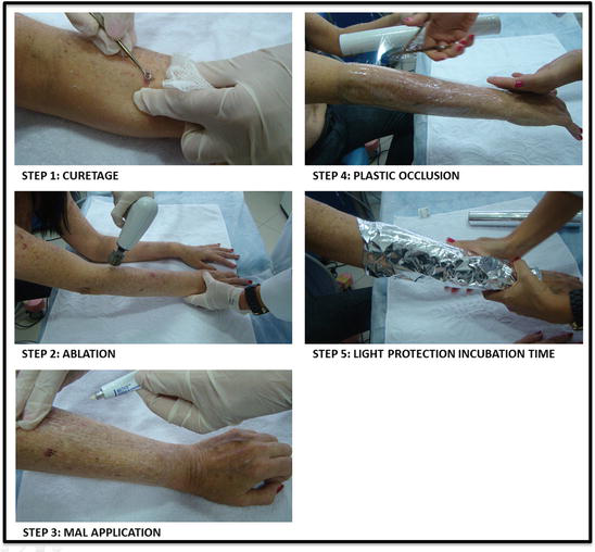
When associating ablative methods and PDT, the protocol is the following: the lesion is prepared with a superficial curettage, and the ablative laser or ablative RF is applied on the area to be treated, with low density. MAL is applied immediately after laser or RF, and it is occluded with plastic film and laminated paper. The incubation time can be reduced for 1–2 h (Issa et al. 2013a). Illumination with red light, dose of 37 J/cm2, is performed, as in the standard conventional PDT protocol (Fig. 1).
Literature Review and Author’s Experience
Some studies have been published about PDT associated with an ablative method for nonmelanoma skin cancer, extramammary Paget’s disease, and porokeratosis treatment with good results (Haak et al. 2012; Fukui et al. 2009). AFXL-assisted MAL-PDT has been studied in clinical practice, showing benefit to immunocompetent patients and immunosuppressed patients with AK and field cancerization (Paasch and Haedersdal 2011; Haedersdal et al. 2011; Togsverd-Bo et al. 2012).
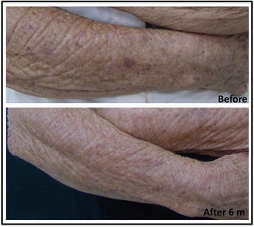
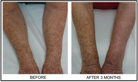
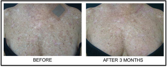
Kassuga and Issa et al. (Kassuga et al. 2011) reported that the incubation time could be reduced for 1 h when associating ablative RF before topical PDT for AK treatment, maintaining the AK cure rate and with better skin rejuvenation (Fig. 2). The authors also have good experience with CO2 laser before PDT for AK field of cancerization treatment (Figs. 3 and 4).
Three groups of AFXLs was described for TED x PDT: CO2 laser (10,600 nm), Er:YAG (erbium/yttrium-aluminum-garnet, 2,940 nm) laser and Er:YSGG (yttrium-scandium-gallium-garnet, 2,790 nm) laser. These lasers create vertical channels that facilitate the penetration of topically applied MAL into superficial and deep skin layers and promote an intensified PDT response (Haak et al. 2012; Paasch and Haedersdal 2011; Haedersdal et al. 2011).
It is reported that once the stratum corneum is disrupted by ablative fractional laser treatment, there will be no further benefit from drilling deeper laser channels for the delivery of topical photosensitizer. For this reason, some authors reported good results with erbium laser, which is not able to promote deep channels as CO2 laser does (Haak et al. 2012). The benefit of deeper channels produced by CO2 laser comparing with erbium lasers is still questioned.
Pretreatment with fractional laser resurfacing may be a new alternative technique to improve the efficacy of PDT for actinic cheilitis by increasing the bioavailability of MAL within the skin and enhancing the PDT response (Choi et al. 2015).
Very recently, daylight photodynamic therapy (DLPDT) was approved for AK treatment and field of cancerization with similar efficacy and less side effects, with few or no pain, comparing to conventional PDT (Morton et al. 2015). Clinical improvement in texture and pigmentation can also be observed after DLPDT treatment and for this reason can be indicated for skin rejuvenation. DLPDT is contraindicated for skin tumors.
The protocol for DLPDT isolated is already established, and a superficial curettage is done similar to the conventional PDT. However, a chemical sunscreen should be applied on the skin 15 min before applying MAL (not covered). Patient should be indoor for maximum 30 min before daylight exposure for 2 h. DLPDT with TED is a very new modality of treatment, and very few data are reported. DLPDT associated with TED seems to have better results for photorejuvenation, comparing with DLPDT isolated.
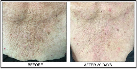
According to authors, experience to both microneedling and laser is safe to be used associated with DLPDT, but it seems that CO2 laser before DLPDT has better results comparing to microneedling associated with DLPDT for field of cancerization treatment (Fig. 5).
Take Home Messages
- PDT allows simple and effective treatment of multiple lesions simultaneously, avoiding numerous surgeries and unaesthetic scars.
- When treating NMSC with conventional PDT, it is supposed that inferior treatment outcomes can occur in thick lesions due to the limited penetration of the photosensitizer.
- Many studies evaluated the effectiveness of associating TED with PDT with the aim to improve photosensitizers delivery through the skin.
- AFXL-assisted MAL-PDT has been studied in clinical practice, showing benefit to immunocompetent patients and immunosuppressed organ transplant recipients with AK and field cancerization.
- TED is also a new option to enhance the delivery of ALA or MAL through the skin when using PDT for skin rejuvenation. For this purpose, TED and PDT have been described using microneedling technique and fractional ablative laser.
- Authors have good experience with ablative RF and CO2 laser before conventional PDT using MAL and red light.
- The association of DLPDT with laser and microneedling is a promising new modality for skin rejuvenation.
References
Aust MC, Fernandes D, Kolokythas P, Kaplan HM, Vogt PM. Percutaneous collagen induction therapy: an alternative treatment for scars, wrinkles and skin laxity. Plast Reconstr Surg. 2008;121(2):1421–9.
Bachhav YG, Summer S, Heinrich A, Bragagna T, Bohler C, Kalia YN. Using laser microporation to improve transdermal delivery of diclofenac: increasing bioavailability and the range of therapeutic applications. Eur J Pharm Biopharm. 2011;78(3):408–14.
Bagazgoitia L, Cuevas Santos J, Juarranz A, Jaén P. Photodynamic therapy reduces the histological features of actinic damage and the expression of early oncogenic markers. Br J Dermatol. 2011;165:144–51.
Baron ED, Harris L, Redpath WS, Shapiro H, Hetzel F, Morley G, Bar-Or D, Stevens SR. Laser-assisted penetration of topical anesthesia in adults. Arch Dermatol. 2003;139(10):1288–90.
Borelli C, Herzinger T, Merk K, et al. Effect of subcutaneous infiltration anesthesia on pain in photodynamic therapy: a controlled open pilot trial. Dermatol Surg. 2007;33:314–8.
Brancaleon L, Moseley H. Laser and non-laser light sources for photodynamic therapy. Lasers Med Sci. 2012;17:173–86.
Choi SH, Kim KH, Song K-H. Efficacy of ablative fractional laser-assisted photodynamic therapy for the treatment of actinic cheilitis: 12-month follow-up results of a prospective, randomized, comparative trial. Br J Dermatol. 2015;173:184–91.
Chung JH, Hanft VN, Kang S. Aging and photoaging. J Am Acad Dermatol. 2003;49:690–7.
Clementoni MT, B-Roscher M, Munavalli GS. Photodynamic photorejuvenation of the face with a combination of microneedling, red light, and broadband pulsed light. Lasers Surg Med. 2010;42:150–9.
Cox NH, Eedy DJ, Morton CA. Guidelines for management of Bowen’s disease: 2006 update. Br J Dermatol. 2007;156:11–21.
Di Nuzzo S, Cortelazzi C, Boccaletti V, et al. Comparative study of trichloroacetic acid vs. photodynamic therapy with topical 5-aminolevulinic acid for actinic keratosis of the scalp. Photodermatol Photoimmunol Photomed. 2015;31(5):233–8.
Dragieva G, Prinz BM, Hafner J, et al. A randomized controlled clinical trial of topical photodynamic therapy with methyl aminolaevulinate in the treatment of actinic keratoses in transplant recipients. Br J Dermatol. 2004;151:196–200.
Ericson MB, Wennberg A-M, Larkö O. Review of photodynamic therapy in actinic keratosis and basal cell carcinoma. Ther Clin Risk Manag. 2008;4(1):1–9.
Friedmann DP, Goldman MP, Fabi SG, Guiha I. The effect of multiple sequential light sources to activate aminolevulinic acid in the treatment of actinic keratoses: a retrospective study. J Clin Aesthetic Dermatol. 2014;7(9):20–5.
Fukui T, Watanabe D, Tamada Y, et al. Photodynamic therapy following carbon dioxide laser enhances efficacy in the treatment of extramammary Paget’s disease. Acta Derm Venereol. 2009;89:150–4.
Gill HS, Prausnitz MR. Coated microneedles for transdermal delivery. J Control Release. 2007;117:227–37.
Haak CS, Farinelli WA, Tam J, et al. Fractional laser-assisted delivery of methyl aminolevulinate: impact of laser channel depth and incubation time. Lasers Surg Med. 2012;44:787–95.
Haedersdal M, Katsnelson J, Sakamoto FH, Farinelli WA, et al. Enhanced uptake and photoactivation of topical methyl aminolevulinate after fractional CO2. Lasers Surg Med. 2011;43(8):804–13.
Hai-yan Zhang, Jie Ji, Yi-mei Tan, Ling-lin Zhang, et al. Evaluation of 5-aminolevulinic acid-mediated photorejuvenation of neck skin. Photodiagnosis Photodyn Ther. 2014;11(4):498–509.
Hartevelt MM, Bavinck JN, Kootte AM, et al. Incidence of skin cancer after renal transplantation in The Netherlands. Transplantation. 1990;49:506–9.
Hsiao CY, Huang CH, Hu S, Ko YS, Sung HC, Chen CC, Huang SY. Fractional carbon dioxide laser treatment to enhance skin permeation of ascorbic acid 2-glucoside with minimal skin disruption. Dermatol Surg. 2012;38(8):1284–93.
Issa MC, Kassuga LE, Chevrand NS, Pires MT, et al. Topical delivery of triamcinolone via skin pretreated with ablative radiofrequency: a new method in hypertrophic scar treatment. Int J Dermatol. 2013a;52:367–70.
Issa MCA, Kassuga LEBP, Chevrand NS, et al. Transepidermal retinoic acid delivery using ablative fractional RF associated with acoustic pressure ultrasound for stretch marks. Lasers Surg Med. 2013b;45:81–8.
Issa MCA, Pires M, Silveira P, et al. Transepidermal drug delivery: a new option for areata alopecia? J Cosmet Laser Ther. 2015;17:37–40.
Kalka K, Merk H, Mukhtar H. Photodynamic therapy in dermatology. J Am Dermatol. 2000;42(3):389–413.
Kassuga LEBP, Issa MCA, Chevrand NS. Transepidermal application of medication combined with photodynamic therapy in the treatment of actinic keratosis. Surg Cosmet Dermatol. 2011;3(4):89–92.
Kaviani A, Ataie-Fashtami L, Fateh M, et al. Photodynamic therapy of head and neck basal cell carcinoma according to different clinicopathologic features. Lasers Surg Med. 2005;36:377–82.
Kennedy JC, Pottier RH, Pross DC. Photodynamic therapy with endogenous protoporphyrins IX: basic principles and present clinical experience. J Photochem Photobiol B. 1990;6:143–8.
Lee WR, Shen SC, Wang KH, Hu CH, Fang JY. The effect of laser treatment on skin to enhance and control transdermal delivery of 5-fluorouracil. J Pharm Sci. 2002;91(7):1613–26.
Lee WR, Shen SC, Al-Suwayeh SA, Yang HH, Yuan CY, Fang JY. Laser-assisted topical drug delivery by using a low-fluence fractional laser: imiquimod and macromolecules. J Control Release. 2011;153(3):240–8.
Lien MH, Sondak VK. Nonsurgical treatment options for basal cell carcinoma. J Skin Cancer. 2011;5:1–6.
Lim HK, Jeong KH, Kim NI, Shin MK. Nonablative fractional laser as a tool to facilitate skin penetration of 5-aminolaevulinic acid with minimal skin disruption: a preliminary study. Br J Dermatol. 2014a;170:1336–40.
Lim HK, Jeong KH, Kim NI, Shin MK, Ko D-Y, Jeon S-Y, Kim K-H, Song K-H. Fractional erbium:YAG laser-assisted photodynamic therapy for facial actinic keratoses: a randomized, comparative, prospective study. J Eur Acad Dermatol Venereol. 2014b;28:1529–39.
Manstein D, Herron GS, Sink RK, Tanner H, Anderson RR. Fractional photothermolysis: a new concept for cutaneous remodeling using microscopic patterns of thermal injury. Lasers Surg Med. 2004;34(5):426–38.
Mikolajewska P, Donnelly R, Garland M, Morrow DIJ, Thakur RS, Juzeniene A. Microneedle pre-treatment of human skin improves 5-aminolevulinic acid (ALA)- and 5-aminolevulinic acid methyl ester (MAL)-induced PpIX production for topical photodynamic therapy without increase in pain or erythema. Pharm Res. 2010;27(10):2213–20.
Moraes AM. Doença de Bowen na região perianal tratada com criocirurgia com nitrogênio líquido. An Bras Dermatol. 2002;77(5):571. [online].
Morton CA, Whitehurst C, Moseley H, et al. Comparison of photodynamic therapy with cryotherapy in the treatment of Bowen’s disease. Br J Dermatol. 1996;135:766–71.
Morton C, MacKie RM, Whitehurst C, et al. Photodynamic therapy for basal cell carcinoma: effect of tumor thickness and duration of photosensitizer application on response. Arch Dermatol. 1998;134:248–9.
Morton C, Horn M, Leman J, et al. Comparison of topical methyl aminolevulinate photodynamic therapy with cryotherapy or fluorouracil for treatment of squamous cell carcinoma in situ: results of a multicenter randomized trial. Arch Dermatol. 2006;142:729–35.
Morton CA, Szeimies R-M, Sidoroff A, et al. European guidelines for topical photodynamic therapy part 1: treatment delivery and current indications – actinic keratoses, Bowen’s disease, basal cell carcinoma. J Eur Acad Dermatol Venereol. 2013a;27:536–44.
Morton CA, Szeimies R-M, Sidoroff A, et al. European guidelines for topical photodynamic therapy part 2: emerging indications – field cancerization, photorejuvenation and inflammatory/infective dermatoses. J Eur Acad Dermatol Venereol. 2013b;27:672–9.
Morton CA, Wulf HC, Szeimies RM, Gilaberte Y, Basset-Seguin N, Sotiriou E, Piaserico S, Hunger RE, Baharlou S, Sidoroff A, Braathen LR. Practical approach to the use of daylight photodynamic therapy with topical methyl aminolevulinate for actinic keratosis: a European consensus. J Eur Acad Dermatol Venereol. 2015;29(9):1718–23.
Moseley H, Allen JW, Ibbotson SH, et al. Ambulatory photodynamic therapy: a new concept in delivering photodynamic therapy. Br J Dermatol. 2006;154:747–50.
Neidecker MV, Davis-Ajami ML, Balkrishnan R, et al. Pharmacoeconomic considerations in treating actinic keratosis. Pharmacoeconomics. 2009;27(6):451–64.
Orringer JS, Hammerberg C, Hamilton T, Johnson TM, Kang S, Sachs DL, Hamilton T, Fisher GJ. Molecular effects of photodynamic therapy for photoaging. Arch Dermatol. 2008;144:1296–302.
Paasch U, Haedersdal M. Laser systems for ablative fractional resurfacing. Expert Rev Med Devices. 2011;8(1):67–83.
Park MY, Sohn S, Lee ES, Chan KY. Photorejuvenation induced by 5-aminolevulinic acid photodynamic therapy in patients with actinic keratosis: a histologic analysis. J Am Acad Dermatol. 2009;62(1):85–95.
Raab O. Über die Wirkung fluoreszierenden Stoffe auf Infusorien. Z Biol. 1900;39:524–46.
Rigel DS, Friedman RJ, Kopf AW. Lifetime risk for development of skin cancer in the U.S. population: current estimate is now 1 in 5. J Am Acad Dermatol. 1996;35(6):1012–3.
Rosen T, Lebwohl MG. Prevalence and awareness of actinic keratosis: barriers and opportunities. J Am Acad Dermatol. 2013;68:S2–9.
Rubel DM, Spelman L, Murrell DF, et al. Daylight photodynamic therapy with methyl aminolevulinate cream as a convenient, similarly effective, nearly painless alternative to conventional photodynamic therapy in actinic keratosis treatment: a randomized controlled trial. Br J Dermatol. 2014;71:1164–71.
Sandberg C, Halldin CB, Ericson MB, et al. Bioavailability of aminolaevulinic acid and methylaminolaevulinate in basal cell carcinomas: a perfusion study using microdialysis in vivo. B J Dermatol. 2008a;159:1170–6.
Sandberg C, Halldin CB, Erickson MB, et al. Bioavailability of aminolevulinic acid and methylaminolevulinate in basal cell carcinomas: a perfusion study using microdialysis in vivo. Br J Dermatol. 2008b;159(5):1170–6.
Sjerobabski Masnec I, Situm M. Photorejuvenation – topical photodynamic therapy as therapeutic opportunity for skin rejuvenation. Coll Antropol. 2014;38(4):1245–8.
Sklar LR, Burnett CT, Waibel JS, Moy RL, Ozog DM. Laser assisted drug delivery: a review of an evolving technology. Lasers Surg Med. 2014;46:249–62.
Svanberg K, Andersson T, Killander D, et al. Photodynamic therapy of non-melanoma malignant tumours of the skin using topical aminolevulinic acid sensitization and laser irradiation. Br J Dermatol. 1994;130:743–51.
Szeimies RM, Ibbotson SH, Murrell DF, et al. A clinical study comparing methyl aminolevulinate photodynamic therapy and surgery in small superficial basal cell carcinoma with a 12-month follow-up. J Eur Acad Dermatol Venereol. 2008;22:1302–11.
Tarstedt M, Rosdahl I, Berne B, et al. A randomized multicenter study to compare two treatment regimens of topical methyl aminolevulinate (Metvix)-PDT in actinic keratosis of the face and scalp. Acta Derm Venereol. 2005;85:424–8.
Togsverd-Bo K, Haak CS, Thaysen-Petersen D, et al. Intensified photodynamic therapy of actinic keratoses with fractional CO2 laser: a randomized clinical trial. Br J Dermatol. 2012;166:1262–9.
Torezan L, Chaves Y, Niwa A, et al. A pilot split-face study comparing conventional methyl aminolevulinate-photodynamic therapy (PDT) with microneedling-assisted PDT on actinically damaged skin. Dermatol Surg. 2013;39:1197–201.
Traianou A, Ulrich M, Apalla Z, et al. Risk factors for actinic keratosis in eight European centres: a case–control study. Br J Dermatol. 2012;167:36–42.
von Tappeiner H, Jesionek H. Therapeutische versuche mit fluoreszierenden stoffen. Munch Med Wochenschr. 1903;47:2042–4.
Wang I, Bendsoe N, Klinteberg CAF, et al. Photodynamic therapy vs. cryotherapy of basal cell carcinoma: results of a phase III clinical trial. Br J Dermatol. 2001;144:832–40.
Wermeling DP, Banks SL, Hudson DA, Gill HS, Gupta J, Prausnitz MR, Stinchcomb AL. Microneedles permit transdermal delivery of a skin-impermeant medication to humans. Proc Natl Acad Sci U S A. 2008;105(6):2058–63.
Zhang H, Zhai Y, Yang X, Zhai G. Breaking the skin barrier: achievements and future directions. Curr Pharm Des. 2015;21(20):2713–24.







