
Microneedling for Transepidermal Drug Delivery on Stretch Marks
Microneedling for Transepidermal Drug Delivery on Stretch Marks
Gabriela Casabona and Paula Barreto Marchese Clinica Vida – Cosmetic, Laser and Mohs Surgery Center, Rua Dr Veiga Filho 350, 01229-000 São Paulo, Brazil
Abstract
Stretch marks (SMs) are a well-recognized, common skin condition that rarely cause any significant medical problems but are often a significant source of distress to those affected. The origins of SM are poorly understood, and a number of treatment modalities (Elsaie et al. 2009) are available for their treatment, yet none of them is consistently effective, and no single therapy is considered to be the consensus for this problem. Multiple sittings of treatments such as chemical peelings, microdermabrasion, nonablative and ablative laser techniques, and light and radiofrequency devices are performed to improve the SM appearance. Microneedling and transepidermal drug delivery together are a new modality of treatment for SM. It is based on stimulation of collagen production and enhancement of the penetration of certain active substances across the skin, with which it is possible to achieve satisfactory results.
Keywords MicroneedlingTransdermal drug deliveryTransepidermal drug deliveryStretch marksCollagen
Introduction
Stretch marks (SMs) are an undesirable cutaneous disorder which causes significant cosmetic problems with an evident psychological impact. There is loss of the normal random collagen distribution to the level of the mid-dermis or deeper (Singh and Kumar 2005). A single treatment modality that is consistently effective with minimal adverse effects does not exist to date. There are several modalities of treatment such as topical tretinoin 0.1% cream, topical 20% glycolic acid, microdermabrasion, lasers, and light and radiofrequency devices.
Microneedling therapy is a new addition to the treatment techniques for SM. It is a simple, inexpensive office procedure with little downtime that allows collagen stimulation and transepidermal drug delivery (TDD). The goal of percutaneous collagen induction is to stimulate collagen production by using the chemical cascade that happens after any trauma. Approximately 5 days after the skin injury, a fibronectin matrix forms with an alignment of the fibroblasts that determines the deposition of collagen (Widgerow 2012). Treatment with skin needling should be able to promote the removal of old damaged collagen and induce more collagen growth beneath the epidermis. The microneedling devices have micron-sized needles that can perforate the skin in a minimally invasive, low-pain manner (Kaushik et al. 2001), thereby creating aqueous transport pathways within the skin referred to as microchannels. Many active substances can be delivered into the skin by microneedling. In this chapter, we will focus on the delivery of three components which are effective in treating stretch marks: vitamin A, vitamin C, and platelet-rich plasma (PRP). The development of microneedling associated with drug delivery seems to be a good option in SM treatment. Combined, they are able to effectively change the collagen organization, transforming SM skin into healthier skin.
Stretch Marks
Stretch marks (SMs) are an undesirable cutaneous disorder which causes significant cosmetic problems with an evident psychological impact. It occurs mainly in adolescence, pregnancy, and obesity (Satish 2009).
The causes of SM are not clear, and a number of theories have been proposed: infection leading to the release of striatoxin that damages the tissues in a microbial-toxic way; the mechanical effect of stretching, which is proposed to lead to rupture of the connective tissue framework (e.g., pregnancy, obesity, weight lifting); normal growth as seen in adolescence and the pubertal spurt that leads to an increase in size of particular body regions; an increase in the levels of adrenocorticotropic hormone and cortisol, which are thought to promote fibroblast activity leading to increased protein catabolism and thus alterations to collagen and elastin fibers (Cushing’s syndrome, local or systemic steroid therapy); and genetic factors (absence of striae in pregnancy in people with Ehlers-Danlos syndrome and their presence as one of the minor diagnostic criteria for Marfan syndrome suggest an important genetic element).
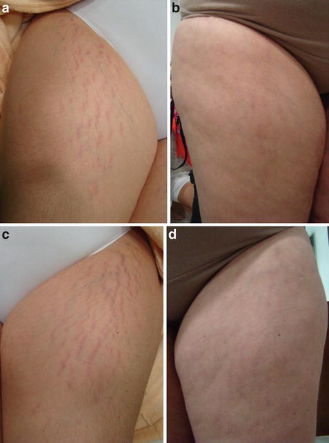
SM can be divided into striae rubrae (Fig. 1a) and striae albae (Fig. 1b). They are distinct, evolutionarily linked forms of SM, and their distinction has therapeutic implications. The color of the SM is related to the stage of evolution and to melanocyte mechanobiological influences.
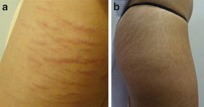
Striae rubra is an erythematous stria; as it matures, it becomes white and finally more atrophic; it is an atrophic dermal scar with overlying epidermal atrophy. Initially, striae appear as flattened areas of thinned skin with a pink-red hue that may be pruritic. They usually increase in length and width and acquire a darker reddish-purple appearance over time. The typical white, depressed appearance of older striae develops with time along the long axis, aligned parallel with the normal lines of skin tension.
Histologically, striae display an atrophic, thinned epidermis with a flattening of the rete pegs. There is loss of the normal random collagen distribution to the level of the mid-dermis or deeper. Elastin stains reveal scarce or absent elastin fibers and reduced fibrillin in the papillary and reticular dermis within affected areas; the elastin fibers present appear tangled and frayed. Those predisposed to developing SM may have an underlying deficiency of fibrillin 54. This histologic appearance probably results from mast cell degranulation and macrophage activation (McDaniel 2002), with resultant destruction of both collagen and elastin fibers (Budamakuntla 2013).
A universal approach to evaluating the severity of SM, and a treatment modality that is consistently effective with minimal adverse effects, do not exist to date. The several modalities of treatment are described below.
Topical Treatments
- Tretinoin 0.1% cream is thought to work through its affinity for fibroblasts and induction of collagen synthesis (Bernard 2002). It has maximal efficacy in striae rubrae and poor, unpredictable responses in striae albae. It seems to decrease the mean length and width of the SM after months of daily use. Studies have demonstrated that improvement in the clinical appearance of striae after treatment with retinoic acid correlates with new fibrillin production (Fisher 1995).
- Creams and lotions that contain actives such as Centella asiatica extract, vitamin E, vitamin A, collagen-elastin hydrolysates, panthenol, and menthol are widely used, mainly by women during pregnancy. Scientific data available are not sufficient to conclude that such creams are effective, and larger studies are needed to determine the efficacy and safety of such products. There is no statistically significant evidence to support their use in the prevention of SM.
- Topical 20% glycolic acid (GA), pure or mixed with other substances such as 0.05% tretinoin or 10% L-ascorbic acid, improves the appearance of striae alba, but the precise mechanism of action of GA is still unknown. It is reported that GA stimulates collagen production by fibroblasts and increases their proliferation, but further investigations and studies are required to prove this theory.
Lasers and Light Devices
Intense pulsed light (515–1,200 nm), a 585 nm flashlamp-pumped pulsed dye laser (PDL), a 308 nm xenon chloride excimer laser, a 577 nm copper bromide laser, a 1,450 nm diode laser, a 1,064 nm Nd:YAG laser, a carbon dioxide (CO2) laser, a fractionated 1,550 nm erbium-doped fiber laser, and fractional photothermolysis are some examples of technologies that can be used in SM treatment (Alster and Handrick 2000, 2005). Before selecting a device, one must correctly analyze the SM and the patient’s Fitzpatrick skin type to diminish the risk of injuries and pigmentary alterations.
Radiofrequency Devices
The effects of dermal heating are well recognized and include immediate effects on collagen structure, with stimulation of dermal fibroblasts inducing synthesis of new collagen fibers (neocollagenesis) and elastic fibers (neoelastogenesis) (Al-Himdani et al. 2014; Gold 2015), improving the appearance and histological findings in SM (Suh et al. 2007).
Microdermabrasion
Microdermabrasion is a skin resurfacing technique using aluminum oxide. It has been reported to increase type I collagen. This technique can be used in combination with intradermal platelet-rich plasma (PRP); in one study, patients were treated with a combination of intradermal PRP and microdermabrasion in the same session. There was significant clinical improvement of SM in patients treated with the combination of PRP and microdermabrasion when compared with patients treated with microdermabrasion alone. A combination of PRP and microdermabrasion in the same session showed better results in the short term (Ibrahim et al. 2015).
Microneedling Therapy
Microneedling therapy is a new addition to the treatment techniques for SM. It is a simple, inexpensive office procedure with little downtime that allows collagen stimulation and transepidermal drug delivery (TDD). Substances such as vitamin A (retinyl palmitate and retinyl acetate), vitamin C, and platelet-rich plasma (PRP) can be used for TDD. In this chapter, we describe the use of the microneedling technique as a way of stimulating collagen and facilitating the delivery of active ingredients to the skin affected by stretch marks.
Microneedling
History
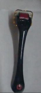
In 1995 Orentreich described collagen percutaneous stimulation with dermal needling for scars. Then, in 1997, Camirand and Doucet described needle dermabrasion using a “tattoo pistol” to treat scars. Only in 2006 did a doctor from South Africa, Dr Desmond Fernandes, develop percutaneous collagen induction therapy with the dermaroller (Dermaroller®) (Henry et al. 1998) (Fig. 2). In 2010 a new electronic pen-shaped device (Dermapen®) (Fig. 3) was developed in Australia for a more cost-effective procedure. Using the pen, one can choose different needle lengths and vibration speeds, providing not only microneedling but also a certain level of dermabrasion depending on the technique used.

Since then, the use of microneedling devices has been growing along with the number of indications. The addition of transepidermal drug delivery to the microneedling technique made it even more interesting for treating certain conditions, such as stretch marks and melasma.
The Devices
Since 2006, when the first dermaroller was used by Dr Fernandes, the device has undergone numerous changes to become more ergonomic and resistant. The standard dermaroller used for acne scars is a drum-shaped roller studded with 192 fine microneedles in 8 rows, 0.5–1.5 mm in length and 0.1 mm in diameter. The microneedles are produced by reactive ion etching techniques on silicon or medical-grade stainless steel. The instrument is sterilized by gamma irradiation. Thinner and larger versions are now available, and needle length can be up to 2.5 mm.
As the roller is supposed to be disposable, if more than one needle length is indicated the doctor has to use more than one roller, which is not cost-effective for the patient. For example, to treat a patient under the eyes and a scar in the malar area, both 1.0 mm and 1.5–2.0 mm needle lengths would be required.
Other electronic pen-shaped microneedle devices were developed with disposable needles. The length and the vibration speed of the exposed needle, in and out, can be controlled. With these devices, one can not only create microtunnels but also achieve a certain level of dermabrasion of the epidermis if that is the goal.
Mechanisms of Action
The microneedle devices create microtunnels that vary from 0.5 to 2.5 mm in depth (Cho 2010). The goal of percutaneous collagen induction is to stimulate collagen production by using the chemical cascade that happens after any trauma. There are three phases in the body’s wound healing process (Tejero-Trujeque 2001), which follow each other in a predictable fashion. This has been well described in The Biology of the Skin by Falabella and Falanga (Falabella et al. 2000). Platelets and eventually neutrophils release growth factors such as connective tissue growth factor, TGF-β1, TGF-β3, platelet-derived growth factor, connective tissue activating protein III, and others that work together to increase production of the intercellular matrix (Lynch 1989; Tran 2004; Faler 2006) (Fig. 4). Only then do monocytes also produce growth factors to increase production of collagen III, elastin, glycosaminoglycans, and so on (Johnstone 2005).
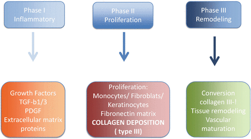
Approximately 5 days after the skin injury, a fibronectin matrix forms with an alignment of the fibroblasts that determines the deposition of collagen. Eventually, collagen III is converted into collagen I, which remains for 5–7 years. Due to this conversion, the collagen tightens naturally over a few months (Martin 2005).
Normally, under the usual conditions of wound healing, scar tissue is formed with minimal regeneration of normal tissue (Fenske 1986). Percutaneous collagen induction causes further tightening of lax skin and smoothing of scars and wrinkles several weeks or even months after the injury (Fernandes 2002). In addition, percutaneous collagen induction has proven to be very effective in minimizing acne and burn scars by promoting replacement of scar collagen with normal collagen and reduction of depressed and contracted scars (Ruszczak 2003).
Treatment with skin needling should be able to promote the removal of old damaged collagen and induce more collagen growth beneath the epidermis. Puncturing the skin multiple times in acne scars increases the amount of collagen and elastin deposition. Thus, it was hypothesized that skin needling would also be useful in SM, because these seem to be dermal scars with epidermal atrophy (Majid 2009).
Microneedling and SM
A study performed in South Korea in 2012 enrolled 16 volunteers with SM, who received three treatments with a microneedling device at 4-week intervals. Clinical response to treatment was assessed by comparing pre- and posttreatment clinical photographs, skin biopsies, and patient satisfaction scores. The general histopathologic features of the lesional specimens collected before treatment showed epidermal thinning with fine dermal collagen bundles arranged in straight lines. After treatment, the epidermis was thickened, and the amounts of dermal collagen and elastic fibers were increased (McCrudden et al. 2015). This study proved that microneedling can be used effectively and safely for SM treatment.
In addition to this technique, transepidermal drug delivery can be associated in order to increase collagen production.
Microneedling and Transepidermal Drug Delivery
The dispersion and effectiveness of active ingredients into the skin without prior perforation are severely limited by the inability of the great majority of drugs to cross the skin at therapeutic rates, due to the great barrier imposed by the skin’s outer stratum corneum layer (Menon et al. 2012).
In 2004 Prausnitz (Prausnitz 2004) described the use of microneedles for transdermal drug delivery. The outstanding motivation for microneedles is that they can provide a minimally invasive means of transporting molecules into the skin (Oh et al. 2015).
As with lasers, the microneedling technique started being used as a way to cross the stratum corneum to deliver active ingredients to the skin in a more efficient way (Brauer et al. 2014).
The stratum corneum (SC), the skin’s outermost layer, is a barrier that prevents molecular transport across the skin. Therapeutic agents such as peptides, proteins, and oligonucleotides have difficulty reaching the deeper layers of the skin by conventional methods or topical delivery (Petchsangsai et al. 2014).
The microneedling technique uses micron-sized needles that can perforate the skin in a minimally invasive, low-pain manner, thereby creating aqueous transport pathways within the skin referred to as microchannels (Park et al. 2012).
Moreover, these microchannels present no limitation regarding the size of molecules that can pass through their tunnels (Kumar and Banga 2012). While the microchannels are in the micron range in terms of size, the macromolecules delivered are typically nanometers in size.
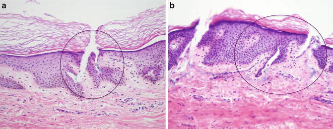
The only question that remains is whether, after microneedling, blood and fibrin will invade the microchannels, creating a new barrier to this penetration (Milewski et al. 2010). Consequently, as shown by the biopsies presented in Fig. 5, we recommend proceeding with an inverse technique, where the drug is applied prior to the microneedling technique and before each step, to guarantee more efficient delivery of the active ingredient.
In the past, local microinjections, the so-called mesotherapy, were introduced in France by Pistor (Pistor 1979). Mesotherapy is a widely used technique in medicine. This technique consists of intradermal or subcutaneous microinjections of 0.05–0.1 mL of highly diluted drug mixtures, or a single drug, on body parts affected by medical or aesthetic problems. Nevertheless, recent studies show that drug delivery with microneedling is more effective than mesotherapy because it not only allows penetration of active ingredients into the skin but also induces the chemical cascade that takes place after trauma, which stimulates collagen production.
Many active substances can be delivered into the skin by microneedling. Depending on the disease to be treated, a number of substances can be chosen, for example steroids (triamcinolone acetonide) for hypertrophic scars and alopecia areata, tranexamic acid for melasma, minoxidil for androgenetic alopecia, and aminolevulinic acid for actinic keratosis.
In this chapter, we focus on the delivery of three components which are effective in treating stretch marks: vitamin A, vitamin C, and platelet-rich plasma (PRP).
Vitamin A
The possibility of using vitamins A and C for percutaneous collagen induction was described by Aust in 2008. Vitamin A, a retinoic acid, is an essential vitamin also considered a hormone for the skin. It expresses its influence on many genes that control proliferation and differentiation of all the major cells in the epidermis and dermis (Rosdahl 1997). Percutaneous collagen induction and vitamin A switch on the fibroblasts to produce collagen and therefore increase the need for vitamin C. Vitamin A may control the release of TGF-β3 in preference to TGF-β1 and TGF-β2, because in general retinoic acid seems to favor development of a regenerative lattice-patterned collagen network rather than the parallel deposition of scar collagen found with cicatrization (Oliveira et al. 2016).
Retinyl palmitate (RP) is an ester of retinol and is the major form of vitamin A found in the epidermis. It has a high molecular weight and a stable formulation. To be active, RP has to be enzymatically converted in the skin to retinol by cleavage of the ester linkage and then converted to tretinoin via oxidative processes. It has been established that topical administration of RP for 14 days in rats resulted in increased protein and collagen and epidermal thickening (Ro et al. 2015).
Ascorbic Acid (Vitamin C)
Ascorbic acid (AA), or vitamin C, is also essential for the production of normal collagen. Percutaneous collagen induction and vitamin A stimulate the fibroblasts to produce collagen and therefore increase the need for vitamin C. Topical vitamins A and C both maximize the initial release of growth factors and stimulate collagen production (Palma 2006).
Apart from its role as a potent antioxidant, it has also been demonstrated that AA functions as an essential cofactor for the enzymes lysyl hydroxylase and prolyl hydroxylase, both of which are required for the posttranslational processing of types I and III collagen (Nusgens 2001).
AA stimulates collagen production in the dermis and can cause a dramatic increase in fibroblast proliferation, potentially resulting in greater collagen production; it might be presumed that AA also possesses the potential to increase collagen production for reduction of SM appearance. AA even plays a role in collagen synthesis at the level of gene expression. It has been shown to upregulate collagen synthesis and increase synthesis of the inhibitor of metalloproteinase-1, which decreases UV-induced collagen degradation.
Platelet-Rich Plasma (PRP)
PRP is a source of numerous growth factors which facilitate repair and healing. It is a potential reservoir of essential growth factors, including platelet-derived growth factor, vascular endothelial growth factor, transforming growth factor-beta 1, and insulin-like growth factor, which play an important role in recovery of the skin (Sonker et al. 2015).
It has been found to accelerate endothelial, epithelial, and epidermal regeneration, stimulate angiogenesis, enhance collagen synthesis, promote soft tissue healing, decrease dermal scarring, enhance the hemostatic response to injury, and reverse the inhibition of wound healing caused by glucocorticoids.
There are different preparation protocols to obtain PRP, and physicians should select proper PRP preparations after considering their biomolecular characteristics and patient indications.
A split-face comparative study published in 2014 of microneedling with PRP versus microneedling with vitamin C in treating atrophic post-acne scars revealed better results with microneedling and PRP. Thirty patients with post-acne atrophic facial scars were offered four sittings of microneedling with PRP on one side and microneedling with vitamin C on the other side of the face at an interval of 1 month. Overall results were better with microneedling and PRP (Chawla 2014).
PRP along with microneedling intensifies the natural wound healing cascade because of the high concentration of the patient’s own growth factors. It acts synergistically with growth factors induced by skin needling in order to enhance the wound healing response. As stretch marks are atrophic scars, we can hypothesize that microneedling and PRP would have good results for them.
Uses and Doses
It is very important to ensure that the substances to be applied on the skin surface before microneedling are substances that could be used intradermally or intravenously. For stretch marks’ treatment, we indicate the use of:
- 15–20% vitamin C (L-ascorbic acid 0.15 g/mL or L-ascorbic acid 0.2 g/mL) ampoules.
- Retinyl palmitate (an ester of retinol) ampoules for intravenous administration can be safely applied topically.
- MTS® ampoules: a combination of palmitoyl tripeptide-28 3% and growth factors 10%.
- PRP preparations (follow proper protocols).
Procedure
During treatment, the needles pierce the stratum corneum and create microconduits (holes) without damaging the epidermis. It has been shown that rolling with a dermaroller (192 needles, 200 mm length and 70 mm diameter) over an area 15 times will result in approximately 250 holes/cm2.
Microneedling is a mild-pain method of drug delivery. Because the skin’s stratum corneum barrier has no nerves, skin anatomy provides the opportunity to pierce needles across the stratum corneum without stimulating nerves. However, the microneedles are inserted only deep enough to reach the superficial dermis, which is not very painful, probably because their small size reduces the odds of encountering a nerve or of stimulating it to produce a strong painful sensation (Figs. 6a, b and 7a, b).

- Take photographs of the area to be treated using a consistent background, position, and lighting; these will be compared with the posttreatment images.
- Anesthetize using a thick application of topical anesthetic cream for about 60 min. Remove it completely before starting the procedure to prevent topical anesthetic intoxication.
- Clean the area with alcoholic chlorhexidine.
- Choose a device: roller or pen.
- Choose the needle depth and the speed of the pen (quicker vibrations lead to less abrasion).
- Choose the active ingredient to use (the use of sterile, injectable products is important to avoid the risk of infection and also diminish the risk of contact dermatitis).
- Apply a thin layer of the liquid with a disposable brush.
- Pass the device and repeat the process over and over. Roll at least 20 passes over the same area in different directions, and with the pen pass at least ten times in each area making circular movements.
- Clean only the excess liquid and blood (leave a thin layer of blood for at least 4 h to function as a natural PRP dressing that helps healing).
- Cover the skin with a sterile active ingredient in an oily vehicle such as glycosaminoglycans (Antiage Flash® Mesoestetic) to prevent water loss through the skin.
- Cover with a thin plastic drape.
- Instruct the patient to follow strict photoprotective measures. Schedule sittings at intervals of 4 weeks.
Other Indications
- Androgenetic alopecia
- Alopecia areata
- Acne scars
- Hypertrophic scars
- Melasma
- Skin regeneration
- Preparation of skin prior to fat graft
Contraindications
- History of contact dermatitis, especially to metals and to the active ingredients that are going to be used
- Chronic urticaria
- Immunosuppression
- Diabetes
- Pustular or nodular rosacea or acne
- Anticoagulant medications
- Pregnancy
- Moles (always verify moles prior to treating the area, and try to avoid using microneedling on top of them)
- Keloids
- Skin infection
- Psoriasis (Koebner phenomenon)
Advantages
- Nonablative.
- Healing process is fast (1–5 days depending on needle length and number of passes) and has less chance of complications compared with ablative techniques.
- The immediate effects are thicker and more resistant skin.
- Can be used in every skin type, even off the face.
- Low cost compared with lasers.
Disadvantages
- Training required.
- If a very high density or coverage is used, healing time and erythema can last longer.
- Does not promote immediate tightening of the collagen fibers.
- Requires more than one session to achieve good results.
Side Effects and Their Management
The microneedling treatment is well tolerated. Minor side effects reported are mild pain at the treated area, erythema, spotty bleeding, and pruritus for 2 or 3 days after the procedure, which resolve without any specific treatment. Moisturizing creams can be applied to keep the area hydrated, which diminishes discomfort.
Infection at the treated area is very unlikely. As the microholes close almost immediately, postoperative infections are rare. If they occur, oral or topical antibiotics can be used for treatment.
Irritant or allergic contact dermatitis can occur depending on the characteristics of the substance applied on the skin surface and the immunological characteristics of the patient. Oral antihistamines and topical steroids can be used if necessary (Soltani-Arabshahi et al. 2014).
Hyperpigmentation is uncommon, but if it happens a topical Kligman formula can be applied. For long-lasting erythema, micropulsed YAG laser, pulsed dye laser (PDL), and intense pulsed light (IPL) devices can be used. The emergence of papules and pustules on the treated area must be treated with the topical or oral antibiotics used in acne and rosacea treatment.
A case report study published in 2014 reports three patients who developed a facial allergic granulomatous reaction and systemic hypersensitivity after microneedling drug delivery therapy. Microneedles are a powerful means of transdermal delivery of drugs. Thus, only chemicals approved for intradermal injection are safe to be used in conjunction with microneedling. Application of various nonapproved topical products before a microneedling procedure can introduce immunogenic particles into the dermis and potentiate local or systemic hypersensitivity reactions (Lima et al. 2013). For facial allergic granulomatous reaction and systemic hypersensitivity, oral steroids and minocycline can be used, but the treatment is not always effective.
Microneedling and Vitamin C Drug Delivery Plus Calcium Hydroxylapatite Fillers: A Pilot Retrospective Study for Stretch Marks
Calcium hydroxylapatite (CaHA) is a white substance made of microspheres (45 μm) of CaHA in a carboxymethyl cellulose gel; it is a filler and biostimulator because it is supposed to induce neocollagenesis during the first 6 months after injection. CaHA can be used either in the subcutaneous layers to volumize a region or in the dermis to correct dermal atrophy. It is made to be applied in deeper planes such as subcutaneously, and when it is injected more superficially it can give a yellowish look. CaHA injection into SM at all depths is intended to improve atrophy (Casabona and Michalany 2014) through stimulation of the production of endogenous collagen, and also to impart a yellowish look to the striae, promoting a more natural appearance matching the color of normal skin (Berlin et al. 2008; Pavicic 2015).
Authors’ Experience
A retrospective study conducted at a private office in São Paulo, Brazil (in the publishing process), over a 3-year period (January 2012 to July 2015), enrolled 35 patients with stretch marks in different regions of the body (gluteus, thighs, knees, abdomen, and breasts) and evaluated the efficacy of a new combined treatment for SM; dermal injection of calcium hydroxylapatite and microneedling with vitamin C drug delivery were combined to enhance the appearance of SM.
Among the 35 patients, there was one male (2.85%) and 34 females (97.14%). Twenty-five (71.42%) had red SM and ten (28.57%) had white SM. Ages ranged between 21 and 34 years, and they were submitted to the same treatment for SM (Table 1).
| Substance | Effect |
|---|---|
| Vitamin A | Growth factor release; regulates differentiation and proliferation of the epidermis and dermis; skin regeneration; increased protein and collagen; epidermal thickening |
| Vitamin C | Stimulates collagen production in the dermis; increases fibroblast proliferation, resulting in greater collagen production |
| Platelet-rich plasma | Enhances collagen synthesis |
Patients had their SM evaluated by a physician observer, and scores were assigned in accordance with a visual analogue scale, the Manchester Scar Scale (Table 2). This evaluation was performed at the beginning and after the end of the treatment sessions. Both scores were compared in order to identify whether there was an improvement in the appearance of stretch marks.
| Side effect | Management |
|---|---|
| Infection | Oral or topical antibiotics |
| Contact dermatitis | Oral antihistamines and topical steroids |
| Hyperpigmentation | Topical Kligman formula |
| Long-lasting erythema | Micropulsed YAG laser, PDL or IPL |
| Acne and rosacea | Oral antibiotics and topical acne treatment |
| Facial allergic granulomatous reaction and systemic hypersensitivity | Oral steroids, minocycline |
| Antibiotic options include lymecycline, minocycline, and tetracycline. | |
Patients were submitted to four treatment sessions with a 4-week interval between them. The first session included dermal injection of calcium hydroxylapatite (Radiesse®) followed by microneedling (Dermapen®) and topical application of 20% vitamin C onto the affected areas. The three remaining sessions included microneedling with vitamin C drug delivery, with no dermal injections.
All 35 patients had better scores according to the Manchester Scar Scale evaluation at the end of the study. Thirty patients were asked about their level of satisfaction with the treatment: 8 patients (27%) were very satisfied, 15 patients (50%) were satisfied, 5 patients (17%) were neither satisfied nor dissatisfied, 2 patients (6%) were unsatisfied, and none answered very unsatisfied.
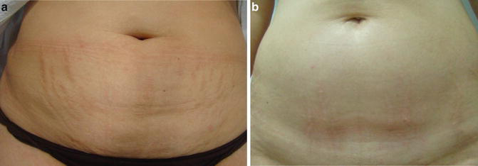
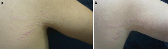
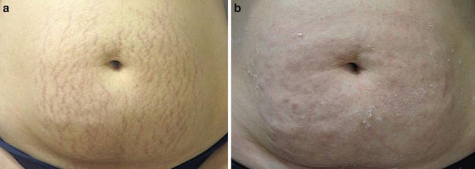
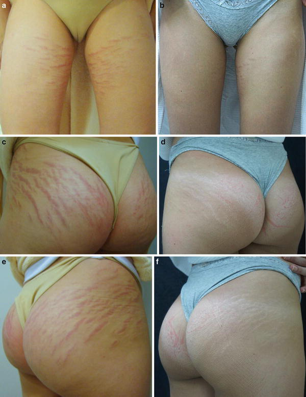
Results are encouraging. With injection of calcium hydroxylapatite and microneedling with topical vitamin C, it is possible to stimulate collagen production in three different pathways at the same time. This combined technique may have better results than those obtained when each technique is performed alone (Figs. 8a, b, 9a, b, 10a–f, and 11a–d).
Conclusions
The improvement in the appearance of SM, rather than its complete removal, is a more realistic clinical goal. Complete disappearance of SM may occasionally be observed; nevertheless, this occurrence is uncommon and should not be presented to patients as a realistic goal.
Using a combination of treatments over a prolonged period of time can improve the appearance of striae. Unless a revolutionary monotherapy becomes available that dramatically improves striae treatment results, the use of multimodal treatments tends to increase consistently (Goldberg et al. 2005).
The development of microneedling associated with drug delivery seems to be a good option in SM treatment. Combined, they are able to effectively change the collagen organization, transforming SM skin into healthier skin.
Take Home Messages
- Stretch marks are often a significant source of distress to those affected, and no single therapy is considered to be the consensus for this problem.
- Microneedling and transepidermal drug delivery together are a treatment option with which it is possible to achieve satisfactory results.
- Skin needling promotes the removal of old damaged collagen, induces more collagen growth beneath the epidermis, and creates aqueous transport pathways within the skin for drug delivery.
- Vitamin A, vitamin C, and platelet-rich plasma are substances that can be used for drug delivery.
- Microneedling with drug delivery is a cost-effective office procedure, well tolerated, with few minor side effects and short downtime, that can be safely used for SM treatment.
- Injection of calcium hydroxylapatite into SM before microneedling with topical vitamin C is an innovative combined technique that may have even better results.
- The improvement in the appearance of SM, rather than its complete removal, is a more realistic goal.
References
Al-Himdani S, Ud-Din S, Gilmore S, Bayat A. Striae distensae: a comprehensive review and evidence-based evaluation of prophylaxis and treatment. Br J Dermatol. 2014;170:527–47.
Alster TS, Handrick C. Laser treatment of hypertrophic scars, keloids and striae. Semin Cutan Med Surg. 2000;19:287–92.
Alster TS, Handrick C. Laser treatment of scars and striae. In: Kuvar ANB, Hruza G, editors. Principles and practices in cutaneous laser surgery. New York: Marcel Dekker; 2005. p. 625–41.
Aust MC, Knobloch K, Reimers K, Redeker J, Ipaktchi R, Altintas ML, Gohritz A, Schwaiger N, Vogt PM. Percutaneous collagen induction therapy: an alternative treatment for burn scars. Burns. 2010;36(6):836–43.
Aust M, et al. Percutaneous collagen induction therapy: an alternative treatment for scars, wrinkles, and skin laxity. Plast Reconstr Surg. 2008;121(4):1421–9.
Berlin AL, Mussarratt H, Goldberg DJ. Calcium hydroxylapatite filler for facial rejuvenation: a histologic and immunohistochemical analysis. Dermatol Surg. 2008;34:S64–7.
Bernard FX. Comparison of gene expression profiles in human keratinocyte monolayer cultures, reconstituted epidermis and normal human skin: transcriptional effects of retinoid treatments in reconstituted human epidermis. Exp Dermatol. 2002;11:59.
Brauer J, et al. Convergence of anatomy, technology, and therapeutics: a review of laser-assisted drug delivery. Semin Cutan Med Surg. 2014;33:176.
Budamakuntla L. A randomised, open-label, comparative study of tranexamic acid microinjections and tranexamic acid with microneedling in patients with melasma. J Cutan Aesthet Surg. 2013;6(3):139.
Camirand A, Doucet J. Needle dermabrasion. Aesthet Plast Surg. 1997;21:48–51.
Casabona G, Michalany N. Ultrasound with visualization and fillers for increased neocollagenesis: clinical and histological evaluation. Dermatol Surg. 2014;40:S194–8.
Chawla S. Split face comparative study of microneedling with PRP versus microneedling with vitamin C in treating atrophic post-acne scars. J Cutan Aesthet Surg. 2014;7(4):209–12.
Cho CW. In vitro characterization of the invasiveness of polymer microneedle against skin. Int J Pharm. 2010;397:201–5.
Elsaie ML, Baumann LS, Elsaaiee LT. Striae distensae (stretch marks) and different modalities of therapy: an update. Dermatol Surg. 2009;35:563–73.
Falabella AF, Falanga V. Wound healing. In: Feinkel RK, Woodley DT, editors. The biology of the skin. New York: Parthenon; 2000. p. 281–99.
Faler BJ. Transforming growth factor-beta and wound healing. Perspect Vasc Surg Endovasc Ther. 2006;18:55.
Fenske NA. Structural and functional changes of normal aging skin. J Am Acad Dermatol. 1986;15:571.
Fernandes D. Percutaneous collagen induction: an alternative to laser resurfacing. Aesthetic Surg J. 2002;22:315.
Fernandes D. Minimally invasive percutaneous collagen induction. Oral Maxillofac Surg Clin North Am. 2006;17:51–63.
Fisher C. Retinoid receptors and keratinocytes. Crit Rev Oral Biol Med. 1995;6:284.
Gold MH. Noninvasive skin tightening treatment. J Clin Aesthet Dermatol. 2015;8(6):14–8.
Goldberg DJ, Marmur ES, Schmults C, Hussain M, Phelps R. Histologic and ultrastructural analysis of ultraviolet B laser and light source treatment of leukoderma in striae distensae. Dermatol Surg. 2005;31:385–7.
Henry S, McAllister DV, Allen MG, Prausnitz MR. Microfabricated microneedles: a novel approach to transdermal drug delivery. J Pharm Sci. 1998;87:922–5.
Ibrahim ZA, El-Tatawy RA, El-Samongy MA, Ali DA. Comparison between the efficacy and safety of platelet-rich plasma vs. microdermabrasion in the treatment of striae distensae: clinical and histopathological study. J Cosmet Dermatol. 2015;14:336.
Johnstone CC. The physiological basics of wound healing. Nurs Stand. 2005;19:59.
Kaushik S, Hord AH, Denson DD, McAllister DV, Smitra S, Allen MG, et al. Lack of pain associated with microfabricated microneedles. Anesth Analg. 2001;92:502–4.
Kumar V, Banga AK. Modulated iontophoretic delivery of small and large molecules through microchannels. Int J Pharm. 2012;434:106–14.
Lima E, Lima M, Takano D. Microneedling experimental study and classification of the resulting injury. Surg Cosmet Dermatol. 2013;5(2):110–4.
Lynch SE. Growth factors in wound healing: single and synergistic effects on partial thickness porcine skin wounds. J Clin Invest. 1989;84:640.
Majid I. Microneedling therapy in atrophic facial scars: an objective assessment. J Cutan Aesthet Surg. 2009;2:26–30.
Martin P. Inflammatory cells during wound repair: the good, the bad and the ugly. Trends Cell Biol. 2005;15:599.
McCrudden MTC, McAlister E, Courtenay AJ, Gonzalez-Vazquez P, Raghu T, Singh R, Donnelly RF. Microneedle applications in improving skin appearance. Exp Dermatol. 2015;24:561–6.
McDaniel D. Laser therapy of stretch marks. Dermatol Clin. 2002;20(1):67–76.
Menon GK, Cleary GW, Lane ME. The structure and function of the stratum corneum. Int J Pharm. 2012;435:3–9.
Milewski M, Brogden NK, Stinchcomb AL. Current aspects of formulation efforts and pore lifetime related to microneedle treatment of skin. Expert Opin Drug Deliv. 2010;7:617–29.
Nusgens BV. Topically applied vitamin C enhances the mRNA level of collagens I and III, their processing enzymes and tissue inhibitor of matrix metalloproteinase 1 in the human dermis. J Invest Dermatol. 2001;116:853.
Oh JH, Kim W, Park KU, Roh YH. Comparison of the cellular composition and cytokine-release kinetics of various platelet-rich plasma preparations. Am J Sports Med. 2015;43:3062.
Oliveira MB, Prado AHd, Jéssica B, Sato CS, Iguatemy Lourenço B, Maria Virgínia S, Gislaine Ricci L, Friberg SE, Chorilli M. Topical application of retinyl palmitate-based drug delivery systems for the treatment of skin aging. Biomed Res Int. 2016;2014:632570, Article ID 632570, 7 pages.
Orentreich DS, Orentreich N. Subcutaneous incisionless (subcision) surgery for the correction of depressed scars and wrinkles. Dermatol Surg. 1995;21:6543–9.
Palma SD. Potential use of ascorbic acid-based surfactants as skin penetration enhancers. Drug Dev Ind Pharm. 2006;32:821.
Park KY, Kim HK, Kim SE, Kim BJ, Kim MN. Treatment of striae distensae using needling therapy: a pilot study. Dermatol Surg. 2012;38:1823–8.
Pavicic T. Complete biodegradable nature of calcium hydroxylapatite after injection for malar enhancement: an MRI study. Clin Cosmet Investig Dermatol. 2015;8:19–25.
Petchsangsai M, Rojanarata T, Opanasopit P, Ngawhirunpat T. The combination of microneedles with electroporation and sonophoresis to enhance hydrophilic macromolecule skin penetration. Biol Pharm Bull. 2014;37(8):1373–82.
Pistor M. Un défi thérapeutique: la mésothérapie. 3rd ed. Paris: Maloine; 1979. p. 1–50.
Prausnitz MR. Microneedles for transdermal drug delivery. Adv Drug Deliv Rev. 2004;56:581–7.
Ro J, Kim Y, Kim H, Park K, Lee K-E, Khadka P, Yun G, Park J, Tai Chang S, Lee J, Jeong JH, Lee J. Pectin micro- and nanocapsules of retinyl palmitate as cosmeceutical carriers for stabilized skin transport. Korean J Physiol Pharmacol. 2015;19:59–64.
Rosdahl I. Vitamin A metabolism and mRNA expression of retinoid-binding protein and receptor genes in human epidermal melanocytes and melanoma cells. Melanoma Res. 1997;7:267.
Ruszczak Z. Effect of collagen matrices on dermal wound healing. Adv Drug Deliv Rev. 2003;55:1595.
Satish D. Microneedling with Dermaroller. J Cutan Aesthet Surg. 2009;2(2):65.
Singh G, Kumar LP. Striae distensae. Indian J Dermatol Venereol Leprol. 2005;71:370–2.
Soltani-Arabshahi R, et al. Facial allergic granulomatous reaction and systemic hypersensitivity associated with microneedle therapy for skin rejuvenation. JAMA Dermatol. 2014;150(1):68–72.
Sonker A, Dubey A, Bhatnagar A, Chaudhary R. Platelet growth factors from allogeneic platelet-rich plasma for clinical improvement in split-thickness skin graft. Asian J Transfus Sci. 2015;9(2):155–8.
Stamford NP. Stability, transdermal penetration, and cutaneous effects of ascorbic acid and its derivatives. J Cosmet Dermatol. 2012;11(4):310–7.
Suh DH, Chang KY, Son HC, et al. Radiofrequency and 585-nm pulsed dye laser treatment of striae distensae: a report of 37 Asian patients. Dermatol Surg. 2007;33:29–34.
Tejero-Trujeque R. Understanding the final stages of wound contraction. J Wound Care. 2001;10:259.
Tran KT. Extracellular matrix signaling through growth factor receptors during wound healing. Wound Repair Regen. 2004;12:262.
Widgerow A. Bioengineered matrices Part 2: focal adhesion, integrins, and the fibroblast effect. Ann Plast Surg. 2012;68(6):574.







