
Transepidermal Drug Delivery with Ablative Methods (Lasers and Radiofrequency)
Transepidermal Drug Delivery with Ablative Methods (Lasers and Radiofrequency)
Maria Claudia Almeida Issa and Paulo Santos Torreão Department of Clinical Medicine – Dermatology, Fluminense Federal University, Praia de Icarai 139, 702 Niterói, RJ, Brazil Hospital dos Servidores do Estado do Rio de Janeiro, RJ, Brazil
Abstract
The skin is almost impermeable for most hydrophilic and charged molecules. A molecular weight (MW) of 500 Da is generally accepted as the upper limit for passive diffusion of lipophilic molecules. Several strategies have been used to improve many drug penetrations into the skin: microneedle, ultrasound, and more recently transepidermal drug delivery (TED). TED is a technique based on applying a medication following an ablative method (CO2 laser, erbium lasers, ablative radiofrequency). So far, there have been many reports about ablative fractional lasers, which create vertical channels to assist the delivery of topically applied drugs into the skin. In this chapter, ablative methods such as radiofrequency, lasers, and microneedling are going to be discussed. See also chapters “Transepidermal Drug Delivery: Overview, Concept, and Applications,” “Transepidermal Drug Delivery and Photodynamic Therapy,” and “Microneedling for Transepidermal Drug Delivery on Stretch Marks,” this volume.
Keywords AblationTransepidermal drug deliveryTransdermal drug delivery laserRadiofrequency, resurfacing, and ablative methods
Introduction
Fractional ablation of the skin is a special laser–skin interaction of photothermolysis first described in 2004. Fractional ablative lasers (erbium:YAG and CO2) or other fractional ablative modalities such as radiofrequency ablate the skin in fractions. Microscopic vertical channels are created providing access pathways for large drug molecules topically applied that would not otherwise traverse the epidermal layer. The penetration of small molecules is also enhanced. The location, diameter, depth, and other characteristics of these channels can be controlled or manipulated by the settings and type of the laser or radiofrequency technology (Mitragotri et al. 1995; Manstein et al. 2004; Alexiades-Armenakas et al. 2008; Haak et al. 2011; Lin et al. 2014; Carniol et al. 2015; Forster et al. 2010).
The use of fractional ablative lasers for TED purpose, such as erbium:YAG and CO2 lasers, has been reported in many different studies (Haerdersdal et al. 2010; Gómez et al. 2008; Stumpp et al. 2005; Wang et al. 2004; Lee et al. 2003; Baron et al. 2003). To form the micro-channels in epidermis, lasers produce vaporization, coagulation, other thermal effects in different proportions depending on the wavelength, and laser settings (Haerdersdal et al. 2010). Apart from lasers, RF device does not use light but another source of electromagnetic energy which is then transformed into thermal energy. To cause ablation with RF, it is necessary to ionize the oxygen, producing microplasma (sparks) on the skin surface. It is possible when there is thin layer (space) between the RF handpiece and the skin; therefore, micro-sparks are formed on the surface, producing micro-channels in the epidermis.
Different drugs can be used for TED and should be chosen accordingly to the disease to be treated, as will be described.
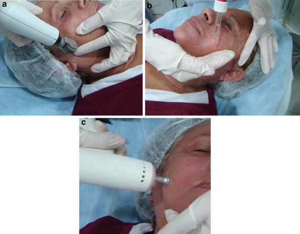
In 2010 a low-frequency and high-pressure US, called impact US, was created in order to enhance the penetration of substances with TED technique. These US waves act by propelling molecules through preformed channels. It depends on the previous use of an ablative method to perform its function, frequently named as ITED (impact US + TED) (Fig. 1a–c) (see chapter “Microneedling for Transepidermal Drug Delivery on Stretch Marks”).
Histopathological Studies
In laser-treated tissues, through hematoxylin and eosin (H&E) staining, it is possible to observe vertical channels created by columns of vaporization. This is true for ablative fractional lasers that create a grid of microscopic treatment zones (MTZ) (Skovbølling Haak et al. 2011). Each MTZ is composed of the ablated channels, the microscopic ablation zones (MAZ), and a border of carbonization surrounded by coagulated tissue, the microscopic thermal zone (Sklar et al. 2014), that result from thermal damage by the light source (Taudorf et al. 2014).
The ideal parameters needed to create the skin channels absolutely vary on the laser type, device model, and settings. Also, the available data involving the best energies and channel depth for skin drug delivery is a bit contradictory. Nevertheless, there is some evidence showing that for hydrophilic and slightly lipophilic molecules, the drug uptake was channel depth-dependent, while lipophilic drugs have shown a channel depth independent uptake (Haedersdal et al. 2016; Wenande et al. 2017).
Haak et al. described the impact of laser treatment density (% of skin occupied by channels) and molecular weight (MW) for fractional CO2 laser-assisted drug delivery. Ablative fractional treatment substantially increased intra- and transcutaneous delivery of polyethylene glycols (PEGs) in a MW that ranged from 240 to 4,300 Da. Increasing laser density from 1% to 20% resulted in augmented intra- and transdermal delivery, but densities higher than 1% resulted in reduced delivery per channel. Mass spectrometry indicated that larger molecules have greater intracutaneous retention than transcutaneous penetration (Haak et al. 2012; Haedersdal et al. 2014).
Skovbølling et al. conducted a histopathological study on an ex vivo animal skin model with a fractional CO2 laser (Medart, Hvidovre, Denmark), with the aim to establish a standard model to document the histological tissue damage profiles after ablative fractional laser treatment. It was shown that the studied laser produced a cone-shaped lesion with the base being the circular epidermal surface lesion and the apex pointing toward the dermis, and a proposed mathematical formula was able to predict cone volume. It was also demonstrated that ablation depth increased in a linear relation with increased laser energies, dermal ablation width increased slightly with increasing energies, and thickness of coagulation zone reached a plateau at a certain energy level (Skovbølling Haak et al. 2011).
Taudorf et al. conducted an animal ex vivo study with a 2,940 nm laser with the aim of establishing the impact of laser parameters, stacked pulses and the tissue effects. It was shown that low pulse energy and high repetition rate required many stacked pulses at the same spot to induce ablation, whereas high pulse energy delivered by decreased pulse repetition rate and fewer stacked pulses led to ablation by less applied total energy. Ablation depth was likewise affected not only by total energy delivered by pulse stacking but also by variations in pulse energy, pulse repetition rate, and pulse duration. Low pulse repetition rate (Hz) and reduced number of stacked pulses are important to avoid progressive accumulation of residual heat by allowing the exposed tissue to cool and ablation plume to be evacuated between pulses, otherwise leading to shallow-wide craters instead of increasing ablation depth. It was also discussed that the advantage of using Er:YAG (2,940) laser instead of CO2 laser is the possibility of creating purely ablated tissue with virtually no coagulation zone. Although the importance of the coagulation zone is not completely established, a thick coagulation zone may pose a significant induced barrier for molecules delivery (Taudorf et al. 2014). Brauer et al. demonstrated that fractional treatment results in untreated areas between the MTZs allowing a more rapid healing response (Brauer et al. 2014).
Banzhaf et al. have published that 100% of the channels created by a fractional ablative CO2 laser were kept opened for the first 30 minutes after the intervention. From this point on, there was a gradual decrease in the percentage of channels opened, with substantial decrease from 6 hours on (Banzhaf et al. 2017). Olesen et al. have shown that liquid based vehicles were better for transepidermal drug delivery purposes than cream or gel (Olesen et al. 2017).
It is not possible to establish the parameters needed to perform the ideal drug delivery. Nevertheless when doing this treatment, it is important to take into account the laser type, the device model, the laser settings, and the desired effect. CO2 lasers may create thicker coagulation zones, when compared to Er:YAG lasers. Treatment with high energy, very short pulse width, and low density may facilitate the penetration of drugs, especially for hydrophilic and slightly lipophilic molecules.
Indications
TED for Photodynamic Therapy
It has already been reported the use of ablative lasers (erbium:YAG or CO2 lasers) prior to the application of methyl aminolevulinate (MAL) for PDT treatment (Haerdersdal et al. 2010; Shen et al. 2006). Some studies also describe the use of microneedling technique applied before the application of the photosensitizer (Donelly et al. 2008; Mikolajewska et al. 2010). See chapter “Transepidermal Drug Delivery and Photodynamic Therapy”.
A recent study was conducted to evaluate and compare clinical effects induced by PDT (MAL-PDT with red light) alone versus PDT (MAL-PDT with red light) associated with TED using fractional ablative RF. The results showed that even reducing the incubation time of the photosensitizing agent (MAL) from 3 to 1 h, improvement of actinic keratosis and skin texture could be observed. TED + PDT was more effective in reducing the number of actinic keratosis lesions in the forearms compared to PDT alone. Moreover, an improvement in the texture and pigmentation was better observed on the side treated with the TED + PDT side (Kassuga et al. 2012).
Scars
Hypertrophic scars and keloids are disorders of the healing process in predisposed individuals in response to different types of injuries to the dermis, such as trauma, inflammation, surgery, burns, and even insect bites (Wolfram et al. 2009). Despite the increasing knowledge in the field of wound repair and collagen metabolism, its treatment remains a challenge for dermatologists and plastic surgeons. Clinically, hypertrophic scars are limited to the original site of injury, while the keloid exceeds this threshold and reaches the adjacent skin. Hypertrophic scars appear after 4 weeks of the triggering event, grow intensely for a few months, and then regress. In keloids, collagen production is 20 times greater than in normal healing (Wolfram et al. 2009) and rarely disappears spontaneously. It can be difficult to distinguish them in the initial growth phase.
Intralesional steroids are the first-line therapy (Al-Attar et al. 2006). The most widely used steroid is triamcinolone acetonide, a synthetic corticosteroid derived from hydrocortisone with potent anti-inflammatory action (Chrousos and Margioris 2003), whose mechanism of action is the inhibition of fibroblast proliferation and collagen synthesis, increase in collagenase production, and reduction of collagenase inhibitors (Al-Attar et al. 2006). It should be applied every 2–6 weeks until clinical improvement of the lesion or the appearance of local side effects that prohibits their use. The use of intralesional corticosteroid is painful and its distribution is not homogeneous.
Garg et al. (2011) demonstrated that the use of CO2 ablative laser alone was not sufficient to permanently treat keloids, requiring intralesional application of triamcinolone every 3–4 weeks for a period of 6 months after ablation.
The use of steroid injections in keloid treatment is the most popular therapy modality for such problem because it is technically easy and less costly for the patient, not to mention the most satisfactory result compared with other proposed methods. On the other hand, injections are painful and sometimes intolerable, and it is also difficult to apply it evenly, causing localized areas of atrophic lesion.
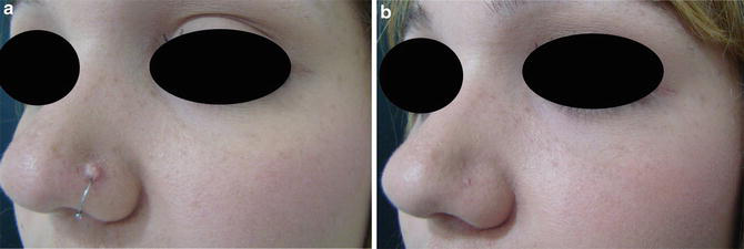
Issa et al. (2012) conducted a study about transepidermal application of triamcinolone assisted by fractional RF associated with the impact US in the treatment of hypertrophic scars. The results showed improvement or complete resolution of the hypertrophic scars with an excellent aesthetic result. This technique was also considered less painful than regular injections (Fig. 2a, b). It is noteworthy that in another study, not published, using the same technique for the treatment of keloids, the same effectiveness was not observed.
Atrophic Striae
Striae are a common and easily recognized condition, which rarely cause significant medical problems, but often are a source of stress for their carriers. The origin of striae is not well known. There are a number of therapeutic modalities, but none of them is considered effective, and no single therapy is considered essential to this problem. With high incidence and poor treatment results, there is a tendency in targeting the research for consensus and optimal treatment. Therapeutic strategies are numerous, and no single therapy has been more consistent than the others. The treatment of the striae remains a problem to dermatologists. Recent striae may be treated with topical medication, such as tretinoin, and show better results after various surgical procedures such as subcisions. Striae with longstanding course do not show the same result. The future of treatment strategies is encouraging, with advances in laser therapy. Many sources reported the use of lasers to diminish the appearance of striae (Elsaie et al. 2009; Bak et al. 2009).
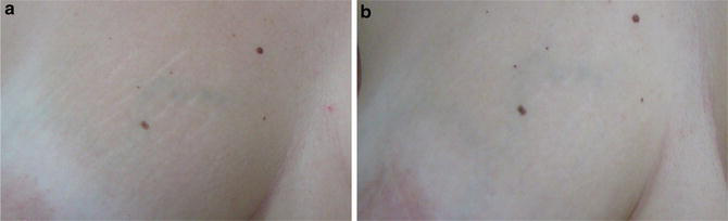
A recent study about TED using 0.05% tretinoin cream after fractional ablative method (RF) (Issa 2013) has shown the efficacy in the treatment of atrophic white striae. When comparing the treatment of stretch marks located in the abdomen with ablative fractional RF alone or TED (RF + 0.05% tretinoin cream + US impact), the latter was more effective. Authors concluded that TED using ablative RF to permeate the tretinoin cream + impact US to increase this permeation is a new interesting method for white old striae, mainly on the breast (Fig. 3a, b).
Alopecia Areata
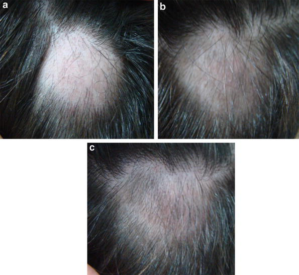
Alopecia areata (AA) is the most common cause of non-scarring alopecia. It is suspected to be an autoimmune disease with a genetic predisposition. Environmental and ethnic factors seem to be involved. It presents commonly as oval or circular patches of non-scarring hair loss. Steroids are widely used to treat AA, and intralesional triamcinolone is a very effective and painful method. Issa et al. (2012) reported five cases of AA treated with TED using ablative fractional resurfacing (CO2 laser or RF) + triamcinolone + acoustic pressure wave US. Triamcinolone solution was dropped above the ablated area, and the impact US was applied just after the medication to push the drug into the skin. In all cases, patients exhibited an excellent improvement, and only one patient did not sustain the result after 12 months of follow-up. In the cases of alopecia areata treated with CO2 laser + triamcinolone + US, excellent results were reached with only one session (Fig. 4a–c). When using ablative RF + triamcinolone + US for AA treatment, good results were also reached, but more than one session was necessary. Authors observed a minimal clinical improvement in the patch treated with fractional ablation CO2 laser + triamcinolone without US, but no clinical improvement was observed on the patch treated with fractional ablation CO2 laser alone, without applying triamcinolone or US.
TED for Photoaging and Melasma
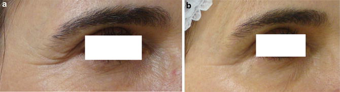
The drugs used for photoaging and melasma treatment include tretinoin 0.05% cream, vitamin C (Farris 2005; Hsiao et al. 2012) 5–10% cream or serum alone or combined with other components such as ferulic acid (Waibel and Wulkan 2013), hyaluronic acid 5% for rejuvenation (Fig. 5a, b), and hydroquinone 4% associated or not with glycolic acid 10% cream for melasma.
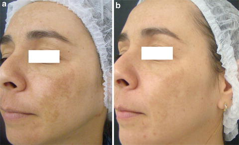
In the case of melasma, CO2 laser should be used at very low energy, only with the aim to produce micro-channels on the skin surface; good results with hydroquinone 4% cream were obtained without post-inflammatory hyperpigmentation (Fig. 6a, b) (study conducted by the author, not published yet).
In a split-face comparison study for photodamaged skin, a CE ferulic acid formula (L’Oréal-SkinCeuticals) was evaluated after fractional laser ablation, showing a decrease in postoperative recovery time and an increase in neocollagenesis in the treated side (Waibel and Wulkan 2013). Topical vitamin C after fractional laser ablation was also assessed for this purpose, but further investigation is needed (Hsiao et al. 2012).
TED for Other Indications
There are some investigations about the use of growth factors for wound healing and the use of antifungal agents and antimicrobials to target infections (Brauer et al. 2014).
Axillary and palmar-plantar hyperhidrosis is another possible indication. It has been evaluated in some patients in a clinical trial in which botulinum toxin was applied through TED on one side and through the standard injectable application on the other for comparison. In the first cases, reduction of sweating was observed in the iodine-starch test with both techniques (study about to be published).
Protocol of Application and Directions
The first step is to clean the skin using a cleanser without soap and chlorhexidine solution. The fractional ablative method is applied immediately before the topical medication, which is chosen according to the disease to be treated. The medication can be presented as cream, solution, or serum, with the same concentrations as the products used topically at home, with no need for high concentrations as in the case of solutions for peelings. If possible, the impact US can be used. This is the last step, with the aim to push the drug into the dermis through the channels preformed by the ablative methods, as a “hammering effect.”
Post-procedure directions include proper hygiene with antiseptic soap, which starts 8 h after the intervention. Avoid friction in the treated area and do not wear tight clothes. The use of a healing moisturizer is advised, three to five times a day, avoiding crust formation. Patients are advised not to remove crusts; these should flake off spontaneously, and to avoid sun exposure throughout the healing time (7–14 days depending on the area). Topical photoprotection should be started on the third day. Oral photoprotection can also be indicated, and it is very important in cases of melasma.
Antiviral prophylaxis is advised at the usual treatment dose, starting 3 days before the procedure when treating the facial region, regardless of herpes simplex history.
Residual hyperpigmentation prevention can be done with the use of depigmenting substances for 3 weeks before the first session and restarting immediately after skin recovery in each session, maintained throughout the treatment period.
Side Effects
For all indications, side effects are usually less intense than those after fractional ablative lasers alone, as a lower density is used for TED. On the other hand, side effects can be related to the lasers and to the drugs applied. Moreover, a systemic side effect is a possibility. Adverse effects include redness, swelling, pain, crusting, and transient residual hyperpigmentation. Usually the topically applied medications, even retinoic acid, do not change the intensity of discomfort caused by the laser or RF previously used. Infections can also occur.
Conclusions
When using fractional ablative methods for TED, it is possible to achieve a more homogeneous and convenient application of intralesional drugs, as well as more effective treatment compared to the use of topical drugs or laser alone. According to the literature, TED with both RF and lasers (CO2 and erbium) can promote good results in many different treatments. It seems that each method has its advantages and disadvantages. RF can be used in all phototypes as it does not cause dyschromia; on the other hand, the CO2 laser can bring better results with fewer sessions.
TED can be considered effective for many diseases and also for cosmetic indications.
Take Home Messages
- TED through ablation methods produces micro-perforations in the epidermis, allowing the permeation of drugs topically applied into the skin through these micro-channels.
- The ideal parameters needed to create the skin channels vary according to the laser type and device model. It has been shown, however, that the best results in drug permeation are achieved with high energy, very short pulse width, and low density.
- Many substances, cosmeceuticals or medicaments, can be used for TED purposes and are chosen according to the disease to be treated.
- TED with ablative methods can be indicated for PDT, scars, striae distensae, alopecia areata, melasma, and photorejuvenation treatment, among others.
- Better clinical results can be reached with TED compared to laser alone.
References
Al-Attar A, Mess S, Thomassen JM, Kauffman CL, Davison SP. Keloid pathogenesis and treatment. Plast Reconstr Surg. 2006;117(1):286–300.
Alexiades-Armenakas MR, Dover JS, Arndt KA. The spectrum of laser skin resurfacing: nonablative, fractional, and ablative laser resurfacing. J Am Acad Dermatol. 2008;58(5):719–37.
Bak H, Kim BJ, Lee WJ, et al. Treatment of striae distensae with fractional photothermolysis. Dermatol Surg. 2009;35(8):1215–20.
Banzhaf CA, Thaysen-Petersen D, Bay C, Philipsen PA, Mogensen M, Prow T, Haedersdal M. Fractional laser-assisted drug uptake: impact of time-related topical application to achieve enhanced delivery. Lasers Surg Med. 2017;49(4):348–54.
Baron ED, Harris L, Redpath WS, Shapiro H, Hetzel F, Morley G, Bar-Or D, Stevens SR. Laser-assisted penetration of topical anesthetic in adults. Arch Dermatol. 2003;139(10):1288–90.
Brauer JA, Krakowski AC, Bloom BS, Nguyen TA, Geronemus RG. Convergence of anatomy, technology, and therapeutics: a review of laser-assisted drug delivery. Semin Cutan Med Surg. 2014;33(4):176–81.
Carniol PJ, Hamilton MM, Carniol ET. Current status of fractional laser resurfacing. JAMA Facial Plast Surg. 2015;17(5):360–6.
Chrousos GP, Margioris AN. Adrenocorticosteroid & cortico-adrenal antagonists. In: Katzung BG. Basic and clinical pharmacology. 8th ed. The McGraw-Hill: United States of America; 2003. p. 574–89.
Donelly RF, Morrow DI, McCarron PA, Woolfson AD, Morrissey A, Juzenas P, Juzeniene A, Iani V, McCarthy HO, Moan J. Microneedle-mediated intradermal delivery of 5-aminolevulinic acid: potential for enhanced topical photodynamic therapy. J Control Release. 2008;129(3):154–62.
Elsaie ML, Baumann LS, Elsaie LT. Striae distensae (stretch marks) and different modalities of therapy: an update. Dermatol Surg. 2009;35(4):563–73.
Farris PK. Topical vitamin C: a useful agent for treating photoaging and other dermatologic conditions. Dermatol Surg. 2005;31(7 Pt 2):814–7.
Forster B, Klein A, Szeimies RM, Maisch T. Penetration enhancement of two topical 5-aminolaevulinic acid formulations for photodynamic therapy by erbium:YAG laser ablation of the stratum corneum: continuous versus fractional ablation. Exp Dermatol. 2010;19(9):806–12.
Garg GA, Sao PP, Khopkar US. Effect of carbon dioxide laser ablation followed by intralesional steroids on keloids. J Cutan Aesthet Surg. 2011;4(1):2–6.
Gómez C, Costela A, García-Moreno I, Llanes F, Teijón JM, Blanco D. Laser treatment on skin enhancing and controlling transdermal delivery of 5-fluorouracil. Laser Surg Med. 2008;40(1):6–12.
Haak CS, Illes M, Paasch U, Haedersdal M. Histological evaluation of vertical laser channels from ablative fractional resurfacing: an ex vivo pig skin model. Lasers Med Sci. 2011;26(4):465–71.
Haak CS, Bhayana B, Farinelli WA, Anderson RR, Haedersdal M. The impact of treatment density and molecular weight for fractional laser-assisted drug delivery. J Control Release. 2012;163(3):335–41.
Haedersdal M, Sakamoto FH, Farinelli WA, Doukas AG, Tam J, Anderson RR. Pretreatment with ablative fractional laser changes kinetics and biodistribution of topical 5-aminolevulinic acid (ALA) and methyl aminolevulinate (MAL). Lasers Surg Med. 2014;46(6):462–9.
Haerdersdal M, Sakamoto FH, Farinelli WA, Doukas AG, Tam J, Anderson RR. Fractional CO2 laser-assisted drug delivery. Laser Surg Med. 2010;42(2):113–22.
Haedersdal M, Erlendsson AM, Paasch U, Anderson RR. Translational medicine in the field of ablative fractional laser (AFXL)-assisted drug delivery: a critical review from basics to current clinical status. J Am Acad Dermatol. 2016;74(5):981–1004.
Hsiao CY, Huang CH, Hu S, Ko YS, Sung HC, Chen CC, Huang SY. Fractional carbon dioxide laser treatment to enhance skin permeation of ascorbic acid 2-glucoside with minimal skin disruption. Dermatol Surg. 2012;38(8):1284–93.
Issa MCA, Kassuga LEBP, Chevrand NS, Pires MTF. Topical delivery of triamcinolone via skin pretreated with ablative radiofrequency: a new method in hypertrophic scar treatment. Int J Dermatol. 2012;52:367–70.
Issa MC, de Britto Pereira Kassuga LE, Chevrand NS, do Nascimento Barbosa L, Luiz RR, Pantaleão L, Vilar EG, Rochael MC. Transepidermal retinoic acid delivery using ablative fractional radiofrequency associated with acoustic pressure ultrasound for stretch marks treatment. Lasers Surg Med. 2013;45(2):81–8.
Kassuga LEBP, Issa MCA, Chevrand NS. Aplicação transepidérmica de medicamento associado a terapia fotodinâmica no tratamento de ceratoses actínicas. Surg Cosmet Dermatol. 2012;4(1):89–92.
Lee WR, Shen SC, Kuo-Hiesen W, Hu CH, Fang JY. Lasers and microdermabrasion enhance and control topical delivery of vitamin C. J Invest Dermatol. 2003;121(5):1118–25.
Lin CH, Aljuffali IA, Fang JY. Lasers as an approach for promoting drug delivery via skin. Expert Opin Drug Deliv. 2014;11(4):599–614.
Manstein D, Herron GS, Sink RK, Tanner H, Anderson RR. Fractional photothermolysis: a new concept for cutaneous remodeling using microscopic patterns of thermal injury. Lasers Surg Med. 2004;34:426–38.
Mikolajewska P, Donnelly RF, Garland MJ, Morrow DI, Singh TR, Iani V, Moan J, Juzeniene A. Microneedle pretreatment of human skin improves 5-aminolevulinic acid (ALA)- and 5-aminolevulinic acid methyl ester (MAL)-induced PpIX production for topical photodynamic therapy without increase in pain or erythema. Pharm Res. 2010;27(10):2213–20.
Mitragotri S, Edwards D, Blankschtein D, Langer R. A mechanistic study of ultrasonically-enhanced transdermal drug delivery. J Pharm Sci. 1995;84:697–706.
Olesen UH, Mogensen M, Haedersdal M. Vehicle type affects filling of fractional laser-ablated channels imaged by optical coherence tomography. Lasers Med Sci. 2017;32(3):679–84.
Shen SC, Lee WR, Fang YP, Hu CH, Fang JY. In vitro percutaneous absorption and in vivo protoporphyrin IX accumulation in skin and tumors after topical 5-aminolevulinic acid application with enhancement using an erbium:YAG laser. J Pharm Sci. 2006;95(4):929–38.
Sklar LR, Burnett CT, Waibel JS, Moy RL, Ozog DM. Laser assisted drug delivery: a review of an evolving technology. Lasers Surg Med. 2014;46(4):249–62.
Skovbølling Haak C, Illes M, Paasch U, Haedersdal M. Histological evaluation of vertical laser channels from ablative fractional resurfacing: an ex vivo pig skin model. Lasers Med Sci. 2011;26(4):465–71.
Stumpp OF, Welch AJ, Milner TE, Neev J. Enhancement of transepidermal skin clearing agent delivery using a 980 nm diode laser. Lasers Surg Med. 2005;37(4):278–85.
Taudorf EH, Haak CS, Erlendsson AM, Philipsen PA, Anderson RR, Paasch U, Haedersdal M. Fractional ablative erbium YAG laser: histological characterization of relationships between laser settings and micropore dimensions. Lasers Surg Med. 2014;46(4):281–9.
Waibel J, Wulkan A. Split face comparison of the effects of vitamin CE ferulic formula serum to decrease postoperative recovery and increase neocollagenesis in fractional ablative laser resurfacing for photodamage. Laser Surg Med. 2013;45 Suppl 25:1.
Wang KF, Fang JY, Hu CH, Lee WR. Erbium:YAG laser pretreatment accelerates the response of Bowen’s disease treated by topical 5-fluorouracil. Dermatol Surg. 2004;30(3):441–5.
Wenande E, Olesen UH, Nielsen MM, Janfelt C, Hansen SH, Anderson RR, Haedersdal M. Fractional laser-assisted topical delivery leads to enhanced, accelerated and deeper cutaneous 5-fluorouracil uptake. Expert Opin Drug Deliv. 2017;14(3):307–17.
Wolfram D, Tzankov A, Pülzl P, Piza-Katzer H. Hypertrophic scars and keloids – a review of their pathophysiology, risk factors, and therapeutic management. Dermatol Surg. 2009;35(2):171–81.







