
Non-ablative Radiofrequency for Cellulite (Gynoid Lipodystrophy) and Laxity
Non-ablative Radiofrequency for Cellulite (Gynoid Lipodystrophy) and Laxity
Bruna Souza Felix Bravo, Carolina Martinez Torrado and Maria Claudia Almeida Issa Instituto de Dermatologia Prof. Rubem David Azulay – Santa Casa da Misericórdia do Rio de Janeiro, Rio de Janeiro, RJ, Brazil Department of Clinical Medicine – Dermatology, Fluminense Federal University, Niterói, RJ, Brazil
Abstract
Cellulite, also known as gynoid lipodystrophy, is a multifactor disorder of the dermis, epidermis, and subcutaneous cellular tissue (Goldman et al. Pathophysiology of cellulite. New York: Taylor & Francis; 2006). In most cases, this alteration occurs in postpubertal women with a prevalence of 80–90% (Goldman and Hexsel. Cellulite: pathophysiology and treatment. 2nd ed. Florida: Editorial Informa, Healthcare; 2010; Emanuele. Clin Dermatol 31(6):725–30, 2013). Clinically, cellulite presents irregularity of the skin surface with depressions, lumps, and nodules associated with laxity. Usually, cellulite is located in the abdomen, buttocks, and lower limbs, but it can also occur on the arms and the back. Histologically, cellulite is caused by subcutaneous herniated fat within the fibrous connective tissue (Rossi and Vergnanini. JEADV 14: 251–62, 2000; Khan et al. J Am Acad Dermatol. 62(3):361–70, 2010; De Peña and Hernández-Pérez. Rev Cent Dermatol Pascua 3:132–5, 2005). An increase in the thickness of the subcutaneous cellular tissue is also observed. Although cellulite is still considered as an unclear etiological condition, there are many hypotheses trying to explain this disorder. Treatments can be invasive and noninvasive. Invasive methods include subcision and mesotherapy, as well as liposuction when patients also want to remove excessive localized fat. Noninvasive treatments include topical treatments, controlled diets, cryolipolysis, focused ultrasound, endermology, laser, and non-ablative radiofrequency. In this chapter we are going to discuss the treatment with non-ablative radiofrequency. Radiofrequency (RF) contracts collagen and stimulates neocollagenesis by thermal heat, promoting thickening of the dermis, avoiding fat herniation. It can also promote improvement in local circulation due to the vasodilatation and lymphatic drainage. Clinically it improves the laxity and the irregularity of the skin surface (Coringrato et al. Radiofrecuencia ablativa en dermatología quirúrgica: Una revisión, Rev. dermatología argentina, vol. XIV, Julio–Septiembre 2008, Número 3; Brightman et al. Lasers Surg Med 41:791–8, 2009).
Keywords CelluliteGynoid lipodystrophyNoninvasive radiofrequencyNeocollagenesisLaxityFat herniationDermis thickness
Introduction
The skin is the most external organ and its appearance contributes to the patient’s personality. Skin diseases and cosmetic problems significantly affect the self-esteem. The psychological effects of the cellulite (gynoid lipodystrophy) are frequently more serious than the physical alteration observed in this dermatosis. It can affect interpersonal relationships in a social, affective, and sexual way. It can affect normal daily activities, such as going to the beach, practicing sports, or using certain type of clothing. In the last years, the idea of having an ideal sculpting body changed the concept of cellulite from a cosmetic problem to a disease. Nowadays, women look for all types of treatments with the hope of reducing cellulite or making it disappear (Goldman and Hexsel 2010; Goldman et al. 2006). Different mechanisms are involved in cellulite’s pathogenesis, and many different treatments are developed with aim to fight against each mechanism. There is no ideal treatment, but there are many therapeutic alternatives to reduce the physical appearance of this condition.
Recently, noninvasive devices such as non-ablative radiofrequency (RF) and the combination of RF with other technologies have gained strength and have been very successful. The most used RFs for cellulite are the unipolar, bipolar, and the combination of RF with infrared light, vacuum, and mechanical massage.
Cellulite (Gynoid Lipodystrophy)
Concept and History
Cellulite, also known as gynoid lipodystrophy, nodular liposclerosis, adiposis edematosa, dermopaniculosis deformans, edematous fibrosclerotic panniculopathy, panniculosis, and cellulite hypodermosis, is a multifactor disorder of the dermis, epidermis, and subcutaneous cellular tissue (Goldman et al. 2006). In most cases, this alteration occurs in postpubertal women with a prevalence of 80–90% (Goldman and Hexsel 2010).
Alquier and Paviot described cellulite for the first time in 1920 (Rossi and Vergnanini 2000). They described a non-inflammatory complex cellular dystrophy of the mesenchymal tissue caused by a water metabolism disorder, which produces the saturation of the adjacent tissues by interstitial fluid (Rossi and Vergnanini 2000). In the same decade, Laguese described cellulite as changes in the subcutaneous tissue characterized by the increased fatty tissue and interstitial edema (Goldman et al. 2006; Laguese 1929). In 1958, Merlem defined cellulite as an angiopathy, and in 1978, Benazzy and Curri suggested the term “sclerotic-fibrous-edematous panniculopathy” (Goldman et al. 2006). In 1978 Nürnberger and Müller (1978) found differences in the structure of the skin and the subcutaneous tissue of men and women that would partially explain the prevalence of cellulite in women (Nürnberger and Müller 1978). Currently, the terms cellulite and gynoid lipodystrophy are the most used terms in medical literature.
Clinical Manifestations
Usually, cellulite is located in the abdomen, buttocks, and lower limbs, but it can also be located on the arms and the back. Cellulite is caused by subcutaneous herniated fat within the fibrous connective tissue, with a typical dimpled appearance (with alternated depressions with lumps) or orange skin appearance (due to swelling of the flat surfaces and dilation of the follicular openings) (Rossi and Vergnanini 2000; Khan et al. 2010; De Peña and Hernández-Pérez 2005). The majority of the injuries have an oval shape, where the axis of the injuries is parallel to the tension lines of the skin (Goldman and Hexsel 2010). Cellulite is more evident with laxity, therefore getting worse with aging. During palpation, pinching the skin in a cellulite area, we can feel an increasing in the thickness of the subcutaneous cellular tissue and in its consistency and a decreasing in its mobility by adhesion (De Peña and Hernández-Pérez 2005).
In general, cellulite is an asymptomatic alteration; however, painful nodules can accompany it in the severe stages, suggesting an inflammation in dermis and adipose subcutaneous underlying tissue (Goldman and Hexsel 2010; Emanuele 2013).
Cellulite is different from obesity; obesity is characterized by hypertrophy and hyperplasia of the adipose tissue that is not necessarily limited to the abdomen or the lower limbs (Rossi and Vergnanini 2000). The presence of cellulite should not be confused with obesity, even though adipogenesis aggravates this condition (Emanuele 2013).
Etiopathogenesis
The etiopathogenesis of cellulite has been widely discussed in the last decades. Although cellulite is still considered as an unclear etiological condition, there are many hypotheses with respect to its origin, such as vascular changes and edema, architectural difference of the skin related to gender, inflammatory alterations, and hormonal variations.
Vascular Changes and Edema
The first document about the pathophysiology of cellulite reported the increasing in the glycosaminoglycan amount in the dermis and extracellular matrix and on the capillary dermal vessels of the skin in the cellulite area. This could explain the amount of water retained in the skin, attracted by glycosaminoglycans. Based on these findings, edema in cellulite was related to the changes in the composition of the extracellular matrix (Emanuele 2013).
Some degree of damage in the skin vasculature with an alteration in the precapillary arteriole sphincters was also reported. Other findings, such as the presence of new capillary formations, telangiectasia, microthrombi, and micro-hemorrhages, related cellulite to a chronic venous insufficiency (Rossi and Vergnanini 2000). These contribute to capillary permeability and excessive retention of fluids in the dermis, as well as in the fat layer, between the adipocytes and the lobular septa, increasing the interstitial pressure due to the hydrophilic properties of glycosaminoglycan.
Vascular alterations, edema, and decreased venous return cause hypoxia of the tissue which leads to the thickening of the fibrous septa in the superficial adipose tissue and deep dermis, which causes the padded aspect of cellulite (Curri and Bombardelli 1994).
Architectural Difference of the Skin Related to Gender
This theory was described by Nürnberger and Müller in 1978 when they reported cellulite as a fat herniation called “papillae adiposae.” In this condition, fat penetrates from the subcutaneous tissue through the interior surface of a weak dermis in the dermo-epidermal interface, which is considered a characteristic of female anatomy (Nürnberger and Müller 1978). This alteration has been confirmed through ultrasound, spectroscopy, and magnetic resonance imaging (MRI) (Bravo et al. 2013). It has been observed that the herniation of the tissue is typical in women because the fibrous septa are different in this gender than in men. In men, septa are distributed in an intersecting way forming small polygons that generate a type of cell that does not move forward facially toward the dermis and are not placed perpendicularly as what occurs in women. This particularity helps to understand why cellulite can occur both in skinny and in obese women (Nürnberger and Müller 1978).
Querleux et al. (2002) reveal the three main orientations of the septa, which are parallel, perpendicular, and angular (approximately 45°). They describe that women with cellulite have a higher percentage of perpendicular septa than women or men without cellulite.
Inflammatory Alterations
Inflammatory alterations were cited as one of the main pathophysiology factors of cellulite. Kligman concluded that cellulite is a blurred appearance of swollen chronic cells (Nürnberger and Müller 1978); also there is evidence of lymphocyte and macrophage infiltration in the fibrous septa, resulting in adipocytes and cutaneous atrophy (Nürnberger and Müller 1978; Bravo et al. 2013). This hypothesis would explain why some patients with cellulite have pain and sensitivity to compression. Other authors do not consider inflammation as part of this process (Piérard et al. 2000).
Hormonal Variations
Other authors suggest that cellulite is a connective tissue abnormality that results from estrogen activity over fibroblasts to produce matrix metalloproteases which damage the connective tissue, degrading collagen fibers in the trabeculae inside adipose tissue (Bravo et al. 2013).
The physical manifestations of cellulite are due to the destruction of the normal architecture of the collagen trabeculae that keep the adipose tissue confined in only two layers: superficial and deep layers of fat (Pugliese 2007). Therefore, the correlation between cellulite and hormones is justified by its appearance after puberty, worsening during pregnancy and with estrogen treatments, as well as in obesity, in which estrogen level is increased.
On diet with excessive fat and carbohydrate, a hyperinsulinemia is produced, increasing lipogenesis and inhibiting the lipolysis, generating fat accumulation. It has also been described that prolactin increases water retention in the adipose tissue generating edema (Isidori 1983).
Other hormones involved are the thyroid hormones that increase lipolysis and reduce the activity of phosphodiesterase and decrease antilipolytic receptor activity. Patients with hypothyroidism have more cellulite due to a reduction of lipolysis (Rosenbaum et al. 1998).
Predisposed Factors
Cellulite occurs in all races, although white women tend to show more cellulite than Asian or black women (Draelos 2001). It is said that Latin women have more cellulite in the hips and gluteus, while Anglo-Saxon and Nordic women have more cellulite in the abdomen, influenced by their biotype.
Diet, as mentioned previously, is also an influential factor. Diets based on high level of carbohydrates, fats, and salt promote cellulite. Tight clothes and high-heel shoes make the venous return difficult, alternating the pumping mechanisms and thus increasing cellulite (Goldman et al. 2006).
Smoking is considered a predisposed factor as it can modify skin’s microcirculation and can increase elastic fiber and collagen fiber degradation (Morita 2007). Some contraceptives and beta blockers may make cellulite worse. There is another series of circumstances that can aggravate cellulite such as obesity, localized fat accumulation, and laxity.
Cellulite can be aggravated or begin after surgeries, mainly liposuction, that cause subcutaneous fibrosis or atrophy (Goldman et al. 2006).
Clinical Evaluation
The patient must be questioned about trauma history, liposuction or injections in the affected area, chronic diseases, pregnancies, surgeries, family history, type of usual diet, and consumption of oral contraceptives or hormones (Goldman and Hexsel 2010).
The physical exam must be conducted with the patient standing and with relaxed muscles. Cellulite is observed better with a pinching test, which consists of taking a piece of the skin between the thumb and the index finger along the natural vertical fold of the skin until forming a skinfold. The palpation must be conducted to assess the elasticity of the skin and the subcutaneous tissue. The venous or lymphatic insufficiency and the degree of obesity or overweight of the patient must be assessed using the body mass index (BMI). Laxity and other aggravating conditions should be evaluated (Goldman and Hexsel 2010; Bertin et al. 2001).
Cellulite Classification
The clinical classification of cellulite is a key element before starting any specific medical treatment, regarding the mechanism of action, number of sessions, and session interval.
Clinical Stages
- Grade I: the patient is asymptomatic and there are no clinical alterations (Goldman and Hexsel 2010; Rossi and Vergnanini 2000).
- Grade II: cellulite is evident only after compression or muscle contraction, paleness, decrease of temperature, and decrease of elasticity.
- Grade III: the mattress appearance of the skin and/or orange skin is evident while resting, fine granulation of palpable sensation in deep levels, pain during palpation, decrease of elasticity, paleness, and decrease of temperature.
- Grade IV: has the same characteristic as the third degree with more palpable, visible, and painful nodules, adhered to deep levels and wavy appearance of the surface of the skin.
Clinical Description
Limited or Hard Cellulite
The skin has a very pronounced thickness and is more prominent in superficial tissues (Goldman et al. 2006; Rossi and Vergnanini 2000; De Peña and Hernández-Pérez 2005). Normally, hard or limited cellulite occurs in young women who practice exercises. Since this cellulite is hard, it is limited and occupies less space. The aspect of cellulite is compact and firm and does not change according to the position. It is usually associated with stretch marks. When a piece of the infiltrated skin is squeezed between the fingers, a roughness similar to the skin of orange with dilated follicular pores will appear on the surface. When rotating a piece of skin between the fingers, small nodules of hard consistency can be seen. Also, there is an impossibility to slide the superficial layer of the skin over a deeper layer. It has a good response to the treatment.
Soft or Diffuse Cellulite
The tissue is not attached to a deeper layer; it’s the most frequent. It usually occurs in women who do not practice exercise. It is associated with muscular hypotonia and laxity. The diagnosis is generally made through visual inspection. It is characterized by a modification of the anatomy, provoking a deformation of the pelvic area. Usually, it is present in women over 40. The skin can have a thickness of 5–8 cm. When standing, the padded appearance can be observed, and the skin shakes with movements and changes in position. When touching, the soft tissue can be felt as well as the presence of small and hard nodules. With respect to mobility, the superficial and deep layers of the skin can slide easier. It can be accompanied by circulatory complications such as varicose veins, ecchymosis, telangiectasias, heavy sensation, fatigue, feet numbness, and night pains, as well as orthostatic hypotension due to blood ectasia in peripheral zones. It is normal to find soft and hard lipodystrophy association.
Edematous Cellulite
Is the most severe and least frequent, usually accompanied by obesity. In this classification the positive Godet sign is observed. In this sign, a pressure on the tissue with the finger will produce a depression on the skin surface, lasting some minutes to disappear. The infiltration is harder compared to the previous one; this characteristic is due to the composition of the interstitial fluid that is viscous and with high molecule weight proteins, with lymphedema appearance. It also associates vascular symptoms. The patient experiences a heavy and painful sensation.
Treatments
The demand for therapeutic options to improve this alteration is very high, and in the past years, the offer of therapeutic alternatives has increased. Among them are invasive treatments such as:
- Liposuction: removing localized fat, not indicated to cellulite (Draelos 2001).
- Subcision is a surgical technique that does not require incisions and leave no scars. Through this technique, fibrous septa are cut and the depressions are reduced.
- Carboxitherapy increases vascular tone and produces active microcirculatory vasodilatation due to the action of CO2 on arteriole smooth muscle cells (Goldman and Hexsel 2010).
- Mesotherapy comprises injections composed by some active substances. These substances have lipolytic action (Goldman and Hexsel 2010).
Noninvasive treatments are the following:
- Topical treatments: which work mainly in four objectives: increasing microvascular flow, reducing lipogenesis and promoting lipolysis, restoring the normal structure of the dermis and the subcutaneous tissue, and preventing or destroying the formation of free radicals (Goldman et al. 2006).
- Oxygen therapy: promotes superficial lipolysis.
- Controlled diets: to avoid overweight.
- Cryolipolysis approved by the FDA to eliminate localized fat, producing a crystallization of the fats in adipose cells at above freezing temperatures, leading to an apoptosis of adipose cells and an inflammatory process that results in the reduction of the fat layer in 2–4 months.
- Focused ultrasound: produces heating of the adipocytes, generating coagulative necrosis and death of the cells in the adipose tissue.
- Endermology: consists of motorized rollers with vacuum suction between them to lift the skin and reach deeper structures, producing stimulation of the metabolism and vascularization with lymphatic drainage (Goldman and Hexsel 2010).
- Laser.
- Ablative and non-ablative radiofrequency.
In this chapter we will discuss in detail the treatment of cellulite with non-ablative radiofrequency.
Radiofrequency
Radiofrequency (RF) is a form of electromagnetic energy (Shapiro et al. 2012). Its frequency ranges from 3 kHz to 300 GHz (Lapidoth and Halachmi 2015). When applied to skin tissue, rapidly oscillating electromagnetic fields cause movement of charged particles within the tissue resulting in heat generation proportional to the tissue’s electrical resistance (Shapiro et al. 2012). The idea of using heat to cauterize comes from Egyptian era. Nevertheless, only in the nineteenth century, a device using a galvanic current was built. Lee de Forest was an American inventor and engineer who created the thermionic valve or vacuum tube that allowed RF amplification (Kotcher Fuller 2005). William Bovie, in 1920 (O’Connor et al. 1996), developed the electrocautery for electrosurgery, which could cut, coagulate, cauterize, and fulgurate tissues. In 1939, the hyfrecator was introduced in the market. It is a device that uses low frequencies and high AC (alternating current) electrical voltage. This device is used for tissue destruction and for bleeding control (Lapidoth and Halachmi 2015).
RF is currently used in medicine in several treatments with different spectrum variations, including aesthetic medicine where it is considered a simple, effective, and minimally invasive method (Lapidoth and Halachmi 2015). RF is used in aesthetic medicine with applications for ablative and non-ablative applications (Lapidoth and Halachmi 2015).
The tissue effects achievable using RF energy depend on the applied energy density; the following are several RF-induced thermal changes of tissue that are commonly used in medicine (Lapidoth and Halachmi 2015):
- Ablation of tissue: this effect is used for cutting or removing tissue and is based on thermal evaporation of tissue.
- Coagulation: this provides hemostasis for controlling bleeding. Coagulation may be applied to soft tissue as well, to induce necrosis.
- Collagen contraction: high temperatures induce immediate transformation in the tertiary structure of proteins. For noninvasive cosmetic procedure, this effect is produced with lower temperatures to avoid skin necrosis.
- Tissue hyperthermia: it is to use subnecrotic temperatures to stimulate natural physiological processes in attempts to modify skin appearance and to reduce subcutaneous fat; an example of this is ThermaCool.
The first device based on non-ablative RF was approved by FDA (Food and Drug Administration) in 2002. It is a monopolar device called ThermaCool (Thermage, Hayward, CA). It was developed to treat photodamaged skin on the face, reducing wrinkles and improving laxity (Lolis and Goldberg 2012; Alexiades-Armenakas et al. 2008).
Non-ablative RF does not absorb or disperse epidermal melanin; therefore, it can be used in all skin phototypes, without risk for epidermis injury. RF contracts collagen fibers and stimulates neocollagenesis by thermal energy, improving laxity and wrinkles. For cellulite treatment, synthesis of new collagen by fibroblasts promotes dermis thickening, improving the clinical aspect of cellulite (Coringrato et al. 2008; Brightman et al. 2009).
Radiofrequency Frequency
The frequency of electrical current characterizes how many times per second an electrical current changes its direction and is reported in hertz. This change in direction is associated with a change of voltage polarity. Frequencies in the range of 200 kHz to 6 MHz are the most common in medicine (Lapidoth and Halachmi 2015).
Radiofrequency Waves
The energy generated by RF can spread in a tridimensional way in the area treated with a controlled depth. The depth of the RF depends on the RF electrode configuration, the device parameters, and the tissue to be treated (Alan and Drover 2010).
The electric conductivity in RF is variable according to the tissue. Tissues with few amount of water have less electric conductivity. Therefore, good results can be reached when RF is used to treat tissues with high water concentration, as sweat or sebaceous glands (Alan and Drover 2010).
The RF energy can be delivered in continuous wave (CW) mode, burst mode, and pulsed mode. For gradual treatment of large areas, the CW mode is most useful as it allows a slow increase in temperature in bulk tissue as in cellulite or skin tightening (Lapidoth and Halachmi 2015). The burst mode delivers RF energy with repetitive pulses of RF energy, and it is used, for example, in blood coagulation (Lapidoth and Halachmi 2015). Pulsed mode is optimal when the goal is to heat a small tissue volume while limiting heat conduction to the surrounding tissue and is effective for fractional skin ablation (Lapidoth and Halachmi 2015).
In RF the impedance matching systems are a combination of several capacitors and inductors (Lapidoth and Halachmi 2015). The challenge in these devices is that they must distinguish the different impedances of different parts of the human body (Lapidoth and Halachmi 2015). The impedance matching system must compensate for these differences (Lapidoth and Halachmi 2015).
An impedance matching system may be variable (Thermage) or broadband (Accent) (Lapidoth and Halachmi 2015).
Radiofrequency Power
The most important characteristics of RF energy are its peak and average power. Peak power is important to estimate the thermal effect produced, while average power affects the speed at which the heating is induced (Lapidoth and Halachmi 2015).
High-power density applied to a large skin surface may create only gentle warming, but when applied through a needle electrode, the small power is applied over a small contact point, leading to high-power density (Lapidoth and Halachmi 2015).
Penetration Depth and Radiofrequency Energy Distribution Between Electrodes
Penetration depth is a parameter broadly used in laser dermatology to mean the distance below the skin, which is heated. In RF current decreases at a distance from the electrode due to the divergence of current lines (Lapidoth and Halachmi 2015). The depth of penetration can be affected by altering the topology of the skin and optimizing the electrode system and can be affected by the anatomical structure of treated area (Lapidoth and Halachmi 2015).
Types of Radiofrequencies
The energy used for RF is calculated with the following formula (Table 1):

| Types of RF | Properties | Mechanism/indication |
|---|---|---|
| Monopolar | Capacitive coupling Active tip + return electrode Heat depth depends on size and geometry of the tip Heat decreases as distance increases from the electrode Depth: 3–6 mm |
Tissue ablation Coagulation Subnecrotic heating (collagen remodeling) Laxity/cellulite Wrinkles |
| Unipolar | No electric current Electromagnetic field is produced by RF (high rotating frequency of water molecules) Penetration depth: up to 20 mm |
Body contour Cellulite |
| Bipolar | Two active electrodes in the tip Low penetration depth: 3 mm |
Promotes fiber contraction: collagen remodeling Cellulite Laxity Wrinkles |
| Tripolar | Combines the effects of monopolar and bipolar in one applicator Heats superficial and deep skin Very low power, safer |
Laxity Cellulite |
Monopolar Radiofrequency
The first monopolar RF (MRF) device was approved by the Food and Drug Administration for the improvement of periorbital rhytides in 2002, followed by full-face wrinkles in 2004, as well as the temporary improvement in the appearance of cellulite when vibration was added to the delivery system (Carruthers et al. 2014). Since then, the monopolar radiofrequency is an important part of the treatment of cellulite (Carruthers et al. 2014).
The MRF devices release energy using a localized dipole in the tip and another in contact with the patient’s skin, acting as a ground or return electrode. The electrode is designed to disperse energy uniformly through the skin by a process called capacitive coupling that creates a zone of higher temperature to controlled depth of 3–6 mm.
The tissue exposed to an electric field promotes resistance to the RF waves, generating heat, which modifies micro- and macromolecular structure of the tissue (Alan and Drover 2010). The electromagnetic energy (RF) is transformed into thermal energy (Fig. 1). The heat promotes collagen denaturation and collagen fiber contraction, stimulates new fibers of collagen synthesis, and increased fibroblast activity, improving laxity and wrinkles. On the other hand, monopolar RF can also be used in electrosurgery because of its ablative potential (Bravo et al. 2013; Carruthers et al. 2014). The amount of newly produced collagen seems to be dependent on the intensity of heating of connective tissue and time that the tissue is heated (Carruthers et al. 2014). Depending on the MRF system used, energy can be delivered in a “stamped” mode, a continuously gliding movement, or internally along the dermal–hypodermal interface through a fiber or electrode (Carruthers et al. 2014).
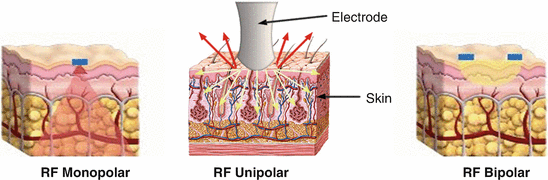
The heat generated by RF current near the active electrode does not depend on the size, shape, or position of the return electrode when the return electrode is much larger in size than the active electrode and is located at a distance which is much greater than the size of the active electrode (Lapidoth and Halachmi 2015). Heating decreases dramatically as distance increases from the electrode (Lapidoth and Halachmi 2015). Monopolar devices have a more deeply penetrating effect than bipolar or unipolar devices (Carruthers et al. 2014). The depth of heating depends on the size and geometry of the treatment tip (Bravo et al. 2013). Typically, the device heats the dermis from 65 °C to 75 °C, the temperature at which collagen denatures (Lolis and Goldberg 2012).
Monopolar devices are most commonly used for tissue cutting (Lapidoth and Halachmi 2015) like the electrical bistoury; this is an electromedical apparatus that uses the heat generated by the passage of high-frequency current, of the order of 1 MHz, between an active, punctiform electrode and an electrode of greater surface area placed in close contact with the skin. Its function is to perform tissue ablation (International Electrotechnical Commission).
Treatment effects with monopolar devices depend on the density of RF energy, which can be controlled with RF power, and the size of active electrode (Lapidoth and Halachmi 2015):
- Tissue ablation: very high-density energy is required.
- Cutting instruments: a needle-type electrode is used to concentrate electrical current on a very small area.
- Coagulation hand pieces have a larger surface area than ablative devices.
- Subnecrotic heating is usually used for treatments related to collagen remodeling.
Monopolar RF can be used on all types of the skin because it is not reflected or absorbed by epidermal melanin or vasculature, as it passes through the skin, making it safer to use (Carruthers et al. 2014); also monopolar RF can be used in different parts of the body such as the thighs, arms, neckline, neck, jowls, eyebrows, eyes, midfacial, cheeks, nasolabial folds, and melolabial folds.
Some devices of monopolar radiofrequency are available in the market, including Thermage ThermaCool, Exilis, and Pellevé S5. Each of them brings different advantage, as speed, vibration, cooling system, and progressive heating (Lapidoth and Halachmi 2015).
The most common adverse effects are burning sensation and heat in the area during the procedure. Edema and erythema usually disappear within hours after the procedure. Less frequent are burns, crust formation, erosion, depigmentation, scar, and dysesthesia (Bravo et al. 2013; Lapidoth and Halachmi 2015).
Unipolar Radiofrequency
The mechanism of action of unipolar RF differs from monopolar radiofrequency. In unipolar RF, no electric current is produced in the tissue (Bravo et al. 2013). Instead, a high-frequency electromagnetic radiation is produced, resulting in a fast alternating polarity of the electromagnetic field, inducing high rotating frequency in water molecules (considered as chromophore). Such superfast oscillations produce heat and later that heat dissipates in the tissue. The electromagnetic wave phase produced by this device is controlled in such a way that it allows for the penetration of heat in the tissue to a depth of up to 20 mm.
The heat produced by the movement of water molecules allows the superficial temperature of the skin to stabilize in approximately 40° centigrade, while the highest temperatures of 50–75° centigrade are obtained in the reticular dermis (Bravo et al. 2013).
Unipolar RF with high frequency is more appropriate to define body contour, as it penetrates deeper (20 mm) and reaches higher temperatures. Unipolar RF is considered more effective than bipolar RF for the treatment of cellulite because of its penetration depth, as the penetration of bipolar RF is from 2 to 4 mm (Alexiades-Armenakas et al. 2008). On the other hand, bipolar RF induces dermal thickening, contributing to the skin irregularity, a component of cellulite.
Bipolar Radiofrequency
Bipolar RF consists of two active electrodes placed within a short distance. In this type of device, the current has a flow between electrodes, which means that the treatment is symmetric and is limited to the tissue between them. The penetration depth is low, approximately half the distance between the two electrodes (Lapidoth and Halachmi 2015).
It can be used in all types of skin, as other RF. The mechanism of action is similar to the monopolar RF. The heat promoted by RF induces extracellular fiber contraction and then stimulates new collagen fiber production in the dermis, increasing dermis thickness, which contributes to avoid fat herniation into the dermis. Clinically it is observed as a smoother skin.
The association of bipolar with unipolar is very useful to treat body contour, laxity, and cellulite by the fact that they can reach different depth. A new technology in which the same device promotes RF energy able to reach different depths of skin at same treatment session was developed, called HD 3D (Alma Lasers, Israel). This device has a special tip in which rollers promote a massage with the rollers, improving local circulation and lymphatic drainage.
The most common adverse effects are hot sensation during the procedure, erythema, and light edema, which last some minutes after the procedure. Burn and post-inflammatory hyperpigmentation are rare and operator dependent.
TriPollar RF
TriPollar RF produces homogeneous and deep volumetric heating of tissue, thus combining the effects of monopolar and bipolar RF modalities in one applicator. The RF current flows between three poles (electrodes). This arrangement of electrodes causes each to act as a common pole, eliminating the need for cooling of the electrodes and skin, optimizing safety and simultaneously heats superficial and deep skin layers at the same time. The densely focused current between three poles results in a high-power density in the treatment area and therefore low-power consumption, providing clinical effects with longer-term results over successive treatment sessions without discomfort (Manuskiatti et al. 2009; McKnight et al. 2015).
This novel technology has a special electrode configuration that produces high density and focused RF energy of approximately 18 W/cm (Goldman et al. 2006) deep into all skin layers (Manuskiatti et al. 2009). The total maximum power of the TriPollar RF powered system is 30 W compared to 200–300 W in unipolar systems. This relatively low-power consumption enables the TriPollar configuration to achieve safe and effective results without any active cooling (Manuskiatti et al. 2009).
Studies demonstrate in ex vivo human skin statistically significant increases in collagen synthesis in the superficial and mid-dermis, a lipolytic activity, a draining activity, and a firming effect on the skin (Boisnic 2008). The adverse effects like erythematous papules, papular urticaria, primary degree burns, blisters, and bruising could be provoked by an inadequate amount of glycerin oil used (Manuskiatti et al. 2009).
Combination of Radiofrequency with Other Technologies
Some devices, as VelaShape (Syneron Medical Ltd.), combine bipolar RF energy, vacuum, and infrared light. The combination improves collagen contraction and neocollagenesis. The vacuum stimulates the lymphatic (Alexiades-Armenakas et al. 2008; Sadick and Magro 2007; Adatto et al. 2014). The effects seem to be long lasting, but new sessions of treatment are required for further improvement (Alster and Tanzi 2005). Temporary side effects include erythema, edema, and bruising. Blisters, peeling, infection, hypopigmentation, and hyperpigmentation can rarely occur (Sadick and Magro 2007).
The mechanical manipulation generated by vacuum and rollers in the special tip increases local and lymphatic drainage (Adatto et al. 2014).
Cellulite and Radiofrequency: Clinical Studies
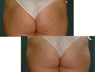
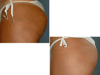
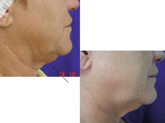
Bravo et al. (2013) conducted a study in eight female patients, with II and III grade of cellulite in buttocks and legs. These patients were submitted for four sessions of unipolar radiofrequency (Accent RF system – Alma Lasers) with 2 weeks interval. Patients were clinically evaluated through comparative before and after pictures, as well as through laboratorial tests and ultrasonography (US) images. Pictures and US images were performed before starting the treatment and 30 days after the last session. Laboratorial tests to evaluate possible side effects were done before and after the first and the last session of treatment. All of the eight patients showed clinical improvement, and US images showed increasing in the thickness of the dermis in seven of the eight patients. No alteration in the laboratorial exams was reported. Authors concluded that this type of unipolar RF was an efficient and safe method in the treatment of cellulite (Figs. 2 and 3). These authors also reported good results for photodamaged skin treatment, as demonstrated in Fig. 4, the improvement of the laxity in the neck.
Hexsel et al. (2011) conducted a pilot study with a device combining bipolar RF technology, infrared light, vacuum, and mechanical massage. They evaluated nine patients that had a body mass index (BMI) of 18–25 kg/m2 and at least 6° in the severity scale of cellulite (CSS) (Hexsel et al. 2009). The scale used in this article was described by Hexsel and consists in five key clinical morphologic features of cellulite: (A) the number of evident depressions; (B) depth of depressions; (C) morphological appearance of skin surface alterations; (D) grade of laxity, flaccidity, or sagging skin; and (E) the classification scale originally described by Nürnberger and Müller (Hexsel et al. 2009). Authors reported reduction on cellulite severity and on corporal circumference of the buttocks in all patients. Poor results were observed in the thighs with the same treatment (Hexsel et al. 2011).
Goldberg et al. (2008) evaluated the efficacy of the unipolar device (Accent RF system – Alma Lasers) in 30 patients with III/IV cellulite grade in the upper part of the thigh. They were treated with six sessions every 15 days and were evaluated 6 months after the treatment. Clinical circumference measurements, skin biopsy, MRI, and blood lipid evaluation were performed. Twenty-seven patients showed clinical improvement. The reduction on the circumference of the thighs was 2.45 cm in average. Histological changes showed dermal fibrosis. No changes in the lipids blood level were observed nor in the MRI of the treated patients.
Sadick and Magro (2007) conducted a study in 20 patients aged from 28 to 59, phototypes I to VI, in which they used different intensities of bipolar RF, infrared light, and levels of vacuum. Sixteen of the 20 patients remained in the study. Twelve sessions of 30 min were performed twice a week, 3 days apart, for 6 weeks. The RF energy, the optic energy, and the levels of vacuum were adjusted from patient to patient to ensure that the optimum parameters of the treatment were reached. Clinical results were evaluated through photographs, circumference measurements of the leg, and dermatological exams by the researchers. A total of 65% of the patients had a reduction in the circumference of the leg; 50% of the patients had an improvement greater than 51%. In most patients, some degree of improvement could be observed in the appearance of cellulite.
Adatto et al. (2014) conducted a study with 35 healthy female patients with skin laxity and subcutaneous fat deposits localized in the abdomen, buttocks, or thighs, using a new high-power radiofrequency technology combined with infrared light and mechanical manipulation. Sixty percent of the patients showed improvement of 24.1%; 27% of the patients showed improvement between 25% and 49%; and 5% showed improvement between 50% and 74%, while only 8% of the patients did not show any improvement.
Take Home Messages
- Cellulite, also known as gynoid lipodystrophy, is a multifactor disorder of the dermis, epidermis, and subcutaneous cellular tissue.
- Although cellulite is still considered as an unclear etiological condition, there are many hypotheses trying to explain this disorder.
- In the last years, the idea of having an ideal sculpting body changed the concept of cellulite from a cosmetic problem to a disease.
- Treatments can be invasive and noninvasive. Noninvasive treatments include topical treatments, controlled diets, cryolipolysis, focused ultrasound, endermology, laser, and non-ablative radiofrequency.
- Radiofrequency (RF) or RF spectrum is the name given to one of the parts of the electromagnetic spectrum, with frequencies from 3 kHz to 300 GHz.
- The energy generated by RF can spread in tridimensional volumes of the tissue, to controlled depths.
- The depth of the RF depends on multiple factors such as the tissue over which it is applied (dermis, epidermis, subcutaneous tissue), the configuration of the electrodes (monopolar, bipolar, tripolar), the programming of the RF waves, and the applied temperature.
- RF does not have absorption or dispersion due to epidermic melanin and therefore cannot be used in all types of skins and generate significant heat without a risk for the epidermis.
- Radiofrequency (RF) contracts collagen and stimulates neocollagenesis by thermal heat, promoting thickening of the dermis and avoiding fat herniation.
- RF can also promote improvement in local circulation due to the vasodilatation and lymphatic drainage. Clinically it improves the laxity and the irregularity of the skin surface.
- New RF devices have been developed associating other technologies to reach different layers of the skin and to improve lymphatic drainage.
- Many studies report efficacy with RF for cellulite and body laxity with few sessions and minimal side effects.
References
Adatto MA, Adatto-Neilson RM, Morren G. Reduction in adipose tissue volume using a new high-power radiofrequency technology combined with infrared light and mechanical manipulation for body contouring. Lasers Med Sci. 2014;29(5):1627–31.
Alan M, Drover JS. Lifting y estiramiento no quirúrgico, part 2, cap 3. USA: Elsevier; 2010.
Alexiades-Armenakas MR, Dover JS, Arndt KA. Unipolar radiofrequency treatment to improve the appearance of cellulite. J Cosmet Laser Ther. 2008;10(3):148–53.
Alster TS, Tanzi EL. Cellulite treatment using a novel combination radiofrequency, infrared light, and mechanical tissue manipulation device. J Cosmet Laser Ther. 2005;7:81–5.
Bertin C, Zunino H, Pittet JC, et al. A double-blind evaluation of the activity of an anti-cellulite product containing retinol, caffeine, and ruscogenine by a combination of several non-invasive methods. J Cosmet Sci. 2001;52:199–210.
Boisnic S. Evaluation du dispositif de radiofréquence tripolaire Regen™ en utilisant un modèle expérimental de peau humaine. Nouv Dermatol. 2008;28:331–2.
Bravo BSF, Issa MCA, Muniz RLS, Torrado CM. Tratamento da lipodistrofia ginoide com radiofrequência unipolar: avaliação clínica, laboratorial e ultrassonográfica. Surg Cosmet Dermatol. 2013;5(2):138–144.
Brightman L, et al. Improvement in arm and post-partum abdominal and flank subcutaneous fat deposits and skin laxity using a bipolar radiofrequency, infrared, vacuum and mechanical massage device. Lasers Surg Med. 2009;41:791–8.
Carruthers J, Fabi S, Weiss R. Monopolar radiofrequency for skin tightening: our experience and a review of the literature. Dermatol Surg. 2014;40 Suppl 12:S168–73.
Coringrato M, Jaled M, De Carli E, Cacabelos M. Radiofrecuencia ablativa en dermatología quirúrgica: Una revisión, Rev. dermatología argentina, vol. XIV, Julio–Septiembre 2008, Número 3.
Curri SB, Bombardelli E. Local lipodystrophy and districtual microcirculation. Cosmet Toilet. 1994;109:51–65.
De Peña J, Hernández-Pérez M. Lipodistrofia ginecoide (celulitis). Rev Cent Dermatol Pascua. 2005;3:132–5.
Draelos ZD. In search of answers regarding cellulite. Cosmet Dermatol. 2001;14(1):55–8.
Emanuele E. Cellulite: advances in treatment: facts and controversies. Clin Dermatol. 2013;31(6):725–30.
Goldberg DJ, Fazeli A, Berlin AL. Clinical, laboratory, and MRI analysis of cellulite treatment with a unipolar radiofrequency device. Dermatol Surg. 2008;34(2):204–9; discussion 209. Epub 2007 Dec 17.
Goldman MP, Hexsel D, editors. Cellulite: pathophysiology and treatment. 2nd ed. Florida: Editorial Informa, Healthcare; 2010.
Goldman MP, Bacci PA, Leisbachoff G, et al. Pathophysiology of cellulite. New York: Taylor & Francis; 2006.
Hexsel D, DalForno T, Hexsel CL. Severity scale of cellulite. J Eur Acad Dermatol Venereol. 2009;23:523–8.
Hexsel DM, Siega C, Schilling-Souza J, et al. A bipolar radiofrequency, infrared, vacuum and mechanical massage device for treatment of cellulite: a pilot study. J Cosmet Laser Ther. 2011;13(6):297–302.
International Electrotechnical Commission. http://www.iec.ch/.
Isidori A. Fattori predisponenti. In: Ribuffo A, Bartoletti C, editors. La celulite. Rome: Sallus; 1983. p. 49–59.
Khan MH, Victor F, Rao B, Sadick NS. Treatment of cellulite: part I. Pathophysiol J Am Acad Dermatol. 2010;62(3):361–70.
Kotcher Fuller J. Surgical technology: principles and practice. 4th ed. St. Louis: Editorial Elsevier; 2005.
Laguese P. Sciatique et infiltration cellulagique. These Méd Lyon; 1929.
Lapidoth M, Halachmi S, editors. Radiofrequency in cosmetic dermatology, Aesthet Dermatol, vol. 2. Basel: Karger; 2015. p. I–VI.
Lolis MS, Goldberg DJ. Radiofrequency in cosmetic dermatology: a review. Dermatol Surg. 2012;38(11):1765–76.
Manuskiatti W, Wachirakaphan C, Lektrakul N, Varothai S. Circumference reduction and cellulite treatment with a TriPollar radiofrequency device: a pilot study. J Eur Acad Dermatol Venereol. 2009;23(7):820–7.
McKnight BB, Tobin R, Kabir Y, Moy R. Improving upper arm skin laxity using a tripollar radiofrequency device. J Drugs Dermatol. 2015;14(12):1463–6.
Morita A. Tobacco smoke causes premature skin aging. J Dermatol Sci. 2007;48:169–75.
Nürnberger F, Müller G. So called cellulite: an invented disease. J Dermatol Surg Oncol. 1978;4:221–9.
O’Connor JL, Bloom DA, William T. Bovie and electrosurgery. Surgery. 1996;119(4):390–6.
Piérard GE, Nizet JL, Piérard-Franchimont C. Cellulite: from standing fat herniation to hypodermal stretch marks. Am J Dermatopathol. 2000;22(1):34–7.
Pugliese PT. The pathogenesis of cellulite: a new concept. J Cosmet Dermatol. 2007;6:140–2.
Querleux B, Cornillon C, Jolivet O, Bittoun J. Anatomy and physiology of subcutaneous adipose tissue by in vivo magnetic resonance imaging and spectroscopy: relationships with sex and presence of cellulite. Skin Res Technol. 2002;8:118–24.
Rosenbaum M, Prieto V, Hellmer J, Boschmann M, Krueger J, Leibel RL, Ship AG. An exploratory investigation of the morphology and biochemistry of cellulite. Plast Reconstr Surg. 1998;101(7):1934–9.
Rossi AB, Vergnanini AL. Cellulite: a review. JEADV. 2000;14:251–62.
Sadick N, Magro C. A study evaluating the safety and efficacy of the Velasmooth™ system in the treatment of cellulite. J Cosmet Laser Ther. 2007;9:15–20.
Shapiro SD, Eros Y, Abrahami Y, Leviav A. Evaluation of safety and efficacy of the TriPollar technology for treatment of wrinkles. Lasers Surg Med. 2012;44(6):453–8. Epub 2012 Jun 29.







