
Q-Switched Lasers for Melasma, Dark Circles Eyes, and Photorejuvenation
Q-Switched Lasers for Melasma, Dark Circles Eyes, and Photorejuvenation
Juliana Neiva, Lilian Mathias Delorenze and Maria Claudia Almeida Issa Brazilian Society of Dermatology (SBD) and American Academy of Dermatology (AAD), Rio de Janeiro, Brazil Hospital Universitário Antonio Pedro, Universidade Federal Fluminense – Niterói, RJ, Brazil Department of Clinical Medicine – Dermatology, Fluminense Federal University, Praia de Icaraí 139, 702, Niterói, RJ, Brazil
Abstract
Melasma is a common and persistent disorder of hyperpigmentation that affects a significant portion of the population, affecting mainly women. It is often a therapeutically challenging disorder. Physical therapies such as chemical peels, dermabrasion, lasers, and intense pulsed light have also been used with varying degrees of success and side effects. Dark circles eyes, also known as periorbital hyperpigmentation, are a common condition that occurs in both sexes with an increasing frequency in females. Aesthetic treatments include microdermabrasion, chemical peels, lasers, radiofrequency, injectable fillers, surgery, fat transfer, and lightening topical products. Clinical signs of photoaging include coarse skin texture, irregular pigmentation, and laxity of skin tone, as well as the appearance of fine lines and wrinkles. Diverse treatment modalities have been used to improve skin wrinkling and laxity, including chemical peeling, soft tissue filler, laser ablation, and facelift surgery. Lasers have revolutionized the treatment of many dermatological conditions. Different types of lasers can be indicated for pigmentary disorders. Recently, Q-switched lasers arose as a successful application in melasma, dark circles eyes, and photorejuvenation due to its low fluence, short pulse, and specific wavelength.
Keywords LasersQ-switched laserMelasmaDark circles eyesPhotorejuvenation
Introduction
Lasers (light amplification by stimulated emission of radiation) are sources of high-intensity monochromatic coherent light that can be used for the treatment of various dermatologic conditions depending on the wavelength, pulse characteristics, and fluence of the laser being used and the nature of the condition being treated (Arora et al. 2012).
Q-switched (QS) lasers deserve special attention due to its recently uprising application in the treatment of melasma, dark circles eyes, and photorejuvenation.
Q-Switched Lasers
Lasers have demonstrated significant efficacy in the treatment of hyperpigmented disorders by selectively destructing pigment cells with a short pulse and low fluence. The effectiveness of laser treatment for pigmented lesions is based on the theory of selective photothermolysis introduced by Anderson and Parrish, which states that when a specific wavelength of energy is delivered over a period of time shorter than the thermal relaxation time (TRT) of the target chromophore (Arora et al. 2012; Anderson et al. 1989; Anderson and Parrish 1983; Jang et al. 2011), heat and injury are restricted to the target, with less damage to the surrounding tissue (Anderson and Parrish 1983; Jang et al. 2011).
The thermal relaxation time for a melanosome with 1-μm diameter ranges from 50 to 100 ns (Polder et al. 2011). Hence, a laser should emit a wavelength that is specific and well absorbed by the particular chromophore being treated.
A selective window for targeting melanin lies between 630 and 1,100 nm, where there is good skin penetration and preferential absorption of melanin over oxyhemoglobin (Stratigos et al. 2000). Absorption for melanin decreases as the wavelength increases, but a longer wavelength allows deeper skin penetration. Shorter wavelengths (<600 nm) damage pigmented cells with lower energy fluencies, while longer wavelengths (>600 nm) penetrate deeper but need more energy to cause melanosome damage (Arora et al. 2012). Besides wavelength, pigment specificity of lasers also depends on pulse width (Chan et al. 2010).
These lasers lead to a photoacoustic mechanical disruption of melanin caused by rapid thermal tissue expansion (Arora et al. 2012).
QS Nd:YAG Laser
The 1,064-nm Q-switched neodymium-doped yttrium aluminum garnet (QS 1,064-nm Nd:YAG) laser is widely used in cosmetic laser dermatology (Arora et al. 2012; Chan et al. 2010) for pigmented and vascular lesions, removal of tattoos, and unwanted hair (Chan et al. 2010).
With a wavelength of 1,064 nm, these devices allow for much deeper energy penetration and minimal melanin absorption compared with the QS ruby laser or QS alexandrite laser. The QS Nd:YAG laser uses a collimated handpiece to deliver a high peak power over very short pulse durations (≤20 ns), maximizing selective photothermolysis of cutaneous melanosomes (Friedmann and Goldman 2015).
The 1,064-nm QS Nd:YAG is well absorbed by melanin, and being a longer wavelength causes minimal damage to the epidermis and is not absorbed by hemoglobin. The deeper skin penetration is also helpful to target dermal melanin. Low-dose QS Nd:YAG laser induces sublethal injury to melanosomes, causing fragmentation and rupture of melanin granules into the cytoplasm (Arora et al. 2012; Anderson and Parrish 1983; Lee 2003). This effect is highly selective for melanosomes, as this wavelength is well absorbed by melanin relative to other structures. There is also subcellular damage to the upper dermal vascular plexus, which is one of the pathogenetic factors in melasma (Kim et al. 2007). The subthreshold injury to the surrounding dermis stimulates the formation of collagen, resulting in brighter and tighter skin (Schmults et al. 2004).
QS Ruby Laser
The Q-switched ruby laser (QSRL) was the first laser reported to be highly efficacious for the treatment of benign epidermal-pigmented lesions (Park et al. 2008; Taylor and Anderson 1993; Nelson and Applebaum 1992).
The 694-nm wavelength of QSRLs is moderately absorbed by melanin, yet poorly absorbed by competing chromophores such as hemoglobin (Friedmann and Goldman 2015; Taylor and Anderson 1993). Rapid delivery of high-intensity energy at this wavelength disrupts melanosomes within keratinocytes, melanocytes, and melanophages, making them ideal for pigmented epidermal and superficial dermal lesions in Fitzpatrick skin types I–II (Friedmann and Goldman 2015; Kopera et al. 1997).
QS Alexandrite Laser
The more deeply penetrating 755-nm wavelength of the Q-switched alexandrite laser (QSAL) has a lower absorption coefficient for melanin and is emitted over a longer pulse duration (50–70 ns) than that of the QSRL, which may serve to decrease adverse events (e.g., postinflammatory hyperpigmentation (PIH)) in dark-skinned patients as a result of gentler melanosomal heating. QSAL treatments of Fitzpatrick skin types IV or lower are typically performed with 3- to 5-mm spot sizes and 4–8 J/cm2. Lower fluences may lead to equal efficacy with decreased PIH (Friedmann and Goldman 2015; Wang and Chen 2012).
A novel QSAL with energy delivered in picoseconds (as low as 550 ps) may produce greater tensile stress on melanosomes than nanosecond pulse durations, enhancing their photomechanical and photothermal destruction. Collateral tissue heating and associated adverse events are minimized owing to the lower fluences required (Friedmann and Goldman 2015; Dover et al. 2012). As a result, potentially all skin types may be treated with this device. A 3–5-mm spot size and 1.5–2.83 J/cm2 fixed fluence are favored (Friedmann and Goldman 2015).
Pretreatment Procedure
A history of keloids, conditions that may impair wound healing, recent oral retinoid use, pregnancy, breastfeeding, photosensitivity, and/or abnormalities localized to the treatment area (active infections, malignant lesions, scarring, or burns) should be ruled out before undertaking any procedure. Prophylactic antiviral therapy for herpes simplex virus is not routinely performed before QS lasers (Friedmann and Goldman 2015). Topical depigmentation products can be used pre- and posttreatment (Arora et al. 2012).
All patients should have photographs and written informed consent obtained upon arrival (Arora et al. 2012; Friedmann and Goldman 2015). Before treatment, the area to be treated should be washed with a neutral cleanser to remove any makeup or other impurities. Topical anesthesia is generally unnecessary given the limited treatment area, and it is usually bearable (Friedmann and Goldman 2015).
QS laser treatment of lower eyelid skin within the borders of the bony orbital rim requires intraocular metal eye shields (Arora et al. 2012).
Melasma and Q-Switched Laser
Melasma is a common and persistent disorder of hyperpigmentation that affects a significant portion of the population, affecting mainly women (Werlinger et al. 2007; Park et al. 2011), being more common in Black, Latin, and Asiatic people (Kauvar 2012). Patients report that the condition has a markedly detrimental effect on their quality of life (Park et al. 2011; Balkrishnan et al. 2003; Dominguez et al. 2006).
Melasma presents as symmetric, hyperpigmented macules and patches on the face, usually on the cheeks, bridge of the nose, forehead, chin, and upper lip (Kauvar 2012; Choi et al. 2010; Grimes 1995; Gupta et al. 2006; Zhou et al. 2011). Typically it affects more women of reproductive age with Fitzpatrick type IV–VI, but can also affect men (Sarkar et al. 2014). The ratio between affected women and men is 9:1 (Kauvar 2012). Although its photogenesis is not fully understood, pregnancy, sunlight exposure, birth control pills, hormone therapy, genetic factors, mild ovarian dysfunction, and autoimmune thyroid disease may be implicated (Jang et al. 2011; Kauvar 2012; Choi et al. 2010; Gupta et al. 2006; Sarkar et al. 2014; Lee et al. 2010). Sun exposure can trigger melasma because it stimulates melanocytes to produce increased melanin, and even a small amount of sun exposure can worsen the condition. Irritation or inflammation of the skin can also stimulate melanin production and worsen melasma (Kauvar 2012; Grimes 1995). In some cases, it can disappear spontaneously, but in general, the condition remains for the rest of the patient’s life (Zhou et al. 2011).
The most widely accepted classification of melasma in recent years is based on the pathological manifestations (Zhou et al. 2011; Rigopoulos et al. 2007), including the epidermal type without melanophages in the dermis, the dermal type with melanophages in the dermis, and the mixed type, in which part of the lesion is of the epidermal type and part of the dermal type (Kauvar 2012; Zhou et al. 2011). By Wood’s light (340–400 nm), epidermal melasma can be visualized as brown patches or macules, while the dermal types normally are not visible except when they appear blue or black (Kauvar 2012; Zhou et al. 2011; Sarkar et al. 2014).
The major clinical feature of melasma is hyperpigmentation in the form of patches or macules, but it has also been observed that some patients have an increased distribution of telangiectatic erythema within the macules (Kim et al. 2007).
On histologic examination, it is possible to observe that the quantity of melanocytes is not increased; however, their size becomes enlarged, with more dendrites, and they are more active. The dermal pigment, when present, is normally found in the middle dermis within the melanophages. These melanophages are usually located near small, increased vessels, with little or no inflammation (Kauvar 2012; Zhou et al. 2011).
It is known that melanocytes respond to angiogenic factors, as they express receptors for vascular endothelial growth factor (VEGF). Kim et al. (2007) demonstrated that the area of melasma has 33,89% more vessels than a nearby area without macules and that 16,28% of these vessels are thicker. It is reported that keratinocytes show increased VEGF in melasma. The increase in the density and size of the vessels in the melasma area is directly related to the increase in pigmentation of the macules. Therefore, treatment of the vessels in melasma should be important (Kim et al. 2007).
Melasma is often a therapeutically challenging disorder for dermatologists. The most important aspect of treating melasma is to understand that there are differences in melanocyte activity between people. Regardless of the treatment, it is very important not to irritate the skin when using acids. Patients should understand the importance of treating melasma while avoiding erythema.
Topical treatment of melasma can be divided into two groups: hydroquinone and non-hydroquinone (kojic acid, azelaic acid, ascorbic acid, or alpha arbutin). However, these kinds of treatment provide only temporary results and can also produce long-term complications (Kauvar 2012; Gupta et al. 2006; Katsambas and Antoniou 1995; Jesitus 2014; Polnikorn 2011). Physical therapies such as chemical peels, dermabrasion, lasers, and intense pulsed light (IPL) have also been used with varying degrees of success and side effects (Park et al. 2011; Kauvar 2012; Katsambas and Antoniou 1995).
Physical methods are the only option to remove the melanin and destroy the melanosomes. Hence, treatment with laser has been the most recommended since 2005, when the use of fractional laser for selective photothermolysis in the treatment of melasma began (Se-Yeong et al. 2008).
Various lasers that have been used for melasma include the following (Arora et al. 2012; Goldberg 1997):
- Green light: flashlamp-pumped pulsed dye laser (PDL) (510 nm), frequency-doubled QS Nd:YAG (Q-switched neodymium:yttrium aluminum garnet – 532 nm)
- Red light: QS ruby (694 nm), QS alexandrite (755 nm)
- Near infrared: QS Nd:YAG (1,064 nm)
The green light lasers do not penetrate as deeply into the skin as the other two groups owing to their shorter wavelengths. They are therefore effective only in the treatment of epidermal melasma (Arora et al. 2012).
Since the green wavelength is also well absorbed by oxyhemoglobin, bruising and purpura may occur following laser irradiation. The purpura resolves in 1–2 weeks after treatment, and the clinical lesions lighten in 4–8 weeks. Occasionally, the bruising can lead to postinflammatory hyperpigmentation. Green light lasers often produce a variable response, and thus test spots may be prudent prior to treating the whole area (Arora et al. 2012; Goldberg 1997).
Red lasers have longer wavelengths and thus may penetrate deeper into the dermis. They can also be used to treat epidermal-pigmented lesions without bruising, as they are not absorbed by hemoglobin (Arora et al. 2012; Stratigos et al. 2000). The pulse duration of the QS ruby laser (QSRL) varies from 20 to 50 ns and that of the QS alexandrite laser (QSAL) from 50 to 100 ns (Arora et al. 2012).
Near-infrared lasers include the QS Nd:YAG laser (1,064 nm), which has a pulse duration of 10–20 ns. Despite lower absorption of this wavelength by melanin compared with the green and red light lasers, its advantage lies in its ability to penetrate more deeply into the skin. In addition, it may prove more useful in the treatment of lesions in individuals with darker skin tones (Arora et al. 2012; Goldberg 1997).
QS Nd:YAG Laser × Melasma
There have been several reports of treating melasma with the QS Nd:YAG laser. The mechanism of clinical improvement is proposed to be its ability to induce a nonspecific dermal wound with a healing response and subsequent neocollagenesis (Chan et al. 2010). Mun et al. (2011) found that treatment of melasma skin with this laser resulted in a decreased number of melanocytic dendrites and an altered ultrastructure of melanosomes (Mun et al. 2011).
The proposal is to achieve a selective and more stable photothermolysis, minimally invasive, to remove melanin and melanosomes. This occurs through the use of low energy during the process (Kauvar 2012; Mun et al. 2011; Kang et al. 2011).
For the mechanism of photothermolysis to be efficient, an appropriate wavelength is necessary (1,064 nm is useful because it reaches both the dermis and the epidermis). Likewise, the thermal damage of the emitted pulse must be sufficient to destroy the melanin, and, finally, the pulse duration must be as short as possible to avoid damage to nearby tissues. The ideal for melasma treatment is to cause minimal thermal damage and the greatest photoacoustic damage possible (Mun et al. 2011).
The QS Nd:YAG is the most widely used laser for the treatment of melasma (Arora et al. 2012; Brown et al. 2011). In general, the fluence used is less than 5 J/cm2, with a spot size of 6 mm and a frequency of 10 Hz. The number of treatment sessions varies from 5 to 10 at 1-week intervals (Arora et al. 2012). Zhou et al. (2011), in their studies, used the QS Nd:YAG laser at low energy levels (fluence of 2.5–3.4 J/cm2) weekly for 9 sessions in the treatment of melasma in 50 patients (Zhou et al. 2011).
Choi et al. (2011) treated melasma lesions in 20 patients older than 30 years with a fluence of 2.0–3.5 J/cm2, a spot size of 6 mm, and a repetition rate of 10 Hz over the whole face. The treatment was performed five times at 1-week intervals (Choi et al. 2010).
Over the last few years, the QS Nd:YAG laser has increasingly been performed as “laser toning” or “laser facial” for non-ablative skin rejuvenation and melasma in Asian countries. In laser toning, multiple passes of low-fluence laser (e.g., 1.6–3.5 J/cm2) are delivered through a large spot size (e.g., 6–8 mm) to optimize energy delivery (Arora et al. 2012; Chan et al. 2010). With a clinical endpoint of erythema plus lesional and hair whitening (Chan et al. 2010; Kauvar 2012), QS Nd:YAG treatments involve 10, 20, or more weekly treatments, with as many as 10–20 laser passes per treatment (Arora et al. 2012; Chan et al. 2010; Kauvar 2012). For melasma, laser toning should be considered a second-line therapy, since this treatment is unlikely to be curative and is not without risk (Chan et al. 2010). Complications from these high-cumulative-fluence procedures include pain, urtication, hyperpigmentation, long-term hypopigmentation (guttate leukoderma), and rebound of melasma (Arora et al. 2012; Chan et al. 2010; Kauvar 2012; Kim et al. 2009, 2010).
Some publications compare the use of different types of laser in melasma treatment. Jalaly et al. (2014) compared a low-energy fractional CO2 laser with the QS Nd:YAG 1,064 nm. On each side of the patients’ faces, one of these two lasers was applied. They were treated over 3 weeks with five weekly sessions. Two months after the end of treatment, the patients were evaluated, and the side of the face treated with the fractional CO2 showed a greater reduction in MASI (Melasma Area and Severity Index) (Jalaly et al. 2014).
There is no relationship between the therapeutic response to the QS Nd:YAG 1,064 nm and the severity of the disease or the patients’ Fitzpatrick skin type (Zhou et al. 2011). Some studies suggest that more epidermal melasma responds better to topical treatment and intense pulsed light, and that patients of Fitzpatrick phototype IV respond better to treatment in comparison with those of phototype III. However, as the QS Nd:YAG 1,064-nm laser reaches the epidermis even in dermal lesions, this relationship is not observed during or after treatment.
In practice, the treatment of melasma consists of two stages: a whitening stage and a maintenance stage.
Whitening of the lesions tends to begin between the fourth and sixth weeks and increases after each treatment session. Zhou et al. (2011) observed, after the nine sessions, a 61.3% decrease in MASI using the low-energy QS Nd:YAG. Seventy percent of the patients showed improvement of at least 50% of the lesions, and 10% had 100% improvement. They concluded that the QS Nd:YAG 1,064-nm laser with low energy and a wide spot is the new method of choice for melasma treatment. The response is fast and satisfactory in whitening the pigment (Zhou et al. 2011).
Sim et al. (2014) evaluated the outcome of therapy in 50 patients treated with the QS Nd:YAG 1,064-nm laser with an 8-mm spot and 2,8 J/cm2 of fluence. The patients were treated weekly for 15 weeks. Both patients and investigators reported improvements of 50–74% of the lesions, which was confirmed by imaging. None of the patients had severe adverse effects during treatment. Thus, they judged the treatment to be safe and efficient with this type of laser (Sim et al. 2014).
Other publications demonstrate that the Q-switched Nd:YAG 1,064-nm laser is safe and effective in the treatment of melasma. Suh et al. (2011) evaluated 33 patients, and the treatment consisted of one session per week, for a total of 10 sessions. The reduction in MASI and whitening of the lesions were perceptible from the seventh week on. Follow-up in the first, second, and third months after the end of therapy demonstrated that the whitening process was still present during this period. In this work, no adverse effects were noted (Suh et al. 2011).
Jeon et al. (2008) published a study evaluating the use of the Q-switched Nd:YAG 1,064-nm laser with a 5-ns pulse in 27 patients. They used a 7-mm spot and 2–2.5 J/cm2 of fluence in 17 patients and 1.6–2 J/cm2 in the other 10 patients. The number of passes varied from three up to ten, until slight erythema was reached. Two months after the end of treatment, 64.7% of the patients had recurrence of the lesions, but the intensity of the pigment was lower than in the initial macules, and in 29.41% there were no recurrences (Se-Yeong et al. 2008).
Thus, treatment of melasma during the whitening stage should be gradual, with one session per week for 10–12 weeks. The largest spot possible (6–8 mm) should be used, with a pulse frequency of 5–10 Hz. The energy should vary from 0.8 up to 1.8 J/cm2 (400–900 mJ). Two to four passes should be applied per area until the erythema lightens, with little overlap (10–15%). For a better result, the erythema should be as homogeneous as possible.
Some studies demonstrate that the use of microdermabrasion before the procedure can improve the response to the laser. One or two passes over the lesions are recommended, followed by application of the QS Nd:YAG 1,064 nm (Kauvar 2012). In clinical practice, this type of procedure seems to help cases of more laser-resistant melasma, since thinning of the corneal layer eases penetration of the laser into the skin.
Alsaad et al. (2014) applied microdermabrasion before the QS Nd:YAG 1,064-nm laser with a 5-ns pulse, 6-mm spot, and 1.8–2.0 J/cm2 of fluence in 10 patients; in another 17 patients, they used the QS Nd:YAG 1,064-nm laser with a 50-ns pulse, 5-mm spot, and 1.6 J/cm2 of fluence, along with the use of a whitening cream at home. The patients were followed up for 3, 6, and 12 months after the end of treatment. Patients who completed three or four treatments had better results than those who completed one or two, all of them maintaining some degree of whitening until completion of the 12 months (Alsaad et al. 2014).
After the procedure, it is recommended to cool the face for 10–15 min with a cold mask or a cold-air device. Afterwards, a medium-potency corticosteroid cream and sunscreen lotion should be applied. The use of corticosteroids over the lesions can be recommended for up to 2 days after the laser session, mainly for lesions that appear more unstable and reactive to the procedure.
The sessions are usually well tolerated, and the recurrent adverse effects are erythema, which can last for 1–3 h, and pruritus. The less frequent effects are purpura, edema, acne, and thinning and whitening of the facial hair.
Postinflammatory hypochromic lesions and guttate-leukoderma-type hypopigmentation are described as consequences of using the laser with high energy and multiple passes.
The reported cases of hyperchromia in some studies can affect up to 10% of treated patients and can occur after few laser sessions, but they are usually due to use of the QS Nd:YAG laser with the goal of rejuvenation (Chan et al. 2010). In these protocols, more passes or higher energy are applied so that the heat can stimulate collagen production in the dermis. Often the protocols exclude the use of QS; hence, the 1,064-nm laser begins to operate with a longer pulse duration (e.g., from 5 to 200 ms). In most cases, the hypopigmentation persists.
Recurrence of melasma is defined as an increase in pigmentation or an increase in the size of the lesion after the final treatment, and it remains a problem for therapy with the QS Nd:YAG 1,064-nm laser. Studies demonstrate that recurrence of the lesions tends to occur 2 to 3 months after the last session, with the characteristics of the lesions the same as at the beginning of treatment. This return of pigmentation in the lesions occurs because the thermal effect on the melanocytes is reversible over time. Thus, association with maintenance therapy is necessary (Zhou et al. 2011; Se-Yeong et al. 2008).
The maintenance phase can include the QS Nd:YAG 1,064-nm laser and/or topical creams and oral medication. Intermittent use of hydroquinone during and after treatment is also described to extend the lightening response of the laser, avoiding association with retinoids.
Wattanakrai et al. (2010) conducted a study in patients with dermal or mixed melasma in which, in addition to the QS Nd:YAG 1,064 nm, they used daily application of hydroquinone 2%, compared with a control group of patients who used only hydroquinone. The patients treated with laser had five treatment sessions using a 6-mm spot and 3–3.8 J/cm2 of fluence, along with cold air. After 12 weeks, 92% of the patients who had hydroquinone combined with laser achieved whitening of the macules. On the other hand, in the control group, only 19,7% of the patients showed improvement of the lesions (Wattanakrai et al. 2010).
The topical application of arbutin, hydroquinone, and vitamin C is convenient and preferred (Zhou et al. 2011).
In cases where the QS Nd:YAG 1,064-nm laser is used for maintenance aimed at increasing the interval between sessions, sessions are initially performed every 15 days for 2 months and then once every 30–60 days, according to maintenance of the whitening. Laser maintenance has been shown to be more effective than isolated topical products, as the melanocytes tend to remain less active and smaller in the lesions with laser treatment.
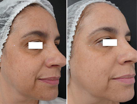
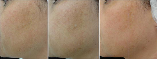
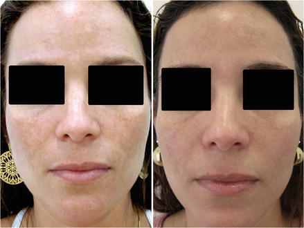
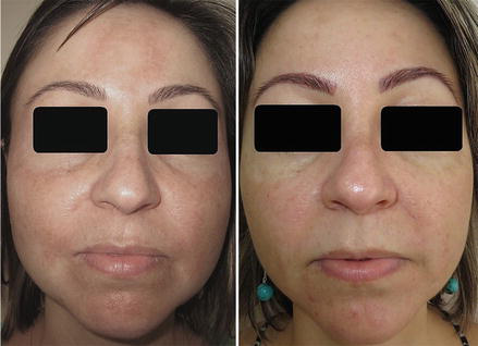
Melasma is a pigmentary disease of the skin with many pathological and aggravating factors involved. Thus, a single treatment does not prove completely effective. The best treatment is a combination of therapies, and, among these, the one that currently presents the best effectiveness and safety in whitening the lesions is the low-energy Q-switched Nd:YAG 1,064-nm laser (Figs. 1, 2, 3, and 4). Its association with whitening creams, microdermabrasion, and peelings makes these results even better and longer lasting.
QS Ruby Laser × Melasma
The efficacy of the QSRL for melasma is still controversial (Arora et al. 2012; Jang et al. 2011). The mechanism is the same as that of the QS Nd:YAG laser; that is, it causes highly selective destruction of melanosomes. The QS ruby laser, with a wavelength of 694 nm, is more selective for melanin than the QS Nd:YAG laser (1,064 nm) (Arora et al. 2012).
So theoretically the QSRL is expected to be more effective than the QS Nd:YAG for melasma (Arora et al. 2012), but severe postinflammatory hyperpigmentation and hypopigmentation occurred in some patients within a short period of time (Choi et al. 2010; Hilton et al. 2013), suggesting that the high-energy mode of QS lasers is likely to be ineffective for melasma and therefore is not a good therapeutic choice for this condition (Zhou et al. 2011; Taylor and Anderson 1994). The role of the QSRL is controversial, with studies showing conflicting results (Arora et al. 2012).
On the other hand, Jang et al. (2011) showed that multiple treatment sessions of low-dose fractional QSRL may be an effective strategy for the treatment of dermal or mixed-type melasma (Jang et al. 2011).
More studies are needed to establish its efficacy and safety in melasma (Arora et al. 2012).
Dark Circles Eyes and Q-Switched Laser
Dark circles eyes are also known as periorbital hyperpigmentation. It is a common condition that occurs in both sexes, with an increasing frequency in females (Friedmann and Goldman 2015; Roberts 2014). It can impart a fatigued and less youthful appearance to the face (Friedmann and Goldman 2015). Globally, patients with skin of color are affected more than Caucasians. There is most likely a familial component, as it may be seen in family members over generations (Roberts 2014).
The formation of dark circles is often multifactorial, with a number of factors reported to play a role. Among these factors are hollowing/shadowing, dermal melanin deposition, postinflammatory hyperpigmentation secondary to atopic or allergic contact dermatitis, a prominent and superficial location of the vasculature, and exogenous causes (penicillamine-induced periorbital pigmentation; bimatoprost-induced periorbital hollowing and hyperpigmentation) (Friedmann and Goldman 2015; Freitag and Cestari 2007; Roh and Chung 2009; Xu et al. 2011). It is important for clinicians, as it may be a sign of an underlying systemic disease, skin disorder, allergic reaction, nutritional deficiency, or sleep disturbance (Friedmann and Goldman 2015; Xu et al. 2011).
Dark circles eyes usually present as bilaterally symmetric hyperpigmented patches around the eyes. One eye may be more involved than the other. It can affect either the upper or lower eyelid or both, and it may extend to involve the glabella and upper nose (Roberts 2014).
Aesthetic treatments include microdermabrasion, chemical peels, lasers, radiofrequency, injectable fillers, surgery, fat transfer, hydroquinone, and topical retinoids (Friedmann and Goldman 2015; Roberts 2014).
Cutaneous melanin has a broad, polychromatic absorption spectrum that peaks in the UV range and declines steadily as a function of increasing wavelength. Although significantly attenuated past 755 nm, energy absorption is still likely with wavelengths up to 1,064 nm, allowing treatment of deeper pigment and darker Fitzpatrick skin types. Given that melanosomes have thermal relaxation times of less than 1 μs, ultrashort pulse durations are required to selectively confine photothermal and photoacoustic effects to these structures (Anderson et al. 1989; Friedmann and Goldman 2015). Many Q-switched lasers with nanosecond (and recently picosecond) pulse durations and wavelengths within the absorption range of melanin are currently available. The typical clinical endpoint of these treatments is immediate lesion whitening without pinpoint bleeding. Lower energy settings should be used initially to minimize the occurrence of PIH (Friedmann and Goldman 2015).
QS Nd:YAG Laser × Dark Circle Eyes
As demonstrated in treating melasma, repeated sessions of low-fluence QS Nd:YAG treatment can decrease stage IV melanosomes, damage melanocytes, and reduce expression of melanogenesis-associated proteins. Higher fluences (4–5 J) can be used with a 3-mm spot size for other types of lower-eyelid hyperpigmentation (Friedmann and Goldman 2015) (Fig. 5).
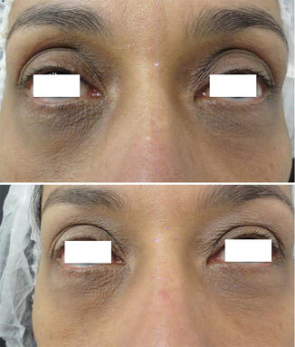
QS Ruby Laser × Dark Circle Eyes
QSRL treatment is performed with 2–4 J/cm2 using a 5-mm spot size (or varied accordingly) at 1.5 Hz. The clinical endpoint with this device is immediate lesion whitening that resolves over 20 min, followed by erythema and edema. Combining the QSRL with topical hydroquinone and tretinoin before and after treatment has also led to significant improvement in this location (Friedmann and Goldman 2015).
Photorejuvenation and Q-Switched Laser
Clinical signs of photoaging include coarse skin texture, irregular pigmentation, and laxity of skin tone, as well as the appearance of fine lines and wrinkles (Hong et al. 2014; Yaghmai et al. 2010).
Diverse treatment modalities have been used to improve skin wrinkling and laxity, including chemical peeling, soft tissue filler, laser ablation, and facelift surgery. Recently, among these modalities, laser ablation has been highlighted because of its relative effectiveness and shorter recovery time compared with other methods. It is classified into ablative and non-ablative lasers. Generally, ablative lasers are considered more effective than non-ablative ones in treating rhytides, but they have a prolonged recovery time and a greater chance of complications, such as postinflammatory hyperpigmentation due to excessive thermal energy transferred to adjacent tissue (Hong et al. 2014).
Non-ablative lasers are thought to work through thermal energy transferred to small vessels and tissues in the upper dermis, which stimulate dermal fibroblasts to induce collagen and elastin regeneration. In line with this hypothesis, long-pulse Nd:YAG lasers (LPND) have the advantage of reaching deeply located blood vessels and transferring energy to collagen adjacent to vessels while bypassing the epidermis. Patients with Fitzpatrick skin type III or higher can be treated with less risk because wavelengths in the infrared range are weakly attracted to melanin (Hong et al. 2014). Although the typical response to this type of treatment is modest clinical improvement in mild to moderate facial rhytides, non-ablative therapy has gained in popularity over the past few years for photoaging therapy because of its little to no downtime. Laser systems traditionally used for this approach rely on thermal induction for tissue change. The introduction of Q-switched laser systems has added a photoacoustic element to the dermal response. The Q-switched Nd:YAG laser has been shown to improve photodamage changes (Yaghmai et al. 2010; Goldberg and Silapunt 2001). With this laser system, relatively lower laser energies are needed, resulting in mild immediate side effects (Yaghmai et al. 2010).
Non-ablative laser therapy remains a very advantageous therapeutic approach for skin rejuvenation. The potential of inducing beneficial textural and pigmentary changes in sun-damaged and aged skin without the need for ablation results in dramatically fewer immediate and long-term postoperative effects. These approaches have resulted in collagen production and subsequent dermal thickening, with a reduction of surface textural changes. In addition, both pigmentary and erythematous changes can be improved. All of this with minor immediate postoperative effects, generally limited to transient erythema and edema. Long-term undesired effects such as fibrosis, scarring, or persistent pigmentary changes have been remarkably diminished relative to ablative procedures (Yaghmai et al. 2010).
The Q-switched Nd:YAG laser has exhibited the ability to achieve clinically desirable improvement in many parameters of skin rejuvenation. Remarkably, this has occurred with a very acceptable adverse-effect profile and, equally important, good patient tolerance and acceptability (Yaghmai et al. 2010) (Fig. 6).
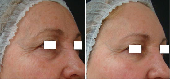
The authors have good experience with the QS Nd:YAG laser. Dr. Issa achieves good results using QS (ClearLift – Alma Lasers) for melasma (Figs. 1 and 2) and dark circles eyes (Fig. 5). This device is a fractional (5 × 5 pixel) QS Nd:YAG 1,064-nm laser with a pulse duration of 20 ns, and its fluence ranges from 500 to 1,200 mJ/p. Regarding photodamaged skin, the best results are observed in the periocular area (Fig. 6). Dr. Neiva has good experience using the QS Nd:YAG (Spectra-Skintech) for melasma (Figs. 3 and 4).
Complications and Post-procedure of Q-Switched Lasers
As low energies are applied, the occurrence of post-procedure complications is very rare. Usually, erythema resolves over minutes to a few hours and edema is not observed. The risk of blistering, PIH, and hypopigmentation is minimal because of the low energy used in treatment.
The authors reinforce the importance of using sunscreen. Lightening creams can be used 24 h after each session during treatment.
Take Home Messages
- Melasma is often a therapeutically challenging disorder for dermatologists. Likewise, dark circles treatment and photorejuvenation are not easy to achieve.
- Lasers have demonstrated significant efficacy in the treatment of hyperpigmented disorders, such as melasma and dark circles, by selectively destroying pigment cells with a short pulse and low fluence.
- Q-switched (QS) lasers, such as the QS Nd:YAG, QS ruby, and QS alexandrite lasers, deserve special attention because of their recently increasing application in the treatment of melasma, dark circles eyes, and photorejuvenation.
- Although QS lasers present the best effectiveness and safety in whitening lesions, combining them with other therapies can improve the treatment of melasma, dark circles, and photorejuvenation.
References
Alsaad SM, Ross EV, Mishra V, Miller L. A split-face study to document the safety and efficacy of clearance of melasma with a 5-ns Q-switched Nd:YAG laser versus a 50-ns Q-switched Nd:YAG laser. Lasers Surg Med. 2014;10:736–40.
Anderson RR, Parrish JA. Selective photothermolysis: precise microsurgery by selective absorption of pulsed radiation. Science. 1983;220:524–7.
Anderson RR, Margolis RJ, Watanabe S, et al. Selective photothermolysis of cutaneous pigmentation by Q-switched Nd:YAG laser pulses at 1064, 532, and 355 nm. J Invest Dermatol. 1989;93:28–32.
Arora P, Sarkar R, Garg V, Arya L. Lasers for treatment of melasma and post-inflammatory hyperpigmentation. J Cutan Aesthet Surg. 2012;5(2):93–103.
Balkrishnan R, McMichael AJ, Camacho FT, et al. Development and validation of a health-related quality of life instrument for women with melasma. Br J Dermatol. 2003;149:572–7.
Brown AS, Hussain M, Goldberg DJ. Treatment of melasma with low-fluence, large spot size, 1064-nm Q-switched neodymium-doped yttrium aluminium garnet (Nd:YAG) laser for the treatment of melasma in Fitzpatrick skin types II–IV. J Cosmet Laser Ther. 2011;13:280–2.
Chan NP, Ho SG, Shek SY, Yeung CK, Chan HH. A case series of facial depigmentation associated with low-fluence Q-switched 1,064-nm Nd:YAG laser for skin rejuvenation and melasma. Lasers Surg Med. 2010;42:712–9.
Choi M, Choi JW, Lee SY, et al. Low-dose 1064-nm Q-switched Nd:YAG laser for the treatment of melasma. J Dermatolog Treat. 2010;21(4):224–8.
Dominguez AR, Balkrishnan R, Ellzey AR, et al. Melasma in Latina patients: cross-cultural adaptation and validation of a quality-of-life questionnaire in Spanish language. J Am Acad Dermatol. 2006;55:59–66.
Dover J, Arndt K, Metelitsa A, et al. Picosecond 755-nm alexandrite laser for treatment of tattoos and benign pigmented lesions: a prospective trial. Lasers Surg Med. 2012;44:6.
Freitag FM, Cestari TF. What causes dark circles under the eyes? J Cosmet Dermatol. 2007;6:211–5.
Friedmann DP, Goldman MP. Dark circles: etiology and management options. Clin Plast Surg. 2015;42(1):33–50.
Goldberg DJ. Laser treatment of pigmented lesions. Dermatol Clin. 1997;15:397–406.
Goldberg DJ, Silapunt S. Histologic evaluation of a Q-switched Nd:YAG laser in the nonablative treatment of wrinkles. Dermatol Surg. 2001;27:744–6.
Grimes PE. Melasma: etiologic and therapeutic considerations. Arch Dermatol. 1995;131(12):1453–7.
Gupta AK, Gover MD, Nouri K, et al. The treatment of melasma: a review of clinical trials. J Am Acad Dermatol. 2006;55:1048–65.
Hilton S, Heise H, Buhren BA, Schrumpf H, Bolke E, Gerber PA. Treatment of melasma in Caucasian patients using a novel 694-nm Q-switched ruby fractional laser. Eur J Med Res. 2013;18:43.
Hong JS, Park SY, Seo KK, et al. Long-pulsed 1064-nm Nd:YAG laser treatment for wrinkle reduction and skin laxity: evaluation of new parameters. Int J Dermatol. 2014;16 Dec.
Jalaly NY, Valizadeh N, Barikbin B, Yousefi M. Low-power fractional CO2 laser versus low-fluence Q-switch 1064-nm Nd:YAG laser for treatment of melasma: a randomized, controlled, split-face study. Am J Clin Dermatol. 2014;15(4):357–63.
Jang WS, Lee CK, Kim BJ, et al. Efficacy of 694-nm Q-switched ruby fractional laser treatment of melasma in female Korean patients. Dermatol Surg. 2011;37(8):1133–40.
Jesitus J. Melasma may require aggressive combination therapy. Dermatol Times. 2014;11 Aug.
Kang HY, Kim JH, Goo BC. The dual toning technique for melasma treatment with the 1064-nm Nd:YAG laser: a preliminary study. Laser Ther. 2011;20(3):189–94.
Katsambas A, Antoniou CH. Melasma: classification and treatment. J Eur Acad Dermatol Venereol. 1995;4:217–23.
Kauvar AN. Successful treatment of melasma using a combination of microdermabrasion and Q-switched Nd:YAG lasers. Lasers Surg Med. 2012;44(2):117–24.
Kim EH, Kim YC, Lee ES, Kang HY. The vascular characteristics of melasma. J Dermatol Sci. 2007;46:111–6.
Kim MJ, Kim JS, Cho SB. Punctate leucoderma after melasma treatment using 1064-nm Q-switched Nd:YAG laser with low pulse energy. J Eur Acad Dermatol Venereol. 2009;23(8):960–2.
Kim T, Cho SB, Oh SH. Punctate leucoderma after 1,064-nm Q-switched neodymium-doped yttrium aluminum garnet laser with low-fluence therapy: is it melanocytopenic or melanopenic? Dermatol Surg. 2010;36(11):1790–1.
Kopera D, Hohenleutner U, Landthaler M. Quality-switched ruby laser treatment of solar lentigines and Becker’s nevus: a histopathological and immunohistochemical study. Dermatology. 1997;194:338–43.
Lee MW. Combination 532-nm and 1064-nm lasers for noninvasive skin rejuvenation and toning. Arch Dermatol. 2003;139:1265–76.
Lee HI, Lim YY, Kim BJ, et al. Clinical pathologic efficacy of copper bromide plus/yellow laser (578 nm with 511 nm) for treatment of melasma in Asian patients. Dermatol Surg. 2010;36:885–93.
Mun JY, Jeong SY, Kim JH, et al. A low-fluence Q-switched Nd:YAG laser modifies the 3D structure of melanocytes and the ultrastructure of melanosomes by subcellular-selective photothermolysis. J Electron Microsc (Tokyo). 2011;60(1):11–8.
Nelson JS, Applebaum J. Treatment of superficial cutaneous pigmented lesions by melanin-specific selective photothermolysis using the Q-switched ruby laser. Ann Plast Surg. 1992;29(3):231–7.
Park JM, Tsao H, Tsao S. Combined use of intense pulsed light and Q-switched ruby laser for complex dyspigmentation among Asian patients. Lasers Surg Med. 2008;40(2):128–33.
Park KY, Kim DH, Kim HK, et al. A randomized, observer-blinded comparison of combined 1064-nm Q-switched neodymium-doped yttrium-aluminium-garnet laser plus 30% glycolic acid peel vs. laser monotherapy to treat melasma. Clin Exp Dermatol. 2011;36(8):864–70.
Polder KD, Landau JM, Vergilis-Kalner IJ, et al. Laser eradication of pigmented lesions: a review. Dermatol Surg. 2011;37(5):572–95.
Polnikorn N. Treatment of melasma, hyperpigmentation, rejuvenation and acne with RevLite. Whitepaper; 2011.
Rigopoulos D, Gregoriou S, Katsambas A. Hyperpigmentation and melasma. J Cosmet Dermatol. 2007;6:195–202.
Roberts WE. Periorbital hyperpigmentation: review of etiology, medical evaluation, and aesthetic treatment. J Drugs Dermatol. 2014;13(4):472–82.
Roh MR, Chung KY. Infraorbital dark circles: definition, causes, and treatment options. Dermatol Surg. 2009;35:1163–71.
Sarkar R, Arora P, Garg VK, Sonthalia S, Gokhale N. Melasma update. Indian Dermatol Online. 2014;5(4):426–35.
Schmults CD, Phelps R, Goldberg DJ. Nonablative facial remodeling: erythema reduction and histologic evidence of new collagen formation using a 300-microsecond 1064-nm Nd:YAG laser. Arch Dermatol. 2004;140:1373–6.
Se-Yeong J, Sung-Eun C, Jae-Bin S, H-Na P, Jee-Ho C, Il-Hwan K. New melasma treatment by collimated low-fluence Q-switched Nd:YAG laser. 2008. http://www.lutronic.com.
Sim JH, Park YL, Lee JS, Lee SY, Choi WB, Kim HJ, Lee JH. Treatment of melasma by low-fluence 1064-nm Q-switched Nd:YAG laser. J Dermatol Treat. 2014;25(3):212–7.
Stratigos AJ, Dover JS, Arndt KA. Laser treatment of pigmented lesions – 2000: how far have we gone? Arch Dermatol. 2000;136:915–21.
Suh KS, Sung JY, Roh HJ, Jeon YS, Kim YC, Kim ST. Efficacy of the 1064-nm Q-switched Nd:YAG laser in melasma. J Dermatol Treat. 2011;22(4):233–8.
Taylor CR, Anderson RR. Treatment of benign pigmented epidermal lesions by Q-switched ruby laser. Int J Dermatol. 1993;32:908–12.
Taylor CR, Anderson RR. Ineffective treatment of refractory melasma and postinflammatory hyperpigmentation by Q-switched ruby laser. J Dermatol Surg Oncol. 1994;20:592–7.
Wang CC, Chen CK. Effect of spot size and fluence on Q-switched alexandrite laser treatment for pigmentation in Asians: a randomized, double-blinded, split-face comparative trial. J Dermatolog Treat. 2012;23:333–8.
Wattanakrai P, Mornchan R, Eimpunth S. Low-fluence Q-switched neodymium-doped yttrium aluminum garnet (1,064-nm) laser for the treatment of facial melasma in Asians. Dermatol Surg. 2010;36(1):76–87.
Werlinger KD, Guevara IL, Gonzalez CM, et al. Prevalence of self-diagnosed melasma among premenopausal Latino women in Dallas and Fort Worth, Tex. Arch Dermatol. 2007;143:424–5.
Xu TH, Yang ZH, Li YH, et al. Treatment of infraorbital dark circles using a low-fluence Q-switched 1,064-nm laser. Dermatol Surg. 2011;37(6):797–803.
Yaghmai D, Garden JM, Bakus AD, et al. Photodamage therapy using an electro-optic Q-switched Nd:YAG laser. Lasers Surg Med. 2010;42(8):699–705.
Zhou X, Gold MH, Lu Z, et al. Efficacy and safety of Q-switched 1,064-nm neodymium-doped yttrium aluminum garnet laser treatment of melasma. Dermatol Surg. 2011;37(7):962–70.







