
Erbium Laser for Photorejuvenation
Erbium Laser for Photorejuvenation
Alexandre de Almeida Filippo, Abdo Salomão Júnior, Paulo Santos Torreão, Lilian Mathias Delorenze and Maria Claudia Almeida Issa Santa Casa de Misericórdia do Rio de Janeiro (Laser Sector), Rio de Janeiro, Brazil Universidade de São Paulo, São Paulo, Brazil Hospital dos Servidores do Estado do Rio de Janeiro, Rio de Janeiro, Brazil Hospital Universitário Antonio Pedro, Universidade Federal Fluminense, Niterói, Rio de Janeiro, Brazil Department of Clinical Medicine – Dermatology, Fluminense Federal University, Praia de Icaraí 139, 702, Niterói, Rio de Janeiro, Brazil
Abstract
The use of ablative lasers for skin resurfacing is considered one of the gold standard treatments for facial rejuvenation. Ablative fractional lasers, mainly erbium and CO2 lasers, were developed to reduce side effects and patient’s downtime. The fractional erbium laser produces a thin ablation zone with little thermal injury in the epidermis and superficial dermis. It is well indicated for skin rejuvenation, improving texture, pigmentation, and fine lines. It can also be used for stretch marks and scars. In this chapter, we are going to describe erbium laser concept, mechanisms of action, protocols of treatment, side effects, and management.
Keywords Erbium:yttrium-aluminum-garnet laserEr:YAG laserFacial rejuvenationResurfacing with ablative lasersFractionated laser technologyRhytidsWrinkleSkin pigmentationSkin texture
Introduction
There are many different treatment options for skin rejuvenation, including medium and deep chemical peels, photodynamic therapy, radiofrequency (see chapters “Non-ablative Radiofrequency for Facial Rejuvenation,” and “Ablative Radiofrequency in Cosmetic Dermatology” in this volume), microneedling techniques and non-ablative and ablative lasers (see chapter Lasers, Lights, and Related Technologies in Cosmetic Dermatology, this volume). Fractional ablative lasers are probably the most efficient treatment available when we take into account cost, safety, and results (Campos et al. 2009; Holcomb 2011; Taudorf et al. 2014).
The fractional laser technology was introduced as a safer alternative, being less operator dependent and with the aim of reducing the patient’s post-operatory restrictions (Sklar et al. 2014; Lee et al. 2012; Kim et al. 2012). The fractional resurfacing method creates microscopic treatment zones (MTZs) with controlled width, depth, and density. These MTZs are surrounded by intact areas of epidermis and dermis allowing for faster tissue repair (Sklar et al. 2014).
Before the development of the pulse variation and the fractional technology, a long recovery time and high frequency of side effects such as bleeding, persistent erythema, edema, and post-inflammatory hyperchromia were common. Those side effects were related to the extent of epidermis ablation and to the almost purely ablative characteristic of the 2940 nm laser (Campos et al. 2009; Holcomb 2011).
Over the years, many operator “modes” have been developed allowing a variety of abilities for the Er:YAG laser that were not present at the beginning. The Er:YAG laser has the highest absorption coefficient for water, which is the chromophore targeted in the resurfacing technique. It is at least ten times higher than the CO2 laser. Biologically, it means that almost all the light energy delivered through the laser will be absorbed by the water present in the skin and will be converted to heat in an exothermic reaction (Holcomb 2011).
Those injuries will be repaired promoting skin rejuvenation (Holcomb 2011). The results with fractional lasers are less evident than those achieved with non-fractionated procedures; however, it is possible to have significant improvement of the photodamaged skin (Kim et al. 2012).
History
The first erbium:yttrium-aluminum-garnet (Er:YAG) laser was approved by the Food and Drug Administration in 1997 for tooth decay. It was later approved for facial rejuvenation. The Er:YAG laser is made of a solid state crystal of yttrium-aluminum-garnet doped with erbium ions (Er3+), and it is pumped by a pulsed broadband flashlamp emitting a wavelength of 2940 nm by the process of laser light amplification (Campos et al. 2009; Diaci and Gaspirc 2012).
The 2940 nm laser was originally conceived as a device to ablate the entire epidermis, completely exposing the dermis. For that reason, several postoperatory side effects were expected and limited the use of the device (Campos et al. 2009; Sklar et al. 2014).
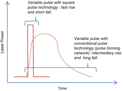
The first generation was an almost purely ablative laser, requiring multiple passes for the end point result and inducing dermal bleeding. The second generation was able to avoid unwanted bleeding by adding a long-pulse or a variable-pulse feature designed to promote tissue coagulation (Holcomb 2011). The variable pulse technology was the ability to extensively control the pulse widths from very short pulses to very long pulses and the possible combinations of short and long pulses at one single laser shot. An even more developed control over the pulses was called the variable square pulse. It is the ability to deliver strictly the same peak power as the average power during the pulse, which means that there is no rise or fall in the curve that measures the power over time but a square shape (Lukac et al. 2007, 2008) (Fig. 1).
More recently, in the beginning of the 2000s, the development of a fractioned version of the 2,940 nm laser was able to reduce the skin surface area injured while preserving the efficacy of the non-fractioned version of the laser (Campos et al. 2009; Holcomb 2011).
Laser and Tissue Interaction
How Does Ablation Work?
In order to choose a laser to promote skin rejuvenation through ablation, the first thing that should be taken into account is the wavelength. Water is a major element present in the skin and is the target for all resurfacing lasers. There are three major laser wavelengths that operate within absorption peaks for water: Er:YAG, Er:YSGG (yttrium-scandium-gallium-garnet), and CO2. When irradiated with those wavelengths, the tissue goes through three basic phases as the energy in the photons is transmitted to the water (Lukac et al. 2007, 2008, 2010):
Phase 1: Direct Heat Within the Optical Absorption Depth
It happens when heat is transmitted directly and almost restrictedly to the skin depth that light is capable of penetrating in the tissue, until all the photons are completely absorbed and converted to heat (Lukac et al. 2008).
The Er:YAG laser penetrates approximately 3 μm into the skin as it has the highest coefficient of absorption for water; the Er:YSGG laser penetrates 10 μm and has the intermediary coefficient of absorption for water; and the CO2 laser penetrates 30 μm into the skin with the lowest coefficient of absorption for water (Lukac et al. 2008, 2010) (Fig. 2).
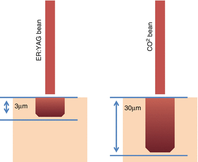
So the higher the affinity for water and the higher the absorption coefficients, the shallower the penetration of the laser, as all the energy within the photons is better absorbed by the water (Lukac et al. 2007, 2008, 2010).
Phase 2: Thermal Diffusion
It happens when the heat is transmitted to deeper lying tissues, beyond the optical absorption depth. The longer the pulse length, the more evident is the result of thermal diffusion, tissue coagulation. Nevertheless, the shorter the pulse length, the less evident is the result of thermal diffusion as the pulse ends faster than the ability of the tissue to transmit the heat within neighboring structures, like in Q-switch devices (Lukac et al. 2008, 2010).
The smallest coagulation zone possible for each laser wavelength is limited by the optical penetration depth, as it is an immutable characteristic of the wavelength determining how much the laser penetrates the skin. But if we manipulate the pulse duration to last longer, heat is accumulated within the tissue, making the coagulation zone thicker through thermal diffusion, beyond the extent of the optical absorption depth. So it is not possible to reduce the coagulation zone limited by laser wavelength and the optical penetration depth, but it is possible to increase the coagulation zone through the thermal diffusion principle by making the pulse width longer (Lukac et al. 2008, 2010).
This is what is done by Er:YAG lasers to emulate the coagulative effect of the CO2 laser, further explained below in the section Ablative/Coagulative Mode.
Phase 3: Tissue Vaporization
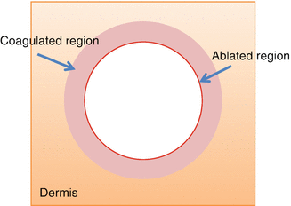
It happens when the top part of the tissue close to the surface is heated to a point where ablation occurs, and material is extruded leaving a small hole on the skin surface, followed by tissue injured by the heat in the surrounding area (Lukac et al. 2008) (Fig. 3).
Fractional vs Non-fractional
The fractional technology was first introduced in 2004 by Dieter Manstein et al., in a pilot study with wavelengths that varied from 1480 to 1550 nm. The ability to reduce the resurfacing side effects while maintaining its efficacy allowed this new technology to be developed into several laser wavelengths: initially the non-ablative lasers, then the fractional CO2 laser, followed by the fractional Er:YAG laser (Manstein et al. 2004).
When skin tissue is treated with fractional lasers, vertical channels are created by columns of vaporization, drawing a grid of microscopic treatment zones, also called microthermal treatment zones or even microscopic thermal zones (MTZ). Each MTZ is composed of the ablated channels, also called microscopic ablation zones (MAZ), and a border of coagulated tissue (Taudorf et al. 2014; Sklar et al. 2014; Skovbolling Haak et al. 2011).
Biologic Effects of the Treatment
Studies evaluating the efficacy of Er:YAG fractioned lasers have shown improvement of collagen arrangement and formation of new collagen types I, III, and VII. Immunohistochemical studies have shown total replacement of the MTZs into new collagen 3 months after treatment. Furthermore, it has been reported that multiple treatments provide continuous new collagen production for more than 6 months, allowing skin rejuvenation with shorter downtime and fewer side effects (Taudorf et al. 2014; Sklar et al. 2014; Lee et al. 2012; Hammami Ghorbel et al. 2014; El-Domyati et al. 2014).
Laser Er:YAG Operator Modes
The Er:YAG laser has several modes of operation and can be used in a non-fractioned or fractioned fashion, produce pure ablation, pure coagulation, and blends of ablation/coagulation at several different ablation depths, and is even capable of emulating a CO2 laser. Variations in pulse length, energy, and fluence, as well as pulse stacking and different spots, promote versatility for this light source. There are five primary operation modes unevenly presented in different devices and industries (Holcomb 2011; Lukac et al. 2007, 2008).
Ablative Mode or Cold Ablation
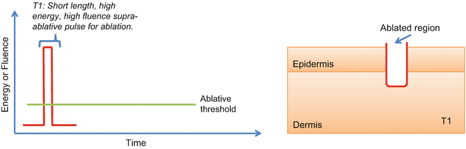
It is the Er:YAG’s native and standard effect. Due to the high affinity for water, the laser produces superficial columns of almost pure ablation, capable of reaching from the epidermis to the superficial dermis (with pulse stacking) (Holcomb 2011; Lukac et al. 2008), as shown in Fig. 4. During the procedure, areas of pinpoint bleeding may be observed, as in this mode there is no coagulation of the blood vessels. It is also used for drug delivery purposes (Jang et al. 2014), mimicking the effect of superficial microneedling due to the superficial and purely ablative laser profile, without barriers of coagulated tissue (Holcomb 2011).
Ablative/Coagulative Mode (Warm or Hot Ablation Mode)
It is the most common mode used in many Er:YAG devices commercially available nowadays. It avoids bleeding by inducing greater thermal effect and coagulation of dermal blood vessels. In this mode, the 2940 nm laser emulates the CO2 laser coagulative effect with the variable pulse or a long pulse operation, generated through a sequence of shots with different energies and pulse durations (Holcomb 2011; Lukac et al. 2007).
There is a train of pulses where the first shot energy is beyond the ablative threshold, and the subsequent energy levels are always below the ablative threshold, accumulating heat in the skin and producing coagulation (Lukac et al. 2007, 2008, 2010) (Fig. 5).
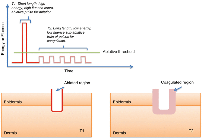
In the warm ablation mode, there is an ablative pulse followed by a sub-ablative long pulse, with a medium-thick coagulation zone. In the hot ablation mode, there is an ablative pulse followed by a longer sub-ablative long pulse, with a thicker coagulation zone (Lukac et al. 2007, 2008, 2010).
This is the best 2940 nm mode for inducing skin rejuvenation as there is greater thermal effect and more collagen remodeling, but it may produce hyperchromias or hypochromias due to the coagulation and heat produced during the procedure (Holcomb 2011; Lapidoth et al. 2008).
It is the authors’ preference to use this mode when rejuvenating the face with high fluences, 3 to 5 pulse stacks and 2 to 3 passes. For non-facial areas: low fluences, 2 stacks, and 1 or 2 passes.
Coagulative Mode or Smooth Mode
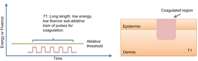
In this mode, only sub-ablative fluences and energies are used during the pulses, emulating the effect of non-ablative lasers (Holcomb 2011). This is also done through the variable-pulse or long-pulse technology. For the coagulative effect, small amounts of energy are delivered within a long pulse, normally a long train of sub-ablative pulses, building heat up in the tissue and promoting coagulation without ablation or with very little ablation (Lukac et al. 2007, 2008, 2010) (Fig. 6).
Stacking Mode
This is not essentially an operator mode as it can also be present within the previously discussed modes, as in the ablative and ablative/coagulative modes. During pulse stacking, a train of high-energy, high-fluence, short-length pulses is delivered in sequence, one on top of the other, without moving the laser handpiece, so the ablation depth of each shot is added to the previous ones, reaching deeper layers in the skin. Again it can appear as an operator mode or it can be hidden within other modes that automatically control the ablation depth (Holcomb 2011; Lukac et al. 2007; Lapidoth et al. 2008).
Non-fractional Mode
In this mode, the laser beam is not split into smaller laser beams. Instead, the entire epidermis is ablated by the laser, leaving no areas of intact skin, as was done originally in the Er:YAG full-field resurfacing treatment regimens (Lukac et al. 2008, 2010).
Indications
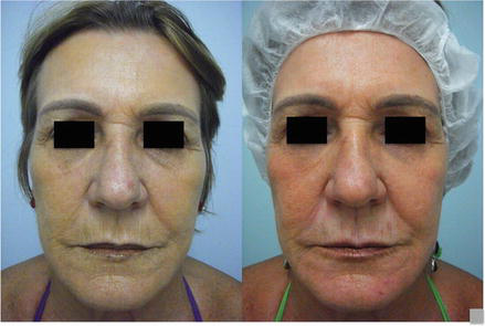
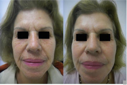
The Er:YAG laser can be used for a variety of lesions especially related to sun damage, including actinic keratoses, solar lentigines, superficial rhytides, mild dyschromia, and Favre–Racouchot disease (Janik et al. 2007). The authors report good results in the treatment of photodamaged skin (Figs. 7 and 8).
It can also be used for colloid milium, angiofibromas, nevi, seborrheic keratosis, xanthelasma, syringomas, sebaceous hyperplasia, rhinophyma, atrophic facial acne scars, trichoepitheliomas, actinic cheilitis, Bowen’s disease, and erythroplasia of Queyrat. It has also been used associated with antineoplastic drugs for drug delivery purposes in the treatment of actinic keratosis and superficial basal cell carcinomas (Janik et al. 2007).
As water is the main target, it is well tolerated by patients with higher phototypes, up to IV, as shown by Lapidoth et al. and Lee et al. (2012).
Pretreatment
Patients should understand the mechanism of action of the proposed treatment, as well as its possible side effects and complications. Dermatologists should explain every step of the process, from wound care to normal timelines for healing, diminishing excessive expectations and worries related to the procedure and post-procedure period (Holcomb 2011).
Patients must understand that it is normal to feel bearable pain during the intervention and that erythema, edema, and crusts are expected side effects. The erythema can be prolonged according to the settings used. Dyschromias are generally rare and tend to be transitory (Holcomb 2011).
The skin rejuvenation promoted by the erbium laser is significant, although several sessions may be necessary to achieve optimal results and some degree of photodamage may always persist (Holcomb 2011).
It is imperative for patients to start antiviral therapy 2 days prior to the procedure (continued for 5–7 days after or until complete reepithelialization), regardless of any previous history of herpes simplex (Lapidoth et al. 2008).
Patients must also be instructed to use 0.05% tretinoin cream nightly for 2–4 weeks prior to laser treatment and should stop it a few days prior to the procedure (Papadavid and Katsambas 2003).
Techniques
Topical anesthetic cream should be applied 30 min before the treatment and removed just before the procedure.
There are several different techniques that change according to the author. In 2008, Lapidoth et al. (2008) reported results from a skin rejuvenation study with a fractionated 2940 nm Er:YAG laser (Pixel, Alma Lasers Ltd.) in patients with Fitzpatrick skin types II–IV. The authors used two to four stacked passes with a 7 × 7 tip (49-dot), which emits 28 mJ/P (per pixel), with the maximum pulse energy output being 1,400 mJ/P. For the first pass, penetration was 20 μm evaporative and 30 μm thermal; for the second pass 35 and 40 μm respectively; for the third pass 50 and 45 μm; and for the fourth pass 60 and 50 μm, with a microzone diameter of 150 μm and a mean number of 3.2 sessions. Sixty days after the final treatment, all patients showed clinical improvement greater than 50%.
Goldberg and Hussain (2011) conducted a study in patients with Fitzpatrick skin types I–III, utilizing a 2940 nm Er:YAG laser with full-face, single-pass treatment at low energy settings of 15–30 mJ/microspot at 40 Hz, with 8–21% coverage, for six sessions. All patients presented clinical improvement in the treated skin; half reported over 50% improvement.
Karsai et al. (2010) studied the 2940 nm laser response in patients with Fitzpatrick skin types I–III by applying a total fluence of 60 J/cm2, with six stacked pulses in a single session. The outcome of this study showed a reduction of approximately 10% in wrinkle depth.
A study performed in Korea by Lee et al. (2012), in patients with Fitzpatrick skin types III–IV treated with an Er:YAG laser (ACTION™, Lutronic, Korea), applied full-face treatment in one or two passes with 12–14 mJ per microspot, a 250 μs pulse width and a mean number of 2.3 sessions. The results showed improvement greater than 26% in 62.5% of subjects.
El-Domyati et al. (2013) showed that both multiple sessions of fractional Er:YAG laser and a single session, with multiple passes, of ablative Er:YAG laser have comparable efficacy clinically and on dermal neocollagenesis, and that multiple sessions of fractional Er:YAG laser resurfacing are effective with higher safety and shorter downtime (El-Domyati et al. 2013).
Posttreatment
When using the pure ablative mode, pinpoint bleeding is expected; therefore, wet gauze compression for several minutes post-procedure is indicated (Campos et al. 2009; Holcomb 2011; Papadavid and Katsambas 2003).
After the laser session, topical semi-occluded dressings (“closed” technique) or topical moisturizers (“open” technique) are applied, as with CO2 laser postoperative care. It is the authors’ preference to apply a D-panthenol or sucralfate healing cream for 8 h and then, after that period, wash the face with a gentle cleanser and reapply the healing cream four times a day for 7–10 days. It is important to tell the patient not to remove crusts or scratch the skin (Campos et al. 2009; Papadavid and Katsambas 2003).
The use of physical sunscreen during the day is advised, starting 48 h after the treatment. Antiviral therapy should be continued for at least 5 days, as mentioned earlier. It is important to give the patient an emergency number in case the post-procedure does not go as planned. Two weeks after laser treatment, an association of topical hydroquinone 2–5%, tretinoin 0.05–0.1%, and hydrocortisone 1% cream is recommended for 2–4 weeks (Papadavid and Katsambas 2003).
Complications
Unexpected side effects and complications include persistent erythema, milia, acneiform eruptions, eczema, infections, impaired healing, scars, and permanent hyper- or hypopigmentation (Janik et al. 2007).
Post-inflammatory hyperpigmentation is transient. Late complications, such as hypopigmentation, are seen in very few patients (4%) treated with the Er:YAG laser. Hypopigmentation seems to be related to ablation depth, which is usually more superficial with the Er:YAG laser. The presence of bleeding forces the surgeon to discontinue laser treatment and therefore prevents serious complications, due to the high absorption coefficient for tissue water and the superficial penetration of this wavelength. The risk of scarring and hypopigmentation could increase if the laser surgeon seeks to achieve considerable ablation depths and more significant clinical improvement.
Conclusion
Er:YAG laser technology has been continuously developed since its beginning and nowadays has become a powerful, versatile tool for the dermatologist. This wavelength is capable of handling conditions from photodamage wrinkles to basal cell carcinomas.
The highest coefficient of absorption for water among ablative lasers assures the smallest penetration depth and the smallest unwanted thermal dissipation effects. Also, variations in pulse width can produce deeper thermal effects when such treatment is desired. The Er:YAG laser is therefore the ideal laser for skin resurfacing.
Take Home Messages
- Resurfacing with ablative lasers is considered one of the gold standard methods for facial rejuvenation.
- It can promote improvement of facial rhytids, skin pigmentation, and skin texture.
- The usual treatment entails short downtime and few side effects.
- It is possible to have significant improvement of facially photodamaged skin after just one session.
- The Er:YAG has the highest coefficient of absorption for water, the smallest penetration depth, and the smallest thermal dissipation effects.
- Deeper thermal effects can be achieved through variations in pulse width.
References
Campos V, de Mattos RA, Filippo A, Torezan LA. Laser no rejuvenescimento facial. Surg Cosmet Dermatol. 2009;1(1):29–36.
Diaci J, Gaspirc B. Comparison of Er:YAG and Er,Cr:YSGG lasers used in dentistry. J Laser Heal Acad. 2012;2012(1):1–13.
El-Domyati M, Abd-El-Raheem T, Abdel-Wahab H, Medhat W, Hosam W, El-Fakahany H, et al. Fractional versus ablative erbium:yttrium-aluminum-garnet laser resurfacing for facial rejuvenation: an objective evaluation. J Am Acad Dermatol [Internet]. 2013;68(1):103–12.
El-Domyati M, Abd-El-Raheem T, Medhat W, Abdel-Wahab H, Al AM. Multiple fractional erbium:yttrium-aluminum-garnet laser sessions for upper facial rejuvenation: clinical and histological implications and expectations. J Cosmet Dermatol. 2014;13(1):30–7.
Goldberg DJ, Hussain M. A study of multiple full-face treatments with low-energy settings of a 2940-nm Er:YAG fractionated laser. J Cosmet Laser Ther. 2011;13(2):42–46.
Hammami Ghorbel H, Lacour J-P, Passeron T. Treatment of inflammatory linear verrucous epidermal nevus with 2940 nm erbium fractional laser. J Eur Acad Dermatol Venereol (JEADV), England. 2014;28:824–5.
Holcomb JD. Versatility of erbium YAG laser: from fractional skin rejuvenation to full-field skin resurfacing. Facial Plast Surg Clin N Am. 2011 May;19(2):261–73.
Jang H-J, Hur E, Kim Y, Lee S-H, Kang NG, Yoh JJ. Laser-induced microjet injection into preablated skin for more effective transdermal drug delivery. J Biomed Opt. 2014;19(11):118002.
Janik JP, Markus JL, Al-dujaili Z, Markus RF. Laser resurfacing. Semin Plast Surg. 2007;1(212):139–46.
Karsai S, Czarnecka A, Junger M, Raulin C. Ablative fractional lasers (CO2 and Er:YAG): a randomized controlled double-blind split-face trial of the treatment of peri-orbital rhytides. Lasers Surg Med. 2010 Feb;42(2):160–7.
Kim SG, Kim EY, Kim YJ, Lee II S. The efficacy and safety of ablative fractional resurfacing using a 2,940-nm Er:YAG laser for traumatic scars in the early posttraumatic period. Arch Plast Surg. 2012;39(3):232–7.
Lapidoth M, Yagima Odo ME, Odo LM. Novel use of erbium:YAG (2,940-nm) laser for fractional ablative photothermolysis in the treatment of photodamaged facial skin: a pilot study. Dermatol Surg. 2008;34(8):1048–53.
Lee HM, Haw S, Kim JE, Won CH, Lee MW, Choi JH, et al. A fractional 2940 nm short-pulsed, erbium-doped yttrium aluminium garnet laser is effective and minimally invasive for the treatment of photodamaged skin in Asians. J Cosmet Laser Ther. 2012;14(6):253–9.
Lukac M, Sult T, Sult R. New options and treatment strategies with the VSP erbium YAG aesthetics lasers. 2007;2007(1):1–9.
Lukac M, Vizintin Z, Kazic M, Sult T. Novel fractional treatments with VSP erbium YAG aesthetic lasers. J Laser Heal Acad. 2008;2008(6):1–12.
Lukac M, Perhavec T, Nemes K. Ablation and thermal depths in VSP Er:YAG laser skin resurfacing. J Laser Heal Acad. 2010;1(1):56–71.
Manstein D, Herron GS, Sink RK, Tanner H, Anderson RR. Fractional photothermolysis: a new concept for cutaneous remodeling using microscopic patterns of thermal injury. Lasers Surg Med. 2004;34(5):426–38.
Papadavid E, Katsambas A. Lasers for facial rejuvenation: a review. Int J Dermatol. 2003 Jun;42(6):480–7.
Sklar LR, Burnett CT, Waibel JS, Moy RL, Ozog DM. Laser assisted drug delivery: a review of an evolving technology. Lasers Surg Med. 2014;46(4):249–62.
Skovbolling Haak C, Illes M, Paasch U, Haedersdal M. Histological evaluation of vertical laser channels from ablative fractional resurfacing: an ex vivo pig skin model. Lasers Med Sci. 2011;26(4):465–71.
Taudorf EH, Haak CS, Erlendsson AM, Philipsen PA, Anderson RR, Paasch U, et al. Fractional ablative erbium YAG laser: histological characterization of relationships between laser settings and micropore dimensions. Lasers Surg Med. 2014 Apr;46(4):281–9.







