
Photodynamic Therapy for Photodamaged Skin
Photodynamic Therapy for Photodamaged Skin
Ana Carolina Junqueira Ferolla and Maria Claudia Almeida Issa Medical Ambulatory of Specialties, Barradas, São Paulo, Brazil Department of Clinical Medicine – Dermatology, Fluminense Federal University, Praia de Icarai 139, 702 Niterói, RJ, Brazil
Abstract
Photodynamic therapy (PDT) is a well-established therapeutic modality for nonmelanoma skin cancer (NMSC). It is based on a photochemical reaction, which causes destruction of the target tissue associated with an inflammatory response. In the last years, PDT has been indicated for some dermatosis in cosmetic dermatology, such as acne, sebaceous hyperplasia, rosacea, and photoaging. Although PDT’s mechanism of action in these dermatoses is not totally clear, some histological studies had shown dermis remodeling induced by PDT in photodamaged skin. In this chapter, we are going to discuss about photoaging and PDT treatment, comparing literature review and authors’ experience.
Keywords Photodynamic therapyPhotosensitizerNonmelanoma skin cancerActinic keratosisBasal cell carcinomaBowen diseasePhotodamaged skinCollagen fibersElastic fibersSkin remodelingDermisAgingPhotoagingPhotorejuvenationRejuvenation
Introduction
Aging is a complex and multifactor process that occurs in all individuals, influenced by environmental, hormonal, and genetic elements, resulting in functional and aesthetical skin changes. Photoaging is clinically manifested by wrinkles, roughness, dryness, sagging, pigmentary spots, telangiectasias, and, in some cases, premalignant and malignant lesions (Ferolla 2007; Issa 2008; Gilchrest 1989).
PDT is based on a photochemical reaction, which causes destruction of the target tissue associated with an inflammatory response. For this reaction, a photosensitizer agent in the target tissue is necessary, a source of light and oxygen. It is approved for NMSC treatment, including actinic keratosis (AK), basal cell carcinoma (BCC), and Bowen disease (squamous cell carcinoma – SCC in situ) (Issa and Patricia-Azulay 2010; Kalka et al. 2000; Szeimies et al. 1996a; Casas et al. 2001; Nestor et al. 2006). The two main photosensitizers are aminolevulinic acid (ALA) and methyl aminolevulinate (MAL), which were approved to be used with light-emitting diode (LED) that emits visible light. PDT with ALA-blue light is very well indicated for superficial lesions, mainly AK. PDT with MAL-red light is recommended for deep lesion treatment because of MAL’s lipophilicity and deeper penetration of the red light in the skin (Hurlimann et al. 1994; Szeimies et al. 1994, 1996b, 2002a, 2005; Fink-Puches et al. 1998; Salim et al. 2003; Lee and Kloser 2013; Babilas et al. 2005).
The global improvement of the skin around AK lesions after PDT called attention to a new possibility for photoaging treatment. Many clinical studies had reported improvement in texture, pigmentation, wrinkles, and laxity in the field of cancerization treated with PDT (Nestor et al. 2006; Hurlimann et al. 1994; Szeimies et al. 1994, 1996b, 2002a, 2005; Fink-Puches et al. 1998; Salim et al. 2003; Lee and Kloser 2013; Babilas et al. 2005; Bruscino and Rossi 2010; Park et al. 2010; Issa et al. 2010; Shamban 2009; Bjerring et al. 2009; Almeida Issa and Piñeiro-Maceira 2009). However, only in the last years, some histological and immunohistochemical studies described some factors involved in the mechanism of skin rejuvenation induced by PDT (Issa et al. 2010; Almeida Issa and Piñeiro-Maceira 2009; Szeimies et al. 2012).
Some other off-label indications include acne, rosacea, sebaceous hyperplasia, vulgar warts, psoriasis, granuloma annulare, necrobiosis lipoidica, and mycosis fungoides (Morton et al. 2013; Alster and Tanzi 2003; Calvazara-Pinton et al. 2010; Bissonnette et al. 2002; Szeimies et al. 2002b; Gold and Goldman 2004a).
Photoaging
Concept
Aging is a continuous process that affects the skin function and its appearance. The chronological aging affects all organs similarly, but environmental (extrinsic) and inherited (intrinsic) processes overlap to produce skin aging. There are evidences that the inherent and extrinsic aging processes have, at least in part, biological, biochemical, and molecular common mechanisms. Chronic sun exposure has been identified as one of the main environmental injuries. Other environmental aggressions, such as smoking, wind, and chemical agent exposure, are also involved (Issa 2008).
Pathogenesis
The UVB radiation (290–300 nm) is responsible for sunburn, while the UVA (320–400 nm), which penetrates more deeply in the skin, promotes collagen and elastic fiber damage. Both UVA and UVB are involved in the pathogenesis of skin cancer and photoaging. The UV radiation causes genetic and molecular changes in the cells of the epidermis, leading to cell atypia (Ferolla 2007; Issa 2008; Gilchrest 1989).
The response to UV radiation involves complex paths, which start in the cell surface receptors that activate transcription factors, AP-1, and NF-kappaB. These factors regulate synthesis of cytokines and interleukins (IL-1, IL-6, IL-8, TNF-alpha) and metalloproteinases, such as MMP-1, MMP-9, MMP-3, and MMP-10. These cytokines and interleukins stimulate an inflammatory process, and on the other hand, they mediate some antioxidant enzymes, balancing the damage. MMP-1 is increased in photodamaged skin and is responsible for breaking collagen types I and III into high molecular fragments of collagen, which change the extracellular matrix, inhibiting synthesis of new collagen. MMP-9 breaks these fragments into smaller fractions of collagen, modifying the relationship between the fibroblasts and the extracellular matrix, allowing neocollagenesis (Kang et al. 2001; Chung et al. 2001; Naderi-Hachtroudi et al. 2002; Dougherty et al. 1998; Fisher et al. 2002).
Clinical and Histopathologic Aspects
The clinical findings about photodamaged skin include wrinkles, roughness, dryness, pigmentation, telangiectasias, and, in some cases, premalignant lesions (actinic keratosis) and malignant lesions (BCC and SCC). In the epidermis, cellular atypia is observed, as well as loss of polarity of keratinocytes and reduced number of Langerhans cells. In the dermis, eosinophilic material (elastosis solar) is observed, as well as thick and curly elastic fibers and thin and flat collagen fibers (Ferolla 2007; Issa 2008; Gilchrest 1989; Kalka et al. 2000).
Treatment
Photoaging treatment includes topical medicaments and cosmeceuticals, oral nutraceuticals, and cosmetic procedures. Chemical peelings, microneedling, transepidermal drug delivery, lasers, botulinum toxin, and fillers are the most important procedures in aesthetic dermatology (discussed in another chapter). If premalignant and malignant lesions are present, a different approach is necessary.
The benefits of PDT for photodamaged skin were realized during the field of cancerization treatment, when the improvement of texture, pigmentation, and wrinkles was observed. PDT’s protocol for photodamaged skin varies a lot in literature with different numbers of sessions, using ALA or MAL and different sources of light (blue and red light-emitting diode (LED), intense pulsed light (IPL), and laser) (Issa 2008; Palm and Goldman 2011; Sanclemente et al. 2012; Bjerring et al. 2002; Karrer et al. 1999a; Dover et al. 2005; Alster et al. 2005; Alexiades-Armenakias and Gernemus 2003; Gold et al. 2006; Togsverd-Bo et al. 2012; Ruiz-Rodriguez et al. 2002).
Photodynamic Therapy
Concept and Mechanism of Action
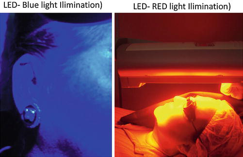
Photodynamic therapy is based on a chemical reaction activated by light, in the presence of a photosensitizer in the target tissue, light, and oxygen. ALA and MAL are the most important photosensitizers used. In fact, they are prodrugs, absorbed by the target cell and then transformed into protoporphyrin IX (Pp IX), inside the cytoplasm and mitochondria (Ferolla 2007; Issa 2008; Gilchrest 1989; Issa and Patricia-Azulay 2010; Kalka et al. 2000; Szeimies et al. 1996a; Casas et al. 2001). Different sources of light can be used, but LED with red or blue light (Fig. 1) is the most important source for premalignant and malignant lesion treatment. The selective destruction of target tissue occurs through a chemical reaction, where the photosensitizer agent in the tissue absorbs the light, becoming excited and then transferring the energy to the oxygen. That sequence of chemical reactions promotes reactive oxygen species (ROS) production, especially the singlet oxygen, which is cytotoxic to malignant cells (Ferolla 2007; Issa 2008; Casas et al. 2001; Hurlimann et al. 1994; Szeimies et al. 1996b, 2005; Palm and Goldman 2011; Sanclemente et al. 2012; Bjerring et al. 2002; Karrer et al. 1999a; Dover et al. 2005; Alster et al. 2005; Alexiades-Armenakias and Gernemus 2003; Gold et al. 2006; Togsverd-Bo et al. 2012; Ruiz-Rodriguez et al. 2002). PDT causes an inflammatory response, activating nuclear factors that control the expression of many cytokine and interleukin genes (IL-1, IL-2, IL-6, IL-10, α-factor of tumoral necrosis – TNF-α) (Ferolla 2007; Issa 2008).
Light Sources
Light-Emitting Diode (LED): Visible Light
PpIX exhibits maximum light absorption (the Soret peak) at 405 nm (blue light) and a weaker absorption band at 635 nm (red light). Blue light is better absorbed, but it penetrates more superficially, around 1–2 mm. Red light penetrates around 4 mm, and it is considered the best choice for deeper lesions (carcinoma) treatment. Both blue and red light can be used for AK lesions and for photoaging treatment (Issa 2008; Naderi-Hachtroudi et al. 2002; Palm and Goldman 2011; Sanclemente et al. 2012).
Other Light Sources
Laser or IPL can be used for PDT treatment, but they are expensive devices without advantages comparing to LED for AK lesion treatment. For this reason, they are better indicated for photorejuvenation and acne treatment, as they can act directly in vessels, pigments, and collagen (Bjerring et al. 2002; Karrer et al. 1999a; Dover et al. 2005; Alster et al. 2005; Alexiades-Armenakias and Gernemus 2003; Gold et al. 2006; Togsverd-Bo et al. 2012; Ruiz-Rodriguez et al. 2002).
Photosensitizers
Aminolevulinic Acid (ALA)
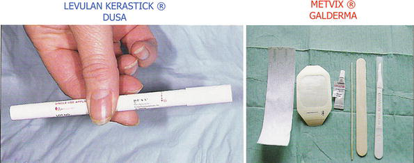
In the USA, ALA was approved by the FDA in 1999 associated with blue light for AK treatment. It is available as a stick, called Levulan Kerastick (DUSA Pharmaceuticals) (Fig. 2), in a solution with 20% of ALA and 48% of ethanol. This stick has two glass bottles inside. One of them contains powder of ALA and the other contains ethanol (solvent). The bottles should be broken with a slight manual pressure in the tube, which is shaken to mix the powder and the solvent. After mixing, ALA is ready to be used through an applicator in one of the stick’s extremities.
The incubation time of ALA on the skin varies in literature, but at least 3 h are necessary for NMSC treatment. When ALA is used for photorejuvenation, 1 or 2 h are enough, but usually more than one session is necessary (Ferolla 2007).
Methyl Aminolevulinate (MAL)
MAL is a methyl ester derivative of ALA. It is lipophilic and therefore has a better permeability through the cell membrane. It is well recommended for NMSC treatment, not only for AK lesions but also for carcinomas (BCC and Bowen disease). For NMSC, an incubation time of 3 h is necessary. Otherwise, when MAL is used for skin rejuvenation, 1 or 2 h are enough (Issa 2008; Issa et al. 2010).
Different from ALA, MAL has its distribution all over the world, including Brazil. Its commercial name is Metvix (Galderma) (Fig. 2), and it is available in a tube containing 2 g of a lipophilic cream. In many countries it is approved for AK and BCC. In the USA, it is approved for AK, and in Brazil and some other countries, it is approved for AK, BCC, and Bowen disease. The cure rate of MAL-PDT for AK ranges from 70% to 100%, up to 95% for superficial basal cell carcinomas, and 70–93% for Bowen disease (Issa 2008; Issa and Patricia-Azulay 2010; Hurlimann et al. 1994; Szeimies et al. 2005; Fink-Puches et al. 1998; Salim et al. 2003).
Procedure
PDT’s protocol is well established for NMSC treatment. Photosensitizer’s incubation time is 3 h with an occlusive dressing (Fig. 3). ALA-blue light is approved for AK treatment, and MAL-red light is approved for AK, BCC, and Bowen disease. One session is recommended for AK lesions, and two sessions with 1-week interval are indicated for BCC and Bowen disease. The protocol for rejuvenation varies a lot in literature, regarding skin preparation, photosensitizer incubation time, source of light, and number of sessions (Ferolla 2007; Issa 2008).
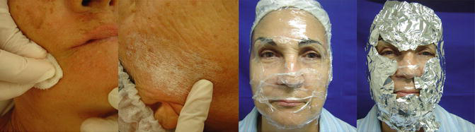
In the standard protocol for NMSC, a slight curettage should be done over the lesion to prepare the skin before applying the photosensitizer. Recently, in order to increase the photosensitizer penetration, some other methods, as ablative lasers and microneedling, have been evaluated. This technique is called transepidermal drug delivery (TED). TED + PDT is an option for photoaging treatment (discussed in another chapter “Transepidermal Drug Delivery and Photodynamic Therapy”) (Kassuga et al. 2012; Torezan et al. 2013).
MAL is ready to be applied 10 mm around the lesion. ALA should be prepared when it is to be used, as explained before. An occlusive dressing is maintained during the photosensitizer’s incubation time to increase the penetration and to avoid ambient light exposure before 3 h. The excessive drug is removed with a physiologic solution before illumination. The time of light exposure depends on the LED to be used. After session, patients should be advised to avoid sun exposure for 48 h and to use sunscreen after this period. Cold compress and moisturizing cream can be used. Analgesics can be prescribed, but steroids should be avoided (Issa 2008).
Side Effects
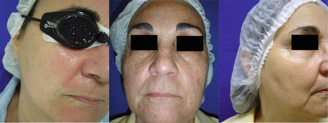
Side effects of topical PDT are limited to the area treated, and it includes pain and burning sensation, during the illumination up to 24–48 h after treatment. Erythema, edema, and crusts occur in the first week (Issa 2008; Issa and Patricia-Azulay 2010; Kalka et al. 2000; Szeimies et al. 1996a; Casas et al. 2001; Nestor et al. 2006) (Fig. 4). The skin peels after 3 days, but is completely recovered after 7–10 days. On extra-facial region, it takes more time to start peeling and to recover. Erythema can be present after 2–4 weeks on the face and up to 3 months on extra-facial region. Dyschromia is very rare, but if it occurs it is temporary. Herpes simplex can occur after 2 or 3 days, and antiviral prophylaxis is recommended for patients with past history of herpes. Bacterial infection is rare, and antibiotic prophylaxis is not necessary. Sterile pustules are described after acne treatment (Ferolla 2007; Issa 2008).
Photodynamic Therapy for Photorejuvenation
Possible Mechanisms Involved
The effects of light on the skin involve complex mechanisms. The balance between the dermal damage and induction of repair seems to define the final effect. Photoaging is mediated by direct absorption of UV radiation and by photochemical reactions mediated by ROS. They play an important role in the pathogenesis of aging and also participate in mechanism of action of PDT (Issa 2008).
Recently, it has been reported that PDT can modulate the expression of interleukins (IL-1, IL-2, IL-6, IL-10) in tumors and in normal tissue; some of them are also produced after UV radiation. These interleukins activate an inflammatory response, causing injury to the tissue, but at the same time they stimulate antioxidant response, limiting the damage and allowing dermis repair (Brenneisen et al. 2002; Karrer et al. 2003; Kolh et al. 2010).
Some authors reported an increase of MMP 1 and 3 and a reduction of collagen type I in an in vitro study in which culture of fibroblasts from normal and scleroderma skin was submitted to PDT (ALA and red light). The result suggests an anti-sclerotic effect of the PDT on the skin (Kolh et al. 2010). This effect is undesirable for skin rejuvenation and motivates new studies to evaluate possible mechanism involved in PDT for skin rejuvenation.
Issa et al. (2008) studied the effects of MAL-red light (two sessions) in patients with photodamaged skin. They evaluated MMPs (1, 3, 7, 9, 12), MMP inhibitors (TIMP 1, TIMP 2), and collagen types I and III. They reported an increase of MMP-9 in the first 3 months, followed by an increase of collagen type I after 6 months, which was statistically significant, measured by morphometry (Issa et al. 2010; Almeida Issa and Piñeiro-Maceira 2009). Authors concluded that MMP-9 degraded the broken collagen (degraded by UV radiation) and changed the extracellular matrix, allowing fibroblasts to synthesize new collagen type I. These histological findings corroborate clinical improvement in texture, wrinkles, pigmentation, and firmness, noticed after 3 months of treatment, with progressive improvement up to 6 months.
Clinical Reports
Ruiz-Rodriguez et al. (2002) treated 17 patients with different degrees of photoaging with AK lesions (a total of 38 lesions) with two sessions of PDT with ALA, with 1-month interval. ALA’s incubation time was 4 h. IPL was used as light source. A total of 33 AK lesions had a complete resolution within 3 months. The technique was well tolerated, and an excellent global improvement was achieved in all patients.
Gold et al. (2006) evaluated ten patients with severe photoaging with AK. The protocol for PDT was application of 20% ALA solution on the face, with an incubation time of 30 min. The light source was IPL. Patients were submitted to three sessions of treatment with 1-month interval. AK cure rate was 85%, and 90% of the patients had a global improvement (texture, pigmentation, and facial erythema).
Alster et al. (2005) compared ALA-IPL and IPL isolated for photoaging treatment and reported a better clinical result, without increasing side effects with ALA-IPL. Marmur et al. (2005) reported clinical improvement with an increase of collagen type I in photodamaged skin after ALA-IPL.
Touma and Gilchrest (2003) compared different time of light exposures and reported improvement in photoaging even with a short period of illumination. In another study, Touma et al. (2004) evaluated the use of 40% urea before PDT with ALA-blue light, but no significant clinical effects were observed.
Alexiades-Armenakias et al. (2003) reported the use of vascular laser 585 nm, after 3–18 h of ALA incubation time in 35 patients with AK. They evaluated 2,561 AK lesions on the face, scalp, and extremities. The cure rate was 99.9% after 10 days, 98.4% after 2 months, and 90.1% after 4 months on the face and scalp. The lesions located on the extremities had a poor response with complete resolution of 49.1% after 2 months. Authors concluded that when using not purpuric parameters of PDL for PDT, it is possible to reach good cure rate with minimum discomfort, low downtime, and excellent aesthetical results.
Palm et al. (2011) treated 18 patients with photoaging, using MAL-PDT, comparing blue light with the red light. There was no statistically significant difference between them.
Sanclemente et al. (2012) studied histopathological findings after PDT treatment with MAL-red light and reported an increase of collagen and elastic tissue, although not statistically significant.
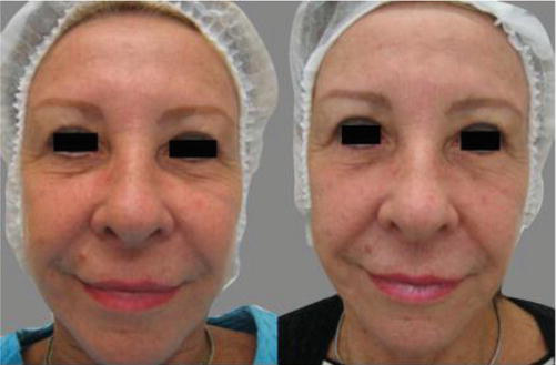
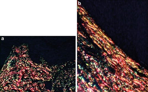
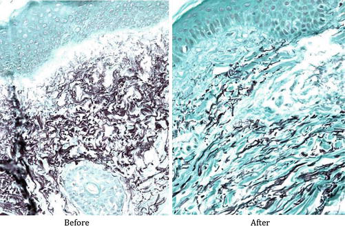
Ferolla et al. (2007) demonstrated the global clinical improvement of skin (pigmentation, fine wrinkles, sagging, AK lesions) after three sessions of ALA-red light (Fig. 5). The incubation time was 2 h, followed by the red light illumination for 20 min. Histological studies showed improvement in collagen fibers organization (Fig. 6a, b).
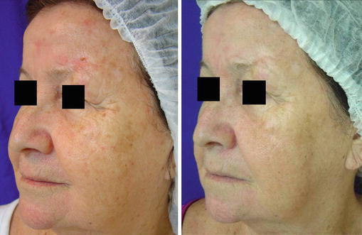
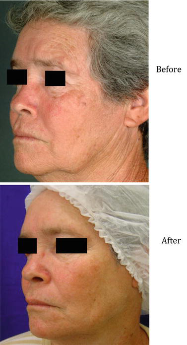
Issa et al. (2008) evaluated the therapeutic response of PDT for photodamaged skin in 14 women with and without AK. Two sessions of MAL-red light were done with 30 days of interval. MAL incubation time was 2 h under occlusion. The light source was a LED (Aktilite – Photocure) with a dose of 37 J/cm2. Clinical results included texture, pigmentation, and wrinkle improvement observed after 3 and 6 months of follow-up (Fig. 7). After 6 months, reduction of skin sagging was more evident (Fig. 8). Side effects included edema, erythema, and crusts. Authors also evaluated histological and morphometric changes in those patients mentioned above and observed a statistically significant increase of collagen and elastic fibers mainly 6 months after treatment (Figs. 9 and 10).
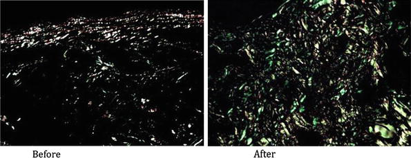
Le Pillouer-Prost and Cartier (2016) reported that PDT is an effective method for photoaging treatment, improving fine wrinkles, skin roughness, and laxity. They consider patients with past history of significant sun exposure and with multiple AK lesions as the best indication for PDT photorejuvenation.
Protocol of Treatment (PDT × Photorejuvenation) and Author’s Experience
- Advise patients regarding the benefits and the limits of the technique.
- Take pictures for further comparison.
- Prophylaxis with aciclovir, if patient has past history of herpes.
- Remove makeup, and then apply alcoholic chlorhexidine to clean the skin.
- Do a slight curettage of AK lesions.
- Apply ALA or MAL on the area to be treated. When using MAL, a thin layer of the cream should be applied throughout the area and it is also recommended to put a thicker layer on the top AK lesions.
- Occlusive dressing with plastic film and aluminum paper can be done or not. ALA or MAL incubation time ranges from 1 to 3 h.
- If AK is visible, it is better to do occlusion for 3 h with one session or for 1 or 2 h within two or three sessions (3–4 weeks of interval).
- LED is better than IPL for AK lesions for a longer follow-up.
- Remove the dressing and the excessive photosensitizer agent, before starting light exposure.
- Time of light exposure depends on the lamp used. Usually it is programmed to deliver light dose and turn off automatically. If IPL or laser is used, protocols are going to vary according to the device based on its irradiance and fluence.
- Analgesic before light exposure and during 24 h, if incubation time is 3 h.
- Avoid sun exposure for 48 h. After this period, patients are advised to use sunscreen.
- The use of moisturizing cream and cold compresses with thermal water is indicated. Corticoid should be avoided, but can be used if necessary.
Take Home Messages
- The light effects on the skin involve complex mechanisms. The balance between dermal damage and induction of repair seems to define the final effect.
- Photoaging is mediated by direct absorption of UV radiation and by photochemical reactions mediated by ROS. They play an important role in the etiopathogeny of photoaging and participate in the mechanism of action of PDT.
- PDT protocols for photodamaged skin are reported with ALA or MAL and with different sources of light: IPL, laser, and LED.
- Protocols vary in literature: different incubation times of ALA or MAL (30 min to 3 h), with or without occlusive dressing, number of sessions (average of 2–3), and interval between the sessions (2–4 weeks).
- Clinically, a global improvement of skin is observed, with the improvement of texture, actinic keratosis, pigmentation, wrinkles, and sagging.
- Histologically, there is an improvement in the organization of elastic and collagen fibers, with a statistically significant increase of those fibers at morphometry.
- A possible mechanism of action related to photorejuvenation involves the increase of MMP-9 after PDT treatment, which modifies the extracellular matrix, inducing dermis remodeling with neocollagenesis.
References
Alexiades-Armenakias MR, Gernemus RG. Laser-mediated photodynamic therapy of actinic keratosis. Arch Dermatol. 2003;139:1313–20.
Almeida Issa MC, Piñeiro-Maceira J, Farias, et al. Immunohistochemical expression of matrix metalloproteinases in photodamaged skin by photodynamic therapy. Br J Dermatol. 2009;161(3):647–53.
Alster TS, Tanzi EL. Photodynamic therapy with topical aminolevulinic acid and pulsed dye laser irradiation for sebaceous hyperplasia. J Drugs Dermatol. 2003;2:501–4.
Alster TS, Tanzi EL, Welsh EC. Photorejuvenation of facial skin with topical 20% 5-aminolevulinic acid and intense pulsed light treatment: a split-face comparison study. J Drugs Dermatol. 2005;4:35–8.
Babilas P, Karrer S, Sidoroff A, et al. Photodynamic therapy in dermatology: an update. Photodermatol Photoimmunol Photomed. 2005;21:142–9.
Bissonnette R, Tremblay J-F, Juzenas P, Boushira M, Lui H. Systemic photodynamic therapy with aminolevulinic acid induces apoptosis in lesional T lymphocytes of psoriatic plaques. J Invest Dermatol. 2002;119:77–83.
Bjerring P, Clement M, Heickendorff L, Lybecker H, Kiernan M. Dermal collagen production following irradiation by dye laser and broadband light source. J Cosmet Laser Ther. 2002;4:39–43.
Bjerring P, Christiansen K, Troilius A, et al. Skin fluorescence controlled photodynamic photorejuvenation. J Lasers Surg Med. 2009;41(5):327–36.
Brenneisen P, Sies H, Scharffetter-Kochanek K. Ultraviolet-B irradiation and matrix metalloproteinases: from induction via signaling to initial events. Ann N Y Acad Sci. 2002;973:31–43.
Bruscino N, Rossi R, Dindelli M, et al. Facial skin rejuvenation in a patient treated with photodynamic therapy for actinic keratosis. Dermatol Ther. 2010;23(1):86–9.
Calvazara-Pinton P, Arisi M, Sereni E, Ortel B. A critical reappraisal of off-label indications for topical photodynamic therapy with aminolevulinic acid and methylaminolevulinate. Rev Recent Clin Trials. 2010;5(2):112–6.
Casas A, Fukuda H, Di Venosa G, et al. Photosensitization and mechanism of cytotoxicity induced by the use of ALA derivatives in photodynamic therapy. Br J Cancer. 2001;85:279–84.
Chung JH, Seo JY, Choi HR, Lee MK, Youn CS, Rhie G, et al. Modulation of skin collagen metabolism in aged and photoaged human skin in vivo. J Invest Dermatol. 2001;117(5):1218–24.
Dougherty TJ, Gomer CJ, Henderson BW, Jori G, Kessel D, Korbelik M, et al. Photodynamic therapy. J Natl Cancer Inst. 1998;90(12):889–905.
Dover JS, Bhatia AC, Stewart B, Arndt KA. Topical 5-aminolevulinic acid combined with intense pulsed light in the treatment of photoaging. Arch Dermatol. 2005;141:1247–52.
Ferolla ACJ. Estudo da pele humana fotoenvelhecida após tratamento com terapia fotodinâmica associada ao ácido 5 delta aminolevulínico tópico: avaliação imunoistoquímica, do colágeno e do tecido elastic [TESE]. São Paulo: Faculdade de Medicina da Universidade de São Paulo; 2007.
Fink-Puches R, Soyer HA, Hofer A, et al. Long-term follow-up and histological changes of superficial nonmelanoma skin cancers treated with topical δ-aminolevulinic acid photodynamic therapy. Arch Dermatol. 1998;134:821–6.
Fisher GJ, Kang S, Varani J, Bata-Csorgo Z, Wan Y, Datta S, et al. Mechanisms of photoaging and chronological skin aging. Arch Dermatol. 2002;138(11):1462–70.
Gilchrest BA. Skin aging and photoaging: an overview. J Am Acad Dermatol. 1989;21:610–3.
Gold MH, Goldman MP. 5-aminolevulinic acid photodynamic therapy: where we have been and where we are going. Dermatol Surg. 2004;30:1077–84.
Gold MH, Bradshow VL, Boring MM, Bridges TM, Biron JA. Split face comparison of photodynamic therapy with 5-aminolevulinic acid and intense pulsed light versus intense pulsed light alone for photodamage. Dermatol Surg. 2006;32:795–801.
Hurlimann AF, Panavizzon RG, Burg A. Topical photodynamic treatment of skin tumours and dermatoses. Dermatology. 1994;3:327.
Issa MCA. Estudo Da Remodelação Dérmica Induzida Pela Terapia Fotodinâmica (MAL-TFD) Na Pele Fotodanificada [Tese]. Rio de Janeiro: Universidade Federal do Rio de Janeiro; 2008.
Issa MCA, Patricia-Azulay M. Terapia Fotodinâmica: revisão da literatura e documentação iconográfica. Anais Bras Dermatol. 2010;85(4):501–11.
Issa MCA, Pineiro-Maceira J, Vieira, et al. Photorejuvenation with topical methyl aminolevulinate and red light: a randomized, prospective, clinical, histopathologic, and morphometric study. Dermatol Surg. 2010;36:39–48.
Kalka K, Merk H, Mukhtar H. Photodynamic therapy in dermatology. J Am Acad Dermatol. 2000;42:389–413.
Kang S, Fisher GJ, Voorhees JJ. Photoaging: pathogenesis, prevention, and treatment. Clin Geriatr Med. 2001;17(4):643–59, v–vi.
Karrer S, Bäumler W, Abels C, Hohenleutner U, Landthaler M, Szeimies R-M. Long-pulse dye laser for photodynamic therapy: investigations in vitro and in vivo. Lasers Surg Med. 1999;25:51–9.
Karrer S, Bossierhoff AK, Weiderer P, Landthaler M, Szeimies RM. Influence of 5-aminolevulinic acid and red light on collagen metabolism of human dermal fibroblasts. J Invest Dermatol. 2003;120(2):325–31.
Kassuga LEBP, Issa MCA, Chevrand NS. Aplicação transepidérmica de medicamento associado a terapia fotodinâmica no tratamento de ceratoses actínicas. Surg Cosmet Dermatol. 2012;4(1):89–92.
Kolh E, Torezan LA, Landthaler M, Szeimies RM. Aesthetics effects of topical photodynamic therapy. J Eur Acad Dermatol Venereol. 2010;24:1261–9.
Le Pillouer-Prost A, Cartier H. Photodynamic photorejuvenation: a review. Dermatol Surg. 2016;42(1):21–30.
Lee PK, Kloser A. Current methods for photodynamic therapy in the US: comparison of MAL/PDT and ALA/PDT. J Drugs Dermatol. 2013;12(8):925–30.
Marmur ES, Phelps R, Goldberg DJ. Ultrastructural changes seen after ALA–IPL photorejuvenation: a pilot study. J Cosmet Laser Ther. 2005;7(1):21–4.
Morton CA, Szeimies RM, Sidoroff A, et al. European guidelines for topical photodynamic therapy part 2: emerging indications: field cancerization, photorejuvenation and inflammatory/infective dermatoses. J Eur Acad Dermatol Venereol. 2013;27(6):672–9.
Naderi-Hachtroudi L, Peters T, Brenneisen P, Meewes C, Hommel C, Razi-Wolf Z, et al. Induction of manganese superoxide dismutase in human dermal fibroblasts: a UV-B-mediated paracrine mechanism with the release of epidermal interleukin 1 alpha, interleukin 1 beta, and tumor necrosis factor alpha. Arch Dermatol. 2002;138(11):1473–9.
Nestor MS, Gold MH, Kauvar ANB, et al. The use of photodynamic therapy in dermatology: results of a consensus conference. J Drugs Dermatol. 2006;5:140–54.
Palm MD, Goldman MP. Safety and efficacy comparison of blue versus red light sources for photodynamic therapy using methyl aminolevulinate in photodamaged skin. J Drugs Dermatol. 2011;10(1):53–60.
Park MY, Sohn S, Lee ES, et al. Photorejuvenation induced by 5-aminolevulinic acid photodynamic therapy in patients with actinic keratosis: a histologic analysis. J Am Acad Dermatol. 2010;62(1):85–95.
Ruiz-Rodriguez R, Sanz-Sanchez T, Cordoba S. Photodynamic photorejuvenation. Dermatol Surg. 2002;28(8):742–4; discussion 744.
Salim A, Leman JA, McColl JH, et al. Randomized comparison of photodynamic therapy with topical 5-fluorouracil in Bowen’s disease. Br J Dermatol. 2003;148:539–43.
Sanclemente G, Correa LA, Garcia JJ, et al. Methyl aminolevulinate plus red light vs. placebo plus red light in the treatment of photodamaged facial skin: histopathological findings. Exp Dermatol. 2012;37(4):379–86.
Shamban AT. Current and new treatments of photodamaged skin. Facial Plast Surg. 2009;25(5):337–46.
Szeimies RM, Torezan LA, Niwa A, et al. Clinical, histopathological and immunohistochemical assessment of human skin field cancerization before and after photodynamic therapy. Br J Dermatol. 2012;16(1):150–9.
Szeimies R-M, Sassy T, Landthaler M. Penetration potency of topical applied δ-aminolevulinic acid for photodynamic therapy of basal cell carcinoma. Photochem Photobiol. 1994;59:73–6.
Szeimies R-M, Calzavara-Pinton PG, Karrer S, et al. Topical photodynamic therapy in dermatology. J Photochem Photobiol. 1996a;36:213–9.
Szeimies RM, Karrer S, Sauerwald A. Photodynamic therapy with topical application of 5-aminolevulinic acid in the treatment of actinic keratoses: an initial clinical study. Dermatology. 1996b;192:246–51.
Szeimies RM, Karrer S, Radakovic-Fijan S, Tanew A, Calzavara-Pinton PG, Zane C, et al. Photodynamic therapy using topical methyl 5-aminolevulinate compared with cryotherapy for actinic keratosis: a prospective, randomized study. J Am Acad Dermatol. 2002a;47:258–62.
Szeimies RM, Landthaler M, Karrer S. Non-oncologic indications for ALA-PDT. J Dermatolog Treat. 2002b;13 suppl 1:S13–8.
Szeimies RM, Morton CA, Sidoroff A, et al. Photodynamic therapy for non-melanoma skin cancer. Acta Derm Venereal. 2005;85:483–90.
Togsverd-Bo K, Haak CS, Thaysen-Petersen D, et al. Intensified photodynamic therapy of actinic keratoses with fractional CO2 laser: a randomized clinical trial. Erratum Br J Dermatol. 2012;167(2):461 [corrected to Hædersdal, M].
Torezan L, Chaves Y, Niwa A, Sanches JA, Festa-Neto C, Szeimies RM. A pilot split-face study comparing conventional methylaminolevulinate-photodynamic therapy (PDT) with microneedling-assisted PDT on actinically damaged skin. Dermatol Surg. 2013;39(8):1197–201.
Touma DJ, Gilchrest BA. Topical photodynamic therapy: a new tool in cosmetic dermatology. Semin Cutan Med Surg. 2003;22:124–30.
Touma D, Yaar M, Whitehead S, Konnikov N, Gilchrest BA. A trial of short incubation, broad-area photodynamic therapy for facial actinic keratoses and diffuse photodamage. Arch Dermatol. 2004;140(1):33–40.







