
Non-ablative Lasers for Stretch Marks
Non-ablative Lasers for Stretch Marks
Luciana Archetti Conrado, Melina Kichler, Priscilla Spina and Isis Suga Veronez São Paulo, SP, Brazil
Abstract
Striae distensae (SD) or stretch marks (SM), a common skin condition, do not cause any significant medical problem; however, they can cause significant distress to those affected. They are dermatologic lesions, usually asymptomatic, that often arise as a result of mechanical stress, certain endocrine conditions, pregnancy (striae gravidarum), or prolonged exposure to steroids. They can be initially erythematous (striae rubra) but over time become atrophic with a white color (striae alba). Although there is no “gold standard” treatment for stretch marks, various laser parameters alone or in association with other treatment modalities have been studied. Over the years, the non-ablative fractional lasers have shown good clinical results and become very popular, especially because they are well tolerated and safe, even in patients with higher phototypes (IV–V). Post-inflammatory hyperpigmentation is the most common complication; however, it is transitory in most cases and its incidence is lower than with ablative lasers. This chapter will approach the use of non-ablative lasers for SD treatment.
Keywords Striae distensaeStretch marksStriae rubraStriae albaNon-ablative fractional laserAblative fractional laserIntense pulsed lightPulsed dye laserPhotothermolysis
Introduction
Striae distensae (SD), also known as stretch marks, striae atrophicans, or striae gravidarum, are common skin lesions which can pose a significant psychological burden for patients. They were first histologically described in 1889 by Troisier and Menetrier, and until now remain a challenge in terms of treatment and prevention (Troisier and Menetrier 1889). Striae distensae are dermatologic lesions that often arise as a result of mechanical stress (rapid weight change, puberty, pregnancy), certain endocrine conditions (like Cushing’s syndrome and Marfan syndrome), or prolonged exposure to medications such as steroids, whether topical or systemic (Hexsel et al. 2012).
It appears that the group at highest risk of developing severe striae distensae is teenagers (Atwal et al. 2006). The prevalence differs among the adolescence, pregnancy, and obesity groups and ranges from 43% to 88% and 6% to 86% in pregnant women and adolescents, respectively. Among obese individuals the prevalence reported is 43% (Al-Himdani et al. 2014). Elbuluk et al. observed interracial differences in the severity of SD and concluded that African-American women were more severely affected than white women within the same geographical region (Elbuluk et al. 2009). However, the authors also noted that there was a difference in body mass index and smoking status between the two groups, which may indicate that factors other than race could be involved (Al-Himdani et al. 2014).
Anatomical regions can vary depending upon sex and age. In adolescent males, the lower back and knees are usually affected, while in female subjects, the thighs and calves are more often involved. Striae gravidarum occur in more than 70% of pregnant women and are commonly found on the abdomen and breasts, usually developing after the 24th week of gestation (Atwal et al. 2006; Al-Himdani et al. 2014). They tend to appear symmetric and bilateral (Hanauer et al. 2011) and are usually asymptomatic, but in the early stages can cause some itching (Hexsel and Dal’Forno 2013).
Striae distensae are linear atrophic scars, most often initially erythematous (striae rubra), but they can also have different colors such as purple and blue and then over time become atrophic and hypopigmented and attain a white color (striae alba) (Alves et al. 2015; Malekzad et al. 2014). Hermanns and Piérard described two additional types of SD, striae nigrae and striae caerulea, which occur in those with darker skin due to increased melanization (Hermanns and Piérard 2006). The color of the SD is related to the stage of evolution and to melanocyte mechanobiological influences (Al-Himdani et al. 2014).
Pathogenesis
A positive family history is an important risk factor. In adolescents, body mass index, childhood obesity, and seborrhea and atopic dermatitis are also reported as influences in developing SD. On the other hand, risk factors in pregnant women may be constitutional or pregnancy related. Constitutional factors include maternal age, smoking, and body mass index. Younger women are more likely to develop SD, implicating a constitutional difference in the stretching ability of the skin. Pregnancy-related factors such as birth weight, gestational age, weight gain, and polyhydramnios support the theory that changes associated with pregnancy can play a role in the development of SD. Other risk factors include medical conditions such as Marfan syndrome, Cushing syndrome, anorexia nervosa, typhoid fever, rheumatic fever, chronic liver disease, surgery, and medications like systemic and topical corticosteroids, HIV therapy, chemotherapy, tuberculosis therapy, contraceptives, and neuroleptics (Al-Himdani et al. 2014).
The pathogenesis of striae is still unknown but probably relates to changes in the components of the extracellular matrix, including fibrillin, elastin, and collagen (Singh and Kumar 2005). Three main theories relating to SD formation have been described: mechanical stretching of the skin, hormonal changes, and innate structural disturbance of the integument. Mechanical stretching of the skin is postulated due to the perpendicularity of SD to the direction of the skin. Hormonal alterations in adrenocorticotrophic hormone and cortisol are thought to promote fibroblast activity, leading to increased protein catabolism and thus decreasing deposition of collagen in the substance of the dermal matrix (Al-Himdani et al. 2014; Khater et al. 2016).
Pregnancy-related hormones are also believed to influence SD formation. Cordeiro et al. described increased estrogen and androgen receptors in the skin exhibiting SD compared with normal skin (Cordeiro et al. 2010). Lower serum relaxin levels were demonstrated in pregnant women with SD compared with those without SD at 36 weeks’ gestation. SD may also present altered gene expression, in a similar way to keloid disease and scleroderma, which are thought to develop due to disordered gene expression of extracellular matrix (Al-Himdani et al. 2014).
Striae rubra and alba are distinct forms of SD. Their distinction has therapeutic implications (Al-Himdani et al. 2014). Usually the lesion matures from rubra to alba in several steps similar to those of the wound repair mechanism, from wounding to scar formation (Ryu et al. 2013; Ud-Din et al. 2016). In early lesion development, there is deep and superficial perivascular lymphocytic infiltration with edema, vascular ectasia, and possible angiogenesis (Alves et al. 2015; Ryu et al. 2013). On electron microscopy, early histological dermal alteration may be visualized by mast cell degranulation and macrophage activation with release of enzymes such as elastases, which leads to elastolysis of the mid-dermis (Sheu et al. 1991). Inflammation resolves over time, but collagen bundles in the reticular dermis stretch parallel to the skin, resulting in flattening of the epidermis and elongation of rete ridges, absence of hair follicles, and reduction of melanocytes leading to leukoderma, followed by loss of collagen and elastic fibers in the substructures (Al-Himdani et al. 2014; Ryu et al. 2013).
Therapeutic Assessment
Because of the prevalence of SD and its impact on the quality of life of patients, there is substantial demand for a reliable treatment option.
Although there is no “gold standard” treatment for stretch marks, various treatment modalities have been tried. Not only does the outcome depend on the type of SD, but the patients’ Fitzpatrick skin type is important as well, since most adverse effects, particularly with laser, occur in patients with a darker skin type (Al-Himdani et al. 2014).
There are a number of treatment options, including topical agents, acid peels, microdermabrasion, radiofrequency, needling, and lasers and light therapies, described in the literature.
Topical agents such as tretinoin are thought to work through their affinity for fibroblasts and induction of collagen synthesis, and they have good efficacy in striae rubra but poor and unpredictable responses in striae alba. Therefore, they offer only modest benefit in the early stages of the clinical course. Topical creams, lotions, and ointments are used especially in pregnant women at risk of developing SD, but there is no statistically significant evidence to support their use for prevention of SD, according to a Cochrane review in 2012 (Brennan et al. 2012). Acid peel treatments such as glycolic acid and trichloroacetic acid are thought to act by increasing collagen synthesis. Microdermabrasion, which is a skin resurfacing technique, has been reported to increase type I collagen with better efficacy on striae alba (Al-Himdani et al. 2014; Aldahan et al. 2016) (see chapter “Microneedling for Transepidermal Drug Delivery on Stretch Marks”).
Treatment with skin needling might be able to induce more collagen and elastin deposition beneath the epidermis, as can microneedling radiofrequency, which can induce growth factor secretion by delivering higher volumetric heating and deeper heat diffusion (Khater et al. 2016; Ryu et al. 2013). However, all of these treatments have inconsistent clinical outcomes (Aldahan et al. 2016).
Lasers and light therapies offer a variety of wavelengths that can target specific chromophores at varying fluences. This poses a theoretical advantage by allowing individualized treatments. Some wavelengths target blood vessels in striae rubra to reduce their appearance, and others induce collagen and elastin production in mature striae. Among the wavelengths that have been studied for striae distensae are UVB, UVA, excimer laser (308 nm), intense pulsed light (565, 590, 645, 650 nm), copper bromide (577 nm), pulsed dye laser (585, 595 nm), infrared, Nd:YAG (1064 nm), diode (1450 nm), Er:Glass (1540, 1550, 1565 nm), Er:YAG (2940 nm), and CO2 (10,600 nm) (Aldahan et al. 2016).
Ablative fractional lasers and intense pulsed light (IPL) represent emerging therapies that have demonstrated some success (Aldahan et al. 2016). Non-ablative lasers have been studied in the treatment of SD as well.
The 308 nm excimer laser is an ultraviolet laser and has been used to treat SD, with improvement of the pigmentation on mature white striae, but studies have shown that this pigmentation is only temporary, requiring many treatment sessions before significant improvement can be seen.
The pulsed dye laser (PDL) can also be indicated. It is commonly used for striae rubra treatment, as its target is dilated blood vessels (Aldahan et al. 2016; Al-Himdani et al. 2014). Similar to the pulsed dye laser, the Nd:YAG laser can be used to improve striae rubra, but does not have the same result in striae alba (Aldahan et al. 2016).
Intense pulsed light is characterized by the emission of incoherent, pulsed, broad-spectrum light (515–1200 nm). It is used to treat the vascularization of rubra striae, but some studies also show clinical improvement and increased thickness of collagen in striae alba. Repeated sessions may be required to maintain the positive effects (Aldahan et al. 2016; Al-Himdani et al. 2014).
There are two types of fractional lasers used in the treatment of striae distensae: ablative and non-ablative. Fractional treatment is achieved through a pattern of microscopic thermal zones (delivering light energy into the tissue through multiple microscopic columns surrounded by untreated areas) produced by the laser beams at specific depths in the dermis. Fractional photothermolysis stimulates epidermal turnover and dermal collagen remodeling (Mattos and Jordão 2012).
Ablative lasers use long wavelengths to target water in the epidermis and dermis, thereby vaporizing the cells. Lasers available in this category that have been studied for treating SD include the CO2 and Er:YAG (see chapter “CO2 Laser for Stretch Marks”). These techniques provide immediate tissue tightening and induce more collagen stimulation than non-ablative lasers. On the other hand, non-ablative fractional devices are associated with minimal side effects and downtime (Aldahan et al. 2016; Alam et al. 2011; Khater 2016; Tannous 2007).
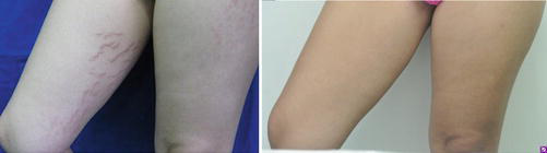
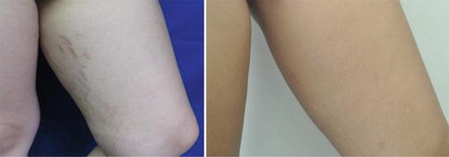
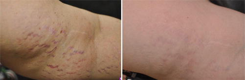
For the treatment of both striae (rubra and alba), the fractional non-ablative Er:Glass (1540, 1550, 1565 nm) is one of the most used (Table 1) (Stotland et al. 2008; de Angelis et al. 2011; Tretti Clementoni and Lavagno 2015); however, other non-ablative lasers are also used with good results. Several sessions are necessary (Figs. 1–3) (see chapter “Erbium Laser for Scars and Striae Distensae”).
| Laser – wavelength (nm) | Parameters | Number of passes |
|---|---|---|
| Erbium glass – 1540 | Energy = 12–55 mJ; Density = 100–320 | 2–3 |
| Erbium glass – 1550 | Energy = 12–18 J; Density = 125–250 | 8–12 |
| Erbium glass – 1565 | Energy = 40–55 J; Density = 150–300 | 2 |
De Angelis et al. treated SD in 51 patients with skin types II–IV, with two to four sessions of laser therapy spaced 4–6 weeks apart. All striae were reported to have at least 50% improvement. Histologically, increased elastic fibers and neocollagenesis were seen in the reticular dermis. Adverse effects were predominantly erythema and edema; however, eight patients developed transient post-inflammatory hyperpigmentation (de Angelis et al. 2011).
Clementoni and Lavagno evaluated the effectiveness and safety of a novel non-ablative fractional 1565 nm laser on the appearance of SD. Good clinical improvement (between 51% and 75%) was observed in all patients. The average pain during treatment was generally defined as tolerable, and the average downtime was 4 days. Transient erythema and severe edema were noted immediately after the procedure, but long-lasting or severe adverse effects were not observed. All patients noted good improvement and were satisfied with the treatment and the results. They thus concluded that treatment with the 1565 nm laser resulted in improved pigmentation, volume, and textural appearance of SD (Tretti Clementoni and Lavagno 2015).
Malekzad et al. studied ten patients with striae alba using a fluence of 50–70 J/cm2 and, among other adverse events, reported one patient who developed acne in the treatment area. Unfortunately, this study demonstrated questionable results, with nine of ten patients showing fair or poor improvement (Malekzad et al. 2014).
Alves et al. reported four patients with corticosteroid-induced striae rubra treated using a 1540 nm Er:Glass laser at 1-month intervals. After three sessions, 50% had marked improvement, and the other two patients achieved similar improvement after four and six sessions, respectively (Alves et al. 2015).
Bak et al. treated Asian patients, with improvement in appearance clinically and histologically. An increase in average epidermal and dermal thickness was seen on post-treatment biopsy, especially in striae alba (Bak et al. 2009).
Stotland treated 14 female patients with a 1550 nm erbium-doped fiber laser; only one patient had striae rubra, and all the others had alba striae. He concluded there was a 26–50% improvement in pigmentation, with transient edema and erythema in most of them (Stotland et al. 2008).
Guimaraes et al. studied the 1550 nm Er:Glass laser in ten patients with striae rubra of the breast, with 4–8 sessions performed at 4-week intervals. Patients who had total improvement received at least six laser sessions (Guimaraes et al. 2009).
Wang et al. compared 1540 nm and 1410 nm non-ablative fractionated lasers in nine patients with abdominal striae. Each patient was treated for six sessions — half of the abdomen with each laser. All subjects demonstrated clinical improvement bilaterally after treatment. Skin biopsies showed an increase in epidermal thickness and in collagen and elastin density when compared with baseline. However, clinical and histological differences between the two lasers were not statistically significant (Wang et al. 2016).
Based on the number of studies alone, it is clear that the treatment of SD with non-ablative fractional lasers is popular, safe, and well tolerated by patients, with minimal adverse events. Mature striae alba have proven to be the most difficult type to treat successfully with fractional lasers (Shin et al. 2011). However, this also occurs with other therapeutic modalities (Tannous 2007).
Post-inflammatory hyperpigmentation (PIH) is the most common complication in patients with Fitzpatrick skin types IV–VI; however, its incidence is lower when compared with ablative lasers. Although melanin does not absorb the 1540–1565 nm wavelengths, pigmentary changes can still occur (Sherling et al. 2010). The degree of PIH has been found to be directly proportional to both the energy and density of the treatment, although density appears to be particularly important (Chan et al. 2007). Thus, it is important to start out using conservative settings in these patients, even when using non-ablative lasers (Shah and Alam 2012).
To reduce the incidence of hyperpigmentation, many dermatologists use bleaching creams like hydroquinone, tretinoin, or glycolic acid (before and/or after the procedure). Although studies are inconclusive as to whether these topical agents are capable of preventing hyperpigmentation, they may be effective when instituted postoperatively as a component of the skin-care regimen (Manuskiatti et al. 2010; Sriprachya-anunt et al. 2002). In combination with non-ablative devices, it is easier to prescribe these bleaching agents, as the epidermis is maintained after the laser (Shah and Alam 2012).
Studies comparing the clinical efficacy of ablative vs. non-ablative fractional photothermolysis systems for the treatment of striae distensae have been conducted.
One of these studies compared the effect of an ablative CO2 fractional laser with that of a non-ablative 1550 nm erbium (ER)-glass fractional laser on SD in Asian patients (Yang and Lee 2011). Although CO2 laser resurfacing might promise better clinical improvement because it may induce more dermal extracellular matrix remodeling than the non-ablative laser treatment, this study failed to prove a statistically significant difference between the two devices. However, treatment with the ablative CO2 fractional laser was considered more painful than treatment with the non-ablative fractional laser and resulted in more post-inflammatory hyperpigmentation and longer post-treatment erythema (Yang and Lee 2011).
When the efficacy of these two methods, ablative vs. non-ablative fractional laser, is compared for the alba type of striae distensae, poor clinical improvement is observed for both laser types. On the other hand, both laser treatments have moderate success for treatment of immature striae distensae (rubra type) (Gungor et al. 2014).
Unfortunately, there are some limitations when comparing laser techniques due to variations between study protocols. Parameters such as fluence, pulse duration, and spot size differ between many devices, making comparison very difficult. Treatment intervals and number of sessions are also important variables to be considered (Aldahan et al. 2016).
We have obtained good results with erbium-glass 1540 nm for SD treatment, mainly for SD rubra. Parameters commonly used are fluence 70 mJ/cm2, pulse duration 15 ms, and three sessions (Figs. 1, 2 and 3).
Conclusion
A variety of laser parameters have been studied either alone or in combination with other modalities for the treatment of SD, such as topicals (retinoic acid and glycolic acid), chemical and mechanical peels (microdermabrasion), intradermotherapy, radio frequency, pulsed dye laser, and intense pulsed light. Although there is no “gold standard” treatment for stretch marks, the treatment of SD with non-ablative fractional lasers is becoming increasingly popular, especially because this approach is generally safe, even in higher phototypes. It is also well tolerated by patients, with few side effects (fewer crusts, erythema, and edema) and fast healing time.
Striae rubra are much more amenable to laser and light therapy, probably because of their predominant vascular components. SD rubra can be successfully treated with non-ablative fractional lasers but also with non-ablative, non-fractional lasers such as the Nd:YAG, pulsed dye laser, and intense pulsed light. Post-inflammatory hyperpigmentation is the most common complication; however, it is transitory in most cases, and its incidence is lower than with ablative lasers.
Combination therapies may be the future for treating SD; however, for further conclusions, it is necessary to standardize study protocols, with a larger number of patients and long-term follow-up evaluation.
Cross-References
- Erbium Laser for Scars and Striae Distensae
- Microneedling for Transepidermal Drug Delivery on Stretch Marks
- CO2 Laser for Stretch Marks
Take Home Messages
- Striae distensae can be caused by mechanical stress, endocrine conditions, pregnancy, or prolonged exposure to steroids.
- They can be rubra or alba.
- The pathogenesis is still unknown but probably relates to changes in the components of the extracellular matrix, including fibrillin, elastin, and collagen.
- There is no “gold standard” treatment, but treatment with non-ablative fractional lasers is becoming increasingly popular, as it can be safely used by patients with high phototypes.
- Better results are achieved for SD rubra treatment.
- Post-inflammatory hyperpigmentation is the most common complication, but is usually transitory and less frequent compared with ablative lasers.
References
Alam M, Dover JS, Arndt KA. To ablate or not: a proposal regarding nomenclature. J Am Acad Dermatol. 2011;64(6):1170–4.
Aldahan AS, Shah VV, Mlacker S, et al. Laser and light treatments for striae distensae: a comprehensive review of the literature. Am J Clin Dermatol. 2016;17(3):239–56. doi:10.1007/s40257-016-0182-8.
Al-Himdani S, Ud-Din S, Gilmore S, Bayat A. Striae distensae: a comprehensive review and evidence-based evaluation of prophylaxis and treatment. Br J Dermatol. 2014;170:527–47.
Alves RO, Boin MFC, Crocco EI. Striae after topical corticosteroid: treatment with nonablative fractional laser 1540 nm. J Cosmet Laser Ther. 2015;17:143–7, early online 1–5.
Atwal GS, Manku LK, Griffiths CE, Polson DW. Striae gravidarum in primiparae. Br J Dermatol. 2006;155:965–9.
Bak H, Kim BJ, Lee WJ, Bang JS, Lee SY, Choi JH, et al. Treatment of striae distensae with fractional photothermolysis. Dermatol Surg. 2009;35(8):1215–20.
Brennan M, Young G, Devane D, et al. Topical preparations for preventing stretch marks in pregnancy. Cochrane Database Syst Rev. 2012;11:CD000066.
Chan HH, Manstein D, Yu CS, Shek S, Kono T, Wei WI. The prevalence and risk factors of post-inflammatory hyperpigmentation after fractional resurfacing in Asians. Lasers Surg Med. 2007;39(5):381–5.
Cordeiro RC, Zecchin KG, de Moraes AM. Expression of estrogen, androgen and glucocorticoid receptors in recent striae distensae. Int J Dermatol. 2010;49:30–2.
de Angelis F, Kolesnikova L, Renato F, Liguori G. Fractional nonablative 1540-nm laser treatment of striae distensae in Fitzpatrick skin types II to IV: clinical and histological results. Aesthet Surg J. 2011;31(4):411–9.
Elbuluk N, Kang S, Hamilton T. Differences in clinical features and risk factors for striae distensae in African American and white women. J Am Acad Dermatol. 2009;60(3 suppl 1):60–2.
Guimaraes PA, Haddad A, Neto MS, Lage FC, Ferreira LM. Striae distensae after breast augmentation: treatment using the nonablative fractionated 1550 nm erbium glass laser. Plast Reconstr Surg. 2009;35(8):1212–20.
Güngör S, Sayilgan T, Gökdemir G, Ozcan D. Evaluation of an ablative and non-ablative laser procedure in the treatment of striae distensae. Indian J Dermatol Venereol Leprol. 2014;80(5):409–12. doi:10.4103/0378-6323.140296.
Hanauer L, Azulay MM, Azulay DR. Atrophic skin diseases and sclerotic. In: Azulay RD, Azulay DR, Azulay, editors. Azulay dermatology, 5th ed. Rio de Janeiro: Guanabara Koogan; 2011. p. 162–69.
Hermanns JF, Piérard GE. High-resolution epiluminescence colorimetry of striae distensae. J Eur Acad Dermatol Venereol. 2006;20:282–7.
Hexsel D, Hexsel CL, Dini TD. Treatment of cellulite and stretch marks. In: Kadunc B, Palermo E, Addor F, Metsavaht L, Rabello L, Mattos R, Martins S, editors. Treaty of dermatologic surgery and laser cosmiatria of the Brazilian Society of Dermatology. Brazil; 2012. p. 416–24.
Hexsel DM, Dal’Forno T. Stretch. In: Azulay L, Hanauer L, Leal F, Azulay DR, Bonalumi A, editors. Atlas of dermatology: symptomatology and diagnosis. Brazil; 2013. p. 393–95.
Khater MH, Khattab FM, Abdelhalim MR. Treatment of striae distensae with needling therapy versus CO2 fractional laser. J Cosmet Laser Ther. 2016;18(2):75–9.
Malekzad F, Shakoei S, Ayatollahi A, Hejazi S. The safety and efficacy of the 1540 nm non-ablative fractional XD probe of the StarLux 500 device in the treatment of striae alba: before–after study. J Lasers Med Sci. 2014;5(4):194–8.
Manuskiatti W, Triwongwaranat D, Varothai S, Eimpunth S, Wanitphakdeedecha R. Efficacy and safety of a carbon-dioxide ablative fractional resurfacing device for treatment of atrophic acne scars in Asians. J Am Acad Dermatol. 2010;63(2):274–83.
Mattos R, Jordão JM. Fractional non-ablative lasers in rejuvenation. In: Kadunc B, Palermo E, Addor F, Metsavaht L, Rabello L, Mattos R, Martins S, editors. Treaty of dermatologic surgery and laser cosmiatria of the Brazilian Society of Dermatology. Brazil; 2012. p. 763–70.
Ryu HW, Kim SA, Jung HA, Ryoo YW, Lee KS, Cho JW. Clinical improvement of striae distensae in Korean patients using a combination of fractionated microneedle radiofrequency and fractional carbon dioxide laser. Dermatol Surg. 2013;39:1452–8.
Shah S, Alam M. Laser resurfacing pearls. Semin Plast Surg. 2012;26(3):131–6. doi:10.1055/s-0032-1329417.
Sherling M, Friedman PM, Adrian R, et al. Consensus recommendations on the use of an erbium-doped 1,550-nm fractionated laser and its applications in dermatologic laser surgery. Dermatol Surg. 2010;36(4):461–9.
Sheu HM, Yu HS, Chang CH. Mast cell degranulation and elastolysis in the early stage of striae distensae. J Cutan Pathol. 1991;18(6):410–6.
Shin JU, Roh MR, Rah DK, Ae NK, Suh H, Chung KY. The effect of succinylated atelocollagen and ablative fractional resurfacing laser on striae distensae. J Dermatolog Treat. 2011;22(2):113–21.
Singh G, Kumar LP. Striae distensae. Indian J Dermatol Venereol Leprol [serial online] 2005; [cited 2016 Mar 27]; 71:370–2. Available from: http://www.ijdvl.com/text.asp?2005/71/5/370/16800-.
Sriprachya-anunt S, Marchell NL, Fitzpatrick RE, Goldman MP, Rostan EF. Facial resurfacing in patients with Fitzpatrick skin type IV. Lasers Surg Med. 2002;30(2):86–92.
Stotland M, Chapas AM, Brightman L, Sukal S, Hale E, Karen J, et al. The safety and efficacy of fractional photothermolysis for the correction of striae distensae. J Drugs Dermatol. 2008;7(9):857–61.
Tannous Z. Fractional resurfacing. Clin Dermatol. 2007;25(5):480–6.
Tretti Clementoni M, Lavagno R. A novel 1565 nm non-ablative fractional device for stretch marks: a preliminary report. J Cosmet Laser Ther. 2015;17(3):148–55.
Troisier E, Menetrier P. Histologie des vergetures. Ann Gynecol. 1889;31:206.
Ud-Din S, McGeorge D, Bayat A. Topical management of striae distensae (stretch marks): prevention and therapy of striae rubrae and albae. J Eur Acad Dermatol Venereol. 2016;30:211–22. doi:10.1111/jdv.13223.
Yang YJ, Lee G-Y. Treatment of striae distensae with nonablative fractional laser versus ablative CO2 fractional laser: a randomized controlled trial. Ann Dermatol. 2011;23(4):481–9. doi:10.5021/ad.2011.23.4.481.
Wang K, Ross N, Osley K, Sahu J, Saedi N. Evaluation of 1540 nm and 1410 nm nonablative fractionated laser for the treatment of striae. Dermatol Surg. 2016;42:225–31.







