
Non-ablative Fractional Lasers for Scars
Non-ablative Fractional Lasers for Scars
Roberto Mattos, Juliana Merheb Jordão, Kelly Cristina Signor and Luciana Gasques de Souza Mogi das Cruzes University, São Paulo, Brazil Skin and Laser Center of Boom, Boom, Belgium Hospital Universitário Evangélico de Curitiba, Curitiba, PR, Brazil
Abstract
Knowledge of the microenvironment of tissue repair enables a better understanding of the healing process and of the techniques currently employed for its correction. There are different types of scars, and the treatment should be chosen according to the lesion. The laser’s mechanism of action on scars is based on two pillars: to reduce blood flow and to reorganize collagen fibers. Devices available for scar treatment include intense pulsed light (IPL) in the vascular mode, non-ablative lasers, and ablative lasers. In this chapter we discuss non-ablative lasers for scar treatment.
Keywords Non-ablative laserNon-ablative scarsKeloidsTissue repairBlood flowCollagen reorganizationSkin remodeling
Introduction
In mammals, wounds that occur in the fetus up to the first trimester of pregnancy heal by regeneration, creating tissue with the same characteristics as the original (Ferguson et al. 1996). After this period, however, the inflammatory process, aimed at controlling infection, results in a scar that differs from the surrounding skin. It can present as an atrophic, hypertrophic, or keloid scar (Ferguson et al. 1996; Mccallion and Ferguson 1996; Ferguson and O’Kane 2004).
Knowing the microenvironment of tissue repair, we can understand the healing process and the techniques currently employed for its correction (Profyris et al. 2012; Martin and Leibovich 2005).
Stages of the Healing Process
Healing is divided into three stages: inflammatory, proliferative, and remodeling. It is a dynamic process, however, so at any point one phase may overlap another (Profyris et al. 2012).
Inflammatory Phase
It begins immediately after the epithelial integrity is broken and lasts 1–3 days. When an injury occurs, the immediate priority is hemostasis, achieved through activation of the extrinsic pathway. A hemostatic plug of fibrin is formed and solidified by the arrival of platelets. It also acts as a mechanical barrier against the invasion of microorganisms and prevents bleeding. It is a temporary matrix for cell migration and a reservoir of cytokines and growth factors (Profyris et al. 2012; Werner and Grose 2003).
Once the risk of bleeding is over, the next priority is to remove dead tissue and prevent infection. During the first 5 days, neutrophils and macrophages reach the injury zone and, through phagocytosis and production of local proteases, eliminate microorganisms and remove dead tissue. They also secrete multiple growth factors, chemokines, and cytokines. These molecules are essential for signaling the events that will occur in the proliferative phase (Profyris et al. 2012; Werner and Grose 2003).
Proliferative Phase
Its function is wound healing. It begins around the fourth day and lasts about 3 weeks (Park and Barbul 2004).
Granulation tissue is the hallmark of the proliferative phase and is so named because of its granular appearance, formed by new vessels (60% of its composition) (Dvorak 2005). It replaces the hemostatic fibrin plug of the inflammatory phase (Park and Barbul 2004).
The proliferative phase can be divided into three steps:
- The first step is the increased vascular permeability induced by cytokines such as vascular endothelial growth factors (VEGF). Increased permeability of microvessels allows the extravasation of macromolecules, such as fibrinogen and other coagulation proteins, which results in extravascular fibrin deposition (Dvorak 2002).
- In the next step, angiogenesis and migration of endothelial cells take place. Many molecules have been implicated in this phase: fibroblast growth factors (FGFs), vascular endothelial growth factors (VEGF), transforming growth factor (TGF)-β, angiogenin, angiotropin, and angiopoietin-1. In addition, platelet-derived growth factor (PDGF) is related to cell migration and fibroblast activation, providing a structural network over which endothelial cells proliferate and produce new vessels (Dvorak 2002).
- Epithelialization is essential for reestablishing tissue integrity. The epithelial coating cells, through the action of specific cytokines, proliferate and migrate from the borders of the wound in an attempt to close it. This process is called reepithelialization. Reepithelialization of a wound by keratinocytes results from the combination of the proliferative stage with the migration of cells near the lesion. Keratinocytes migrate from the remaining skin at the lesion toward its extremities (Profyris et al. 2012; Dvorak 2002; Martin and Leibovich 2005). TGF-β is the main cytokine involved in this step (Werner and Grose 2003; Santoro and Gaudino 2005).
Remodeling Phase
It begins in the third week, lasts up to 1 year, and must be seen as an attempt to restore the normal tissue (Santoro and Gaudino 2005). The third phase of healing consists of remodeling, which begins 2–3 weeks after the onset of the lesion and can last 1 year or more. The core aim of the remodeling stage is to achieve maximum tensile strength through reorganization, degradation, and resynthesis of the extracellular matrix. In this final stage of healing, an attempt to recover the normal tissue structure occurs, and the granulation tissue is gradually remodeled, forming scar tissue that is less cellular and vascular. It exhibits a progressive increase in its concentration of collagen fibers. At this stage, the matrix is deposited and its composition subsequently changes. With closure of the wound, type III collagen undergoes degradation and synthesis of type I collagen increases (Profyris et al. 2012; Mattos et al. 2009).
The main reason for failure to reproduce morphologically identical tissue during the healing process is the architectural layout of the new collagen in parallel bands instead of the network of normal skin; in addition, the absence of hair follicles, sebaceous glands, and sweat glands is remarkable (Profyris et al. 2012).
Scars
The clinical classification of a scar guides the therapeutic choice (Mattos et al. 2009; Mutalik 2005). For treatment purposes, scars can be divided mainly into three types (Osório and Seque 2012):
- Hypertrophic
- Keloids
- Atrophic
Hypertrophic scars are erythematous, raised, firm nodular lesions. The growth of hypertrophic scars is limited to the sites of the original tissue injury, unlike keloids, which proliferate beyond the boundaries of the initial wounds and often continue to grow without regression (Osório and Seque 2012; Sobanko and Alster 2012).
Keloid scars present as reddish-purple papules and nodules, often on the anterior chest, shoulders, and upper back. They are more common in darker-skinned people and, like hypertrophic scars, may be pruritic, anesthetic, and cosmetically disfiguring. While the histology of hypertrophic scars is indistinguishable from other scarring processes, the histology of keloids can be recognized by thickened bundles of hyalinized collagen, haphazardly arranged in whorls and nodules (Sobanko and Alster 2012; Osório and Seque 2012; Mattos et al. 2009; Mutalik 2005).
Atrophic scars, on the other hand, are dermal depressions that result from the aforementioned acute inflammatory processes. The inflammation associated with atrophic scars leads to collagen destruction with dermal atrophy. Atrophic scars are initially erythematous and become increasingly hypopigmented and fibrotic over time. The most common causes are acne, postsurgical injury, and burns (Osório and Seque 2012; Sobanko and Alster 2012).
Acne scarring results from a deviation in the orderly pattern of healing and can have profound psychosocial implications for patients; for this reason, it should be treated whenever possible. Atrophic acne scars are divided into three types: ice pick, rolling, and boxcar. Ice-pick scars are narrow, V-shaped epithelial tracts that extend into the deep dermis or subcutaneous tissue. Rolling scars are wide and undulating because they are tethered by the dermis below. Finally, boxcar scars are sharply delineated epithelial tracts that extend into the dermis but, unlike ice-pick scars, do not taper at the base. This classification system allows treatments to be indicated for the specific type of scarring (Osório and Seque 2012).
Treatment of unsightly scars is a major challenge for the dermatologist. To date, no treatment is considered ideal for scars. Silicone gels or tapes are commonly used and recommended for recent scars. Intralesional corticosteroid infiltration is the most common option for keloids and hypertrophic scars. Peels, subcision, dermabrasion, and surgical excision are traditional options for atrophic scars (Mattos et al. 2009; Mutalik 2005).
Incomplete scar removal, scar worsening, tissue fibrosis, and permanent pigmentary alteration have limited the clinical utility of these treatments. Advances in laser technology have led researchers to study their potential use for this therapeutically difficult condition. Laser scar revision is a well-tolerated procedure with clinically demonstrable efficacy and minimal adverse effects, and it may be used in combination with the scar treatments mentioned above (Sobanko and Alster 2012).
In addition to its indication for treating established scars, recent studies have raised a new therapeutic possibility: the prevention of hypertrophic scars in predisposed patients (Mattos et al. 2009; Mutalik 2005).
Lasers
Much has been studied about how laser systems act in the treatment and prevention of scars. The mechanism of action of these technologies on scars is still poorly elucidated. The therapeutic effect is believed to be based on two pillars:
- Reducing blood flow: since scars have four times greater blood flow than normal tissue, maintained by angiogenesis and VEGF production, the equipment should have at least some ability to produce vascular injury (Mattos et al. 2009; Mutalik 2005).
- Reduction, reorganization, and remodeling of collagen (Profyris et al. 2012).
Three categories of lasers have been shown to improve scars, creating thousands to millions of microscopic thermal wounds distributed throughout the dermis, with or without some epidermal injury. First, millisecond-domain pulsed-dye lasers (PDLs) and similar devices produce selective photothermolysis of small blood vessels. Second, nanosecond-domain Q-switched Nd:YAG lasers produce selective photothermolysis of microvessels and pigmented cells. Third, ablative and non-ablative fractional lasers (NAFLs) produce arrays of nonselective microscopic thermal damage zones throughout the epidermis and dermis (Profyris et al. 2012; Anderson et al. 2014; Kauvar 2014).
Equipment Available for Scar Treatment
- Intense pulsed light (IPL) in the vascular mode
- Non-ablative lasers:
- Pulsed-dye laser (PDL), 585 nm or 595 nm
- Nd:YAG 1064 nm, long pulse
- Nd:YAG 1064 nm, ultrapulsed (Genesis)
- Nd:YAG 532 nm
- Diode, 800 nm or 1340 nm
- 755 nm alexandrite
- Erbium glass: 1550 nm and 1540 nm
- Nd:YAP 1340 nm
- Thulium 1927 nm
- Ablative lasers:
- CO2
- Erbium 2940 nm
Within the scope of this chapter, we discuss treatment preferably with non-ablative lasers, extending to vascular lasers given the importance of the vessel approach, as stated earlier.
To determine which laser system is best for scar treatment, it is necessary to know the type and severity of the scar as well as the patient’s tolerance and expectations. Dyschromia (erythema, hyperpigmentation, or hypopigmentation), scar type (hypertrophic, flat, or atrophic), body location (face, neck, or leg), and patient characteristics (skin phototype and comorbid conditions) should be considered (Profyris et al. 2012; Sobanko and Alster 2012; Anderson et al. 2014).
Flashlamp-Pumped Pulsed-Dye Laser (585 and 595 nm)
There is no consensus on the precise mechanism by which the PDL exerts its effect on scars. The PDL has been shown to reduce transforming growth factor-β expression, fibroblast proliferation, and collagen type III deposition. Other plausible explanations include selective photothermolysis of the vasculature; released mast cell constituents (such as histamine and interleukins) that could affect collagen metabolism; and the heating of collagen fibers and breaking of disulfide bonds with subsequent collagen realignment (Mattos et al. 2009; Mutalik 2005; Sobanko and Alster 2012).
In fact, the PDL has been successful in improving the depth of moderately atrophic facial acne scars, probably through stimulation of collagen remodeling. For hypertrophic scars, most evidence exists for the 585 nm PDL, indicating that it is the best-investigated device for this treatment. As a consequence of this research, the laser of choice for treating hypertrophic, erythematous acne scars and keloids is the vascular-specific 585 nm PDL (Cynergy®). For the 595 nm PDL (VBean®), moderate efficacy (34–66% improvement) has been found (Gira et al. 2004; Sobanko and Alster 2012; Vrijman et al. 2011).
When applying the PDL, the entire surface of the injury should be treated with adjacent, non-overlapping shots in a single pass, or until purplish erythema is reached (end point) (Gira et al. 2004).
Minimally purpuric settings reduce erythema with minimal risks. Non-purpuric settings also improve erythema but appear to be associated with less redness reduction per treatment session (Anderson et al. 2014).
Suggested parameters are (Mattos et al. 2009):
- Fluence of 6–7.5 J/cm2 with a 5–7 mm spot.
- Fluence of 4.5–5.5 J/cm2 with a 10 mm spot.
- The most used pulse durations are 0.45 and 1.5 ms.
- Reduce the fluence by 0.5 J/cm2 in patients with higher phototypes, in delicate areas such as the neck, chest, and eyelids, or if vesicles and crusts were present after the previous session.
- The fluence may be raised in subsequent sessions if the previous session was suboptimal.
When selecting the best treatment option, scar size is very important, since for very large scars laser treatment may become impractical because of the higher cost of disposables (Gira et al. 2004; Mattos et al. 2009).
Q-Switched Frequency-Doubled Nd:YAG 532 nm
This laser is the only device that specifically targets pigmentation. One clinical trial with a small number of patients and a high risk of bias reported low improvement on the Vancouver General Hospital scale (Vrijman et al. 2011; Osório and Seque 2012).
Diode Laser (Laser-Assisted Skin Healing, 810 nm)
Its mechanism of action has not yet been fully established. It generates heat that causes the release of cytokines and the synthesis of proteins involved in the healing process (Osório and Seque 2012).
Suggested parameters are:
- 4 mm spot.
- Fluence: 80–120 J/cm2 — burns are reported with higher fluences.
Non-ablative Fractional Lasers
Non-ablative fractional lasers have a superior safety profile compared with ablative ones (Tziotzios 2012). This type of laser is better tolerated and less invasive than non-fractionated lasers, with good results in several indications (Osório and Seque 2012). It is considered safe even in higher skin types and is the first choice for these patients.
Non-ablative fractional lasers comprise an array of devices — around 30 in total — whose wavelengths lie in the infrared and whose target is water. The aims are collagen stimulation, normalization of color, and scar remodeling (Tziotzios 2012).
They cause coagulative, fractional, localized thermal injury and epidermal necrosis in microscopic treatment zones (MTZs), separated by areas of normal skin. From this intact tissue, epidermal cells migrate into the damaged area to bring about healing (Tziotzios et al. 2012).
The persistence of a histologically intact stratum corneum characterizes it as a non-ablative laser: it allows re-epithelialization within 48 h, preserves the epidermal barrier function, and minimizes side effects (Mattos and Jordão 2012).
In an attempt to elucidate its mechanism of action, Laubach et al. reported in 2006 that these lasers produce microscopic epidermal necrotic debris (MEND) carrying deep melanin to the surface, which explains the improvement in skin color. Hantash et al. showed the elimination of elastic fibers through MEND. Finally, Goldberg et al. showed that new melanocytes and viable skin keratinocytes repopulate the region (Mattos and Jordão 2012).
Amann et al. studied ex vivo NAFL effects on skin morphology and molecular effects on gene regulation. Human three-dimensional (3D) organotypic skin models were irradiated with non-ablative fractional erbium glass laser systems, allowing qRT-PCR, microarray, and histological studies at the same and at different time points. A decreased mRNA expression of matrix metalloproteinases (MMPs) 3 and 9 was observed 3 days after treatment. MMP3 also remained downregulated at the protein level, whereas the expression of other MMPs such as MMP9 recovered or was even upregulated 5 days after irradiation. Inflammatory gene-regulatory responses, measured by the expression of chemokine (C-X-C motif) ligands (CXCL1, 2, 5, 6) and interleukin expression (IL8), were predominantly reduced. Epidermal differentiation markers such as loricrin, filaggrin-1, and filaggrin-2 were upregulated by both tested laser optics, indicating potential epidermal involvement. These effects were also demonstrated at the protein level in immunofluorescence analysis. This study reveals erbium-glass-laser-induced regulation of MMP and interleukin expression. The authors speculate that these alterations at the gene-expression level could play a role in dermal remodeling, anti-inflammatory effects, and increased epidermal differentiation (Amann et al. 2016).
A number of studies have demonstrated mild to moderate improvement in atrophic acne scars with these non-ablative lasers. In this setting, only patients with boxcar and rolling acne scars — which are distensible scars — are excellent candidates for laser resurfacing. Most ice-pick scars with deep bases respond better to punch excision than to fractional lasers, whereas rolling scars may require subcision followed by fractional laser treatment. Because acne scars are usually a mixture of ice-pick, boxcar, and rolling scars, the final effect of fractional lasers depends more on the predominant scar type than on the fractional laser used (Tziotzios 2012; Sobanko and Alster 2012; Sardana et al. 2014).
Recent studies have shown increasing evidence for the role of NAFL in hypertrophic scars. Scars younger than 1 year have the best results. Scar pigmentation and elasticity are the parameters with the best response; vascularization and elevation show the most subtle results. Several authors suggest beginning laser treatment of scars about 2–3 weeks after surgery (Tziotzios 2012). Some authors suggest the use of fractional ablative or non-ablative lasers to prevent the development of hypertrophic scars after surgery. Jang et al. compared the use of fractional ablative and fractional non-ablative lasers on recent thyroidectomy scars. After four laser treatment sessions both types of fractional laser treatment were successful; however, the results indicated that greater effectiveness may be obtained with the ablative and non-ablative lasers for hypertrophic scars and early erythematous scars, respectively (Jang et al. 2016). Keloid scars should not be treated with non-fractional ablative lasers because of the risk of worsening (Sobanko et al. 2015).
Laser Devices on the Market
- 915 nm diode: recently introduced on the market, this laser combines a fractional non-ablative laser with radiofrequency in the same shot. It is widely used in localized areas (periocular and perilabial) for greater warming and improved outcomes. In bony areas, radiofrequency energy should be decreased because both the intensity of heat and the risk of side effects increase. Erythema is fleeting (Sobanko and Alster 2012).
- Long-pulse 1320 nm Nd:YAG: it is combined with an epidermal temperature sensor and a cryogen spray for skin protection. It has a high rate of heat diffusion in the dermis. It is applied in stamp mode, and spots vary from 3 to 10 mm. Prospective studies with the 1320 nm Nd:YAG laser for atrophic acne scars have shown modest efficacy without notable adverse events (Sobanko and Alster 2012).
- 1340 nm Nd:YAG: it has the lowest water absorption coefficient, which increases penetration into the dermis. Pulse duration ranges from 3 to 10 ms; the handpiece density varies from 100 to 400 MTZs. Laser energy is delivered to the dermis in stamp mode. An epidermal cooler is mandatory to protect the epidermal layer from burns. The most important advantage of this technology is the absence of consumables (Mattos and Jordão 2012).
- 1440 nm Nd:YAG: this very fast handpiece carries an increased risk of burns and postinflammatory hyperpigmentation. Pulse duration ranges from 3 to 10 ms; handpiece options are 10, 12, and 15 mm; energy ranges between 2 and 80 mJ/microbeam. The tip has a cooling system to ensure epidermal protection. This technology has no consumables (Mattos and Jordão 2012).
- 1450 nm diode: its fluence ranges from 8 to 25 J/cm2; handpiece options are 4 and 6 mm; it is coupled to a cryogen spray. This diode laser showed a greater clinical effect with fewer adverse effects (Sobanko and Alster 2012).
- 1540 nm erbium glass laser: it is applied in stamp mode; pulse duration ranges from 10 to 100 ms; energy varies from 20 to 100 mJ/cm2. It has a sapphire-cooled tip, a slower rate of shots, and no consumables (Mattos and Jordão 2012).
- 1440 and 1540 nm in the same device: it provides a 20–50% increase in thermal damage depth. It has a cooled sapphire tip, which increases safety (Mattos and Jordão 2012).
- 1550 nm erbium glass: it allows automatic control of the density, width, and depth of the coagulation column. Its administration is quick but painful. This NAFXL can deliver up to 70 mJ of energy to a depth of 300–1400 μm; MTZ density can be selected, adjusting the percentage of skin treated to 5–48%. Several passes can be applied in different directions with cooling intervals to avoid side effects. Considering that the estimated facial skin depth (forehead, nose, medial and lateral cheeks, lips, chin) is approximately 2196 μm, consisting of the epidermis (105 μm), papillary dermis (105 μm), and reticular dermis (1986 μm), a recent review examined the correlation between depth and energy for all fractional lasers. For the 1550 nm fractional laser, the review found that for every mJ the depth of coagulation increased by roughly a factor of 10 μm (10 mJ/100–150 μm). Thus, with a dose of 70–100 mJ, a depth of 700–1000 μm can be achieved, which would be sufficient to ameliorate most superficial and some deep atrophic scars (Sardana et al. 2014).
- Consensus guidelines on NAFXL (non-ablative fractional laser) treatment parameters for acne scars have been proposed for different skin phototypes. Using a 1550 nm erbium glass laser, for lighter skin phototypes (I–III) the recommended settings are an energy of 30–70 mJ, treatment level 7–11, and 8–12 passes. For darker skin phototypes (IV–VI), energy settings of 30–70 mJ are advocated with fewer passes and lower treatment density in order to decrease postinflammatory hyperpigmentation (Sobanko and Alster 2012).
- 1927 nm thulium laser and 1550 nm erbium glass laser combined in the same handpiece: the shots are quick, in scanner mode. The integrated cooling system provides comfort, and the system allows selection of the percentage of coverage and aggressiveness. The number of passes is calculated by the device and requires different spot sizes according to the selected energy. Combining the two wavelengths allows effective treatment of both the skin surface and the dermis (Mattos and Jordão 2012).
Treatment of Atrophic Scars
Atrophic scars are dermal depressions that result from a relative paucity of collagen after an injury, or that are associated with conditions such as acne. The goal of laser treatment in this setting is to stimulate neocollagenesis and remodeling within the atrophic areas. New collagen synthesis is strongly stimulated by fractional laser therapy, and previous studies have demonstrated consistent efficacy for atrophic scars resulting from acne and trauma. For relatively atrophic, shallow, or flat scars, the NAFXL appears to achieve results similar to those of the AFXL (ablative fractional laser) (Vrijman et al. 2011; Anderson et al. 2014).
For acne scars in phototypes IV, V, and VI, including Asian skin, NAFXL is an effective and safe option compared with placebo (Yang et al. 2016). Although NAFXL is generally better tolerated than AFXL, more than one session may be required (Vrijman et al. 2011).
Another option for atrophic scars is the PDL, with satisfactory results in improving the depth of moderately atrophic facial acne scars, probably through stimulation of collagen remodeling (Vrijman et al. 2011; Sobanko and Alster 2012).
Treatment of Hypertrophic Scars
On the basis of available data from randomized controlled trials, silicone gel or sheeting is the preferred first-line therapy for linear hypertrophic scars. In cases where a 2-month course of silicone gel or sheeting does not prove effective, or when the scar is severe, pruritic, or both, adjunctive intralesional corticosteroid injection or 5-fluorouracil (5-FU) is indicated. When these initial steps fail, laser therapy should be considered (Gold et al. 2014).
The first lasers used in the treatment of hypertrophic scars were ablative (CO2 laser) and showed recurrence rates of 90% and higher (Vrijman et al. 2011). The incidence of adverse events was also quite high, so other alternatives were developed (Tziotzios et al. 2012). Fractionated lasers and intense pulsed light (IPL) are relatively new techniques used to treat scars.
Another category used for hypertrophic scars consists of lasers with a wavelength directed against oxyhemoglobin, such as the 585 nm or 595 nm PDL, the 1064 nm long-pulse laser, and potassium-titanyl-phosphate (KTP) 530 nm. These lasers injure the blood vessels, decreasing erythema, telangiectasias, and the inflammatory process. Through thermal damage, the PDL acts by modifying collagen fibers. These synergistic effects make the PDL the ideal treatment for hypertrophic scars and keloids (Mattos et al. 2009; Gira et al. 2004).
These technologies are known to have a variable response in scars, depending on (Mattos and Jordão 2012):
- Thickness: a better response in less thick scars
- Etiology: a better response in organized scars, such as those resulting from surgical procedures
- Maturity: superior results in recent scars
To improve the response of resistant hypertrophic scars, a combination of silicone gel and laser therapy is suggested. The concomitant use of intralesional corticosteroids or fluorouracil with the PDL has been shown to provide additional benefit in proliferative scars. Intralesional injections of corticosteroids (20 mg/mL triamcinolone) are more easily delivered immediately after, rather than before, PDL irradiation, because the laser-irradiated scar becomes edematous, making needle penetration easier. An additional consideration is that when corticosteroid injection is performed before laser irradiation, the skin blanches and becomes less amenable to vascular-specific irradiation (Osório and Seque 2012).
Burn Scar Treatment
Burn scar treatment is complex and often requires combined or alternative therapies, including silicone gel sheeting; individualized pressure therapy; massage and/or physical therapy; corticosteroid application; and surgical procedures. Massage, hydrocolloids, and antihistamines may be added to the therapeutic regimen to relieve pruritus (Michael et al. 2014).
The most recent clinical consensus on burn scar treatment holds that fractional lasers are significantly more effective for burn scar improvement than PDL or Q-switched Nd:YAG lasers. Few controlled, prospective studies have evaluated the comparative efficacy of various fractional devices, but early reports and our clinical experience suggest that the AFXL can induce a more robust remodeling response than the NAFXL. Current AFXL devices have a significantly greater potential depth of thermal injury than NAFXL devices (approximately 4.0 and 1.8 mm, respectively) (Mattos and Jordão 2012).
Most patients report significant improvement in pain, itch, and physical mobility within days to weeks after each treatment. Rapid improvement is typically seen in depigmentation, followed by gradual improvement in texture and possibly range of motion. Pulsed-dye and fractional lasers have distinct and possibly synergistic roles in burn scar treatment. Inflammatory, erythematous scars encountered within the first few years of injury are most amenable to PDL treatment. Ablative fractional lasers typically produce the greatest improvement for hypertrophic and contracted scars, with or without the addition of intralesional or topical medications (e.g., corticosteroids) (Michael et al. 2014).
Non-ablative fractional lasers are effective and approximately equivalent to AFLs for the treatment of atrophic or flat, mature scars. Pigmentary abnormalities (hypopigmentation, hyperpigmentation, or depigmentation) seem to improve more rapidly than textural abnormalities during a course of fractional laser treatments (Mattos and Jordão 2012).
Early intervention (within weeks to months of injury) may be advantageous in mitigating scar contracture formation and progression, with significant benefits in patient rehabilitation, representing a potential breakthrough in the treatment of traumatic scarring (Mattos and Jordão 2012).
The optimal time to begin fractional laser treatment is undetermined. In our opinion, however, AFXL and NAFXL appear to be well tolerated significantly earlier than 1 year after injury. Treatment of freshly healing wounds with unstable epidermal coverage during the first 1–3 months after injury may lead to unpredictable and potentially harmful outcomes. Clinical experience suggests, though, that wounds that are epithelialized and relatively mature, with focal erosions and ulcerations, may heal more rapidly after AFXL treatment. Younger scars (within the first year of injury) are often less tolerant of aggressive treatment than more mature scars, and laser variables should be selected judiciously with regard to settings and combination therapies. A minimum interval of 1 month between fractional laser treatments is suggested, and treatments are continued until a therapeutic plateau or the treatment goals are achieved (Mattos and Jordão 2012).
Keloid Scar Treatment
First-line therapy for minor keloids consists of the combination of silicone gel or sheeting with monthly intralesional corticosteroid injections. Contact or intralesional cryotherapy is a potentially useful adjunct for these lesions, but it has yet to reach widespread use in keloid management in clinical practice. An oral analgesic and intralesional local anesthesia can be used to reduce the pain experienced during cryotherapy. If improvement with conservative therapy is not observed within 8–12 weeks, 5-FU combined with intralesional corticosteroids and, ultimately, laser therapy or surgical excision may be considered (Michael et al. 2014).
Keloids have an unpredictable response to any treatment. They may fail to respond to laser treatment, showing a reduced response at each session, and they are prone to relapse, especially when sessions are not performed at regular intervals (Mattos et al. 2009).
Procedure
Before Treatment
After choosing the laser, it is recommended that photographs be taken before and after treatment to evaluate the clinical response. The patient should sign an informed consent form. A history of medications and chronic diseases should also be obtained (Gira et al. 2004).
Everyone in the procedure room must wear protective eyewear.
The skin is cleaned, and a topical anesthetic solution should be applied about 1 h before the procedure. Reducing the laser application speed and using cooling equipment may minimize patient discomfort (Degitz 2015).
Parameters
The energy level depends on the device used and on the color of the lesion (amount of chromophore). The redder the lesion, the more vessels there are, resulting in abundant targets and greater heat production. In these cases, less energy should be used in the first session. Energy should be gradually increased over the course of the sessions (Michael et al. 2014).
High-energy settings and multiple passes have been shown to promote better clinical results for rejuvenation and atrophic scars. For hypertrophic scars, the opposite applies: high-energy settings and high density may worsen the scar (Anderson et al. 2014).
High density is more likely to result in an increased incidence and severity of erythema, edema, and hyperpigmentation. For any equipment, lower parameters should be used for patients with a high skin phototype (Degitz 2015).
Safe treatment is based on avoiding excessive thermal injury. General principles applicable to laser selection and treatment technique for fractional laser therapy include minimizing the number of concurrent therapies, applying fractional treatments at low densities, using a narrow beam diameter, applying a short pulse width, and minimizing the number of passes. Higher pulse energies require a concomitant decrease in treatment density to minimize the risk of worsening scarring. Results are frequently optimized with a series of treatments. Although individual treatment courses vary widely, patients most commonly receive a series of three to six treatments (Michael et al. 2014).
Results
After the first session, improvement is very subtle, and results become more visible after two to five sessions (Figs. 1, 2, and 3). Hypertrophic scars may show considerable improvement in the second session, but the response of keloids is often unpredictable. Improvement can be observed in thickness, erythema, flexibility, texture, and scar itching (Michael et al. 2014).
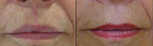
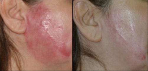
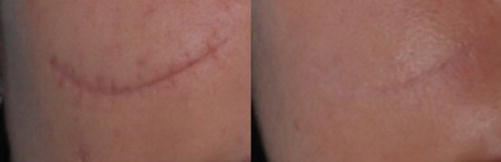
As with other treatment modalities, better results are achieved with a combination of technologies, such as combining laser and IPL (Degitz 2015).
Some studies have demonstrated better results when the laser is combined with triamcinolone or 5-FU infiltration compared with either treatment alone (Degitz 2015).
Post-Procedure
Edema and erythema are expected. The swelling usually improves within 48 h, and ice packs can be used. Anti-inflammatories and oral steroids are indicated for intense edema (Anderson et al. 2014).
Erythema usually improves within 3–5 days and, in extrafacial regions, can persist up to 7 days. Recently, the use of LED (light-emitting diode) immediately after NAFXL has shortened the duration of erythema (Anderson et al. 2014).
The most common adverse effect of PDL treatment is postoperative purpura, which often persists for several days (Anderson et al. 2014).
Complications
Complications are inherent in any technique. Focal hemorrhages can occur between 12 and 24 h, with spontaneous resolution and no sequelae (Ha et al. 2014).
Superficial erosions may occur from inappropriate contact of the tip with the skin surface and may resolve without scarring (Sobanko and Alster 2012).
Burns (bullous and indurated erythematous areas) are rare but can occur when excessive energy is selected or a technical error occurs. Hypo- or hyperpigmentation can occur as a consequence of burns (Ha et al. 2014).
Post-inflammatory hyperpigmentation usually resolves spontaneously within a few months and can be prevented by preparing the skin with bleaching agents (a combination of hydroquinone with retinoic acid) and appropriate photoprotection. It is more common in high phototypes, in the eyelid area, or in areas treated with high fluence (Kim and Cho 2009).
Hypochromia is an unusual, late-onset complication appearing after a few weeks. It is usually related to high fluence, especially in high phototypes, and is commonly preceded by the appearance of crusts. It is reversible with steroids, tacrolimus initially, and retinoic acid at a later stage. For resistant lesions, phototherapy can be a good alternative (Ha et al. 2014).
Infections during the first week carry the risk of delayed wound healing. The most common pathogen is herpes simplex, which can be prevented with prophylaxis started 1 day before the procedure and continued for 5–7 days. Authors diverge regarding herpes prophylaxis for all patients, only those with a history of herpes, or only when an ablative laser is used (Kim and Cho 2009). In our practice, herpes prophylaxis is routinely recommended for patients with a previous history of herpes when treatment is with an ablative laser.
Take Home Messages
- No treatment is considered ideal for scars.
- The mechanism of action of lasers on scars is based on two pillars: reducing blood flow and the reorganization and remodeling of collagen.
- The laser system chosen depends on the type and severity of the scar.
- Non-ablative fractional lasers have a superior safety profile compared with ablative ones.
- Non-ablative lasers are the first choice in higher skin types.
- Non-ablative fractional lasers appear to achieve results similar to those of ablative fractional lasers for relatively atrophic, shallow, or flat scars.
- Lasers have a variable response in hypertrophic scars. A better response is seen in less thick scars, in organized scars such as those from surgical procedures, and in recent scars.
- Keloids have an unpredictable response to any treatment.
References
Anderson R, et al. Laser treatment of traumatic scars with an emphasis on ablative fractional laser resurfacing: consensus report. JAMA Dermatol. 2014;150(2):187–93.
Degitz K. Non-ablative fractional lasers: acne scars and other indications. Hautarzt. 2015;66(10):753–6.
Dvorak HF. Angiogenesis: update 2005. J Thromb Haemost. 2005;3:1835–42.
Dvorak HF. Vascular permeability factor/vascular endothelial growth factor: a critical cytokine in tumor angiogenesis and a potential target for diagnosis and therapy. J Clin Oncol. 2002;20:4368–80.
Ferguson MW, O’Kane S. Scar-free healing: from embryonic mechanism to adult therapeutic intervention. Philos Trans R Soc Lond B Biol Sci. 2004;359:839–50.
Ferguson MW, Whitby DJ, Shah M, Armstrong J, Siebert JW, Longaker MT. Scar formation: the spectral nature of fetal and adult wound repair. Plast Reconstr Surg. 1996;97:854–60.
Gira AK, et al. Keloids demonstrate high-level epidermal expression of vascular endothelial growth factor. J Am Acad Dermatol. 2004;50:850–3.
Gold M, et al. Updated international clinical recommendations on scar management: part 2 – algorithms for scar prevention and treatment. Dermatol Surg. 2014;40:825–31.
Martin P, Leibovich SJ. Inflammatory cells during wound repair: the good, the bad and the ugly. Trends Cell Biol. 2005;15:599–607.
Mattos R, Jordão JM. Lasers fracionados não ablativos no rejuvenescimento. In: Kadunk B, et al., editors. Tratado de Cirurgia Dermatológica, Cosmiatria e Laser da Sociedade Brasileira de Dermatologia. Rio de Janeiro: Elsevier; 2012. p. 763–70.
Mattos R, Torezan L, Osório N. Tratamento de cicatrizes hipertróficas e quelóides. In: Osório N, Torezan L, editors. Laser em Dermatologia: Conceitos básicos e aplicações. São Paulo: ROCA; 2009. p. 237–49.
Mccallion RL, Ferguson MWJ. Fetal wound healing and development of antiscarring therapies for adult wound healing. In: Clark RA, editor. The molecular and cellular biology of wound repair. 2nd ed. New York: Plenum Press; 1996. p. 561–90.
Michael HE, et al. Updated international clinical recommendations on scar management: part 1 – evaluating. Dermatol Surg. 2014;40:817–24.
Mutalik S. Treatment of keloids and hypertrophic scars. Indian J Dermatol Venereol Leprol. 2005;71:3–8.
Osório N, Seque CA. Cicatrizes atróficas e hipertróficas. In: Kadunk B, et al., editors. Tratado de Cirurgia Dermatológica, Cosmiatria e Laser da Sociedade Brasileira de Dermatologia. Rio de Janeiro: Elsevier; 2012. p. 839–45.
Park JE, Barbul A. Understanding the role of immune regulation in wound healing. Am J Surg. 2004;187:S11–6.
Profyris C, Tziotzios C, Do Vale I. Cutaneous scarring: pathophysiology, molecular mechanisms, and scar reduction therapeutics. Part I. The molecular basis of scar formation. J Am Acad Dermatol. 2012;66:1–10.
Santoro MM, Gaudino G. Cellular and molecular facets of keratinocyte reepithelization during wound healing. Exp Cell Res. 2005;304:274–86.
Sardana K, et al. Which type of atrophic acne scar (ice-pick, boxcar, or rolling) responds to non-ablative fractional laser therapy? Dermatol Surg. 2014;40:288–300.
Sobanko JF, Alster TS. Management of acne scarring, part I: a comparative review of laser surgical approaches. Am J Clin Dermatol. 2012;13(5):319–30.
Sobanko JF, Vachiramon V, Rattanaumpawan P, Miller CJ. Early postoperative single treatment ablative fractional lasing of Mohs micrographic surgery facial scars: a split-scar, evaluator-blinded study. Lasers Surg Med. 2015;47(1):1–5.
Tziotzios C, Profyris C, Sterling J. Cutaneous scarring: pathophysiology, molecular mechanisms, and scar reduction therapeutics. Part II. Strategies to reduce scar formation after dermatologic procedures. J Am Acad Dermatol. 2012;66:13–24.
Vrijman C, et al. Laser and intense pulsed light therapy for the treatment of hypertrophic scars: a systematic review. Br J Dermatol. 2011;165:934–42.
Werner S, Grose R. Regulation of wound healing by growth factors and cytokines. Physiol Rev. 2003;83:835–70.
Kauvar AN. Fractional non-ablative laser resurfacing: is there a skin tightening effect? Dermatol Surg. 2014;40:157.
Ha JM, Kim HS, Cho EB, Park GH, Park EJ, Kim KH, Kim LS, Kim KJ. Comparison of the effectiveness of non-ablative fractional laser versus pulsed-dye laser in thyroidectomy scar prevention. Ann Dermatol. 2014;26:615–20.
Kim S, Cho KH. Clinical trial of dual treatment with an ablative fractional laser and a non-ablative laser for the treatment of acne scars in Asian patients. Dermatol Surg. 2009;35:1089–98.
Jang J, et al. Comparison of the effectiveness of ablative and non-ablative fractional laser treatments for early stage thyroidectomy scars. Arch Plast Surg. 2016;43(6):575–81.
Yang Q, Huang W, Qian H, Chen S, Ma L, Lu Z. Efficacy and safety of a 1550-nm fractional laser in the treatment of acne scars in Chinese patients: a split-face comparative study. J Cosmet Laser Ther. 2016;18(6):312–6.
Amann PM, Marquardt Y, Steiner T, Hölzle F, Skazik-Voogt C, Heise R, Baron JM. Effects of non-ablative fractional erbium glass laser treatment on gene regulation in human three-dimensional skin models. Lasers Med Sci. 2016;31(3):397–404.







