
Non-ablative Lasers for Photorejuvenation
Non-ablative Lasers for Photorejuvenation
Maria Angelo-Khattar Aesthetica Clinic, Dubai, UAE
Abstract
Photoaging of the skin principally depends upon the amount of melanin in the skin and the degree of exposure to ultraviolet radiation. Solar damage to DNA leads to a reduction in the skin’s collagen content and ultimately to a deficit in the structural integrity of the skin. This manifests as clinically visible skin atrophy, lines and wrinkles, and dyschromias such as telangiectasias and pigmented lesions. Photorejuvenation entails an improvement in the tone, texture, and pigmentation of the skin. Various laser technologies are available that rejuvenate skin by resurfacing the uppermost layers and allow for the regeneration of new skin cells. The myriad of laser systems includes ablative and non-ablative lasers in both fractionated and nonfractionated or conventional forms. In varying degrees, all of these lasers treat pigmented lesions, soften wrinkles, and reduce the appearance of scars. Although the ablative technologies yield more effective results in terms of the overall reduction of photoaging, the non-ablative lasers allow for swift healing and are rarely associated with any complications or downtime. Furthermore, non-ablative lasers offer a wider spectrum of clinical indications, since they can be used to treat vascular lesions such as telangiectasia, generalized erythema, and rosacea that are commonly associated with aging skin.
Apart from photorejuvenation, non-ablative lasers have multiple applications including the reduction in sebum secretion in acne and the treatment of a variety of scars. However, this review aims to give an overview and highlight the clinical advantages of both fractionated and nonfractionated non-ablative laser platforms in current use for the treatment of photodamaged skin.
Keywords PhotoagingPhotodamaged skinSkin atrophyAging skinWrinklesDyschromiasPigmented lesionsPhotorejuvenationResurfacingNon-ablative lasersFractionated laser
Introduction
The appearance of the skin in humans is integral to the perception of both health and beauty. Hence, in the vast majority of cases, it has a great bearing on the individual’s overall well-being and self-esteem.
Skin aging is a complex biological process influenced by a combination of factors and may be broadly categorized as chronological or intrinsic aging and photoaging. Unlike intrinsic aging, which is determined by the individuals’ genetic predisposition, photoaging depends mainly on the degree of repeated sun exposure and the amount of melanin in the skin. In fact, skin injury by the sun’s ultraviolet (UV) rays accounts for over 70% of skin aging (Kligman and Kligman 1986).
Chronological and photoaging can be easily distinguished clinically, but they share important molecular features. The clinical characteristics of chronologically aged skin include fine wrinkles, laxity, xerosis, and the development of benign lesions such as solar lentigines, cherry angiomas, and seborrheic keratosis (Krutman and Gilchrest 2006).
The features of photoaging include rough skin, wrinkles, deep furrows, pigmented lesions, telangiectasias, poikiloderma of Civatte, solar elastosis, precancerous lesions, and skin cancers (Yaar et al. 2002; Gilchrest 1990). Sun-exposed areas, such as the face, neck, décolleté, hands, and forearms, are most frequently affected. Furthermore, fair-skinned individuals with a history of intensive sun exposure are more susceptible to severe photodamage.
The Mechanism of Ultraviolet Damage to the Skin
Ultraviolet rays induce numerous changes in the skin including melanogenesis, angiogenesis, immune suppression, and degradation of connective tissue, DNA mutations, and oncogenesis.
Advances in skin biology have increased our understanding of the mechanism whereby UV radiation contributes to photoaging and cutaneous disease.
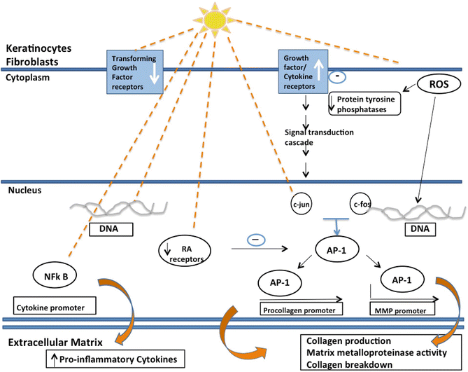
UVB (280–320 nm) rays are reported to have direct cytotoxic and mutagenic effects on skin cells, while UVA (320–400 nm) rays exert their damaging effects predominantly through the production of reactive oxygen species (ROS). A number of transcription factors and inflammatory cytokines are released in response to ultraviolet rays, which result in an increase in matrix metalloproteinase (MMP) production and ultimately accelerated degradation of the dermal matrix.
The widely accepted pathway is via the upregulation of activator protein-1 (AP-1) and nuclear factor kappa B (NF-κB). The latter plays a key role in the signaling pathway leading to the activation of the inflammatory cascade that stimulates the expression of pro-inflammatory cytokines such as tumor necrosis factor alpha. In addition, activation of NF-κB upregulates the expression of MMP-1, and consequently dermal collagen is degraded. Furthermore, NF-κB downregulates type I collagen synthesis (Rijken and Bruijnzeel 2009) (Fig. 1).
Ultraviolet radiation can also upregulate c-Jun, a component of AP-1, and can downregulate retinoic acid (RA) receptors, which decrease RA inhibition of AP-1. This ultimately results in the loss of dermal bulk and skin thinning (Krutmann 2000).
Over the past several years, a plethora of both energy-based and nonenergy-based anti-aging strategies have been developed to retard and reverse skin aging.
Both treatment categories work on the principle of “controlled wounding,” whereby the induction of skin trauma initiates a wound healing response and ultimately results in collagen remodeling and dermal repair (Rivera 2008).
Nonenergy-based treatments include chemical peels, dermabrasion, intradermal injection of growth factors from autologous platelet-rich plasma, fibroblast, and, more recently, stem cell transfer. However, the energy-based devices, which selectively target skin chromophores, have become the preferred treatment modality for many skin conditions including pigmented lesions, telangiectasias, skin laxity, lines and wrinkles, as well as surgical scars, atrophic acne scars, and stretch marks. The energy-based systems include the full spectrum of ablative and non-ablative lasers, intense pulsed light systems, a variety of radiofrequency devices, and high-intensity focused ultrasound systems.
The focus of this chapter will be the full spectrum of non-ablative lasers that are used for photorejuvenation.
History of Non-ablative Lasers
The myriad applications of lasers in dermatology burgeoned with the milestone publication by Anderson and Parrish on selective photothermolysis in 1983 (Anderson and Parrish 1983).
Since then, over the last 30 years, there has been a tremendous evolution in laser technology and design, allowing better control of laser parameters and a consequent increase in safety and efficacy of laser treatments.
Non-ablative lasers are of two types: nonfractional or conventional lasers that act on the entire surface area of the skin and fractional technologies that produce only small columns of thermal injury interspersed among predominantly intact skin.
Historically, the use of lasers in the management of photoaged skin began with the introduction of fully ablative skin resurfacing procedures. The mid-1990s saw the introduction of the carbon dioxide (CO2) and erbium-doped:yttrium-aluminum-garnet (Er:YAG) lasers (Alster 1999). These high-energy pulsed devices create full-thickness wounds and yield impressive clinical outcomes (Alster and West 1996). To date, full resurfacing remains the gold standard for photodamaged skin. However, the procedure is associated with prolonged recovery and poor patient tolerability.
In the late 1990s, Hsu and his colleagues first described non-ablative dermal remodeling due to an incidental observation of the reduction of wrinkles in areas treated with the conventional pulsed dye laser (PDL) for telangiectasias (Hsu et al. 2005). Hence, this led to a move toward minimally invasive procedures, giving rise to a generation of non-ablative resurfacing lasers such as the 1450-nm diode, 1064-nm and 1320-nm neodymium:yttrium-aluminum-garnet (Nd:YAG) lasers, and 1440-nm, 1540-nm, and 1550-nm erbium-glass lasers (Tanzi and Alster 2004). However, this first generation of non-ablative systems showed only moderate efficacy. In 2004, the introduction of fractional photothermolysis (FP) by Manstein and his colleagues (2004) revolutionized the field of laser skin resurfacing and has resulted in the development of numerous non-ablative and ablative fractional laser devices. The popularity of these technologies is due to their ease of operation, lower risk of side effects, good patient tolerability, and relatively quick patient recovery. It is widely accepted that the fractional ablative lasers offer superior efficacy in terms of skin rejuvenation; however, the gentler fractional non-ablative lasers have the advantage of quicker healing and reduced downtime. An increased number of treatment sessions with the non-ablative lasers may ultimately result in equivalent efficacy to ablative lasers.
Non-ablative Laser Photorejuvenation: Mechanism of Action
Photorejuvenation consists of two essential factors:
Removal of Epidermal Signs of Photodamage by Selective Photothermolysis
Microscopic structures such as blood vessels and pigmented cells are photocoagulated by selectively absorbed photons of light, hence resulting in the attenuation of solar lentigines, ephelides, and telangiectasias.
Laser energy absorbed by oxyhemoglobin results in erythrocyte coagulation and the destruction of ectatic vessels. This has been demonstrated by the histological examination of port wine stains after pulsed dye laser. Fibrin, thrombin, and agglutinated red blood cells have been found in the superficial dermal blood vessels (Stratigos and Dover 2000). As a consequence, neovascularization occurs with replacement of the destroyed abnormal vessels.
The precise molecular events involved in the destruction of melanosomes by lasers are unknown (Santiagos et al. 2000), but it is believed that both photothermal and photomechanical mechanisms are responsible (Ara et al. 1990). A sudden rise in temperature results in the vaporization and expansion of target tissues with the consequent destruction of pigmentary lesions. Clinically, frosting is observed immediately after pulsed lasers. This serves as a treatment endpoint and is proposed to be due to the formation of gas bubbles following a sudden increase in temperature of the target tissue (Santiagos et al. 2000). Histologically, destroyed melanosomes, vacuolization, and pigment leakage from cells have been shown (Dover et al. 1989a).
Dermal Remodeling Resulting from Patterns of Microscopic Thermal Injury to the Dermis
The reduction in fine lines and wrinkles and overall improvement in skin texture and tightening are due to new collagen formation and dermal thickening. The mechanism of dermal remodeling by non-ablative lasers centers upon the fact that the non-ablative resurfacing laser wavelengths are absorbed to varying extents by intradermal water. Hence, this results in thermal injury to the dermis that is dependent upon both the amount of energy delivered and the exposure duration. A critical intradermal temperature of 60–80 °C is required for effective denaturation of collagen and initiation of the wound healing response (Paul et al. 2011). Temperatures above this range can lead to complete denaturation of collagen and result in scarring.
The non-ablative lasers treat photodamaged skin without physically removing or vaporizing the skin, as opposed to ablative procedures that literally vaporize a fraction or all of the epidermis and layers of the dermis.
All of these lasers selectively utilize technology to cool and protect the epidermis while creating controlled thermal injury to dermal structures.
Non-ablative Nonfractional Lasers
The first generation of non-ablative conventional laser modalities entered the market in the late 1990s primarily for the use of skin rejuvenation. These included lasers that emit light in the visible, near-infrared, and far-infrared spectrum (Table 1). This diverse group of laser technologies includes the potassium titanyl phosphate (KTP; 532 nm), pulsed dye (PDL; 585 nm, 595 nm), Q-switched (QS) ruby (694 nm), QS alexandrite (755 nm), long-pulsed and QS Nd:YAG (1064 nm), long-pulsed Nd:YAG (1320 nm), long-pulsed diode (1450 nm), and erbium-glass (1440 nm and 1540 nm) lasers.
| Visible light lasers | Near-infrared lasers | Mid-infrared lasers |
|---|---|---|
| 532-nm KTP | 1064-nm long-pulsed and Q-switched nanosecond and picosecond Nd:YAG | 1320-nm Nd:YAG |
| 585-/595-nm pulsed dye | 1450-nm diode | |
| 694-nm Q-switched ruby | 1540-nm erbium-glass | |
| 755-nm Q-switched nanosecond and picosecond alexandrite |
Wavelengths within the visible light spectrum and specifically the QS lasers are mainly indicated for skin dyschromias, whereas the longer wavelength near-infrared and mid-infrared lasers are better suited for dermal remodeling.
Although the reversal of epidermal dyschromias with the nonfractionated devices is relatively effective and predictable, the improvement in skin texture is much more subtle. Hence, non-ablative nonfractional skin resurfacing is ideal for the patient with mild to moderate photodamage and early signs of skin aging.
Visible Light Lasers
These include the 532-nm KTP, 585-/595-nm pulsed dye, 694-nm Q-switched ruby, and Q-switched 755-nm alexandrite lasers.
These visible light lasers are commonly used to treat vascular and pigmented lesions since their wavelengths are well absorbed by the various chromophores in the skin, such as oxyhemoglobin and melanin. The lasers improve skin color by reducing dyschromias including solar lentigines, ephelides, telangiectasias, and generalized erythema. The visible light spectrum lasers should be used with great caution in darker skin types due to the risk of thermal damage of these shorter wavelengths.
An improvement in skin texture has also been repeatedly demonstrated with the visible light spectrum lasers. The absorption of the 585-nm wavelength by oxyhemoglobin results in a thermal insult to the microvasculature and hence initiates an inflammatory response that stimulates fibroblast activity and ultimately dermal collagen synthesis. Bjerring et al. showed a 48% increase in type II collagen synthesis only 2 weeks after a single session of pulsed dye laser (Bjerring et al. 2002). Orringer et al. reported an increase in dermal remodeling due to an increase in type I procollagen mRNA, and Zelickson et al. also showed that only one pass with the 595-nm pulsed dye laser was sufficient to improve dermal collagen (Orringer et al. 2005; Zelickson et al. 1999).
Potassium Titanyl Phosphate Laser 532 nm (Cynosure MedLite C6, RevLite S1, and PicoSure; Syneron-Candela Alex TriVantage; Alma Harmony XL Pro)
Current Q-switched KTP lasers include those in the 5–15 ns range and the novel 350 picosecond lasers. The devices incorporate an Nd:YAG (1064 nm) laser as the main source of light that is converted by a KTP crystal to emit the halved wavelength of 532 nm. This green light is well absorbed by hemoglobin and melanin; hence, the KTP can be used for the treatment of both unwanted vessels and pigment. The short wavelength of the KTP laser is very well absorbed by epidermal melanin; hence, caution should be exercised in darker skin types (Weiss et al. 2005). The KTP laser has only mild effects on the improvement of skin texture (DeHoratius and Dover 2007).
The 532 KTP laser has interestingly been used for lip color lightening, which is a common concern of darker-skinned individuals (Somyas et al. 2001).
Pulsed Dye Laser 580–595 nm (Syneron-Candela VBeam)
In the flashlamp-pumped pulsed dye laser, a flashlamp is used to excite electrons in an organic dye, rhodamine, to emit light of yellow color. The original pulsed dye lasers had a wavelength of 577 nm and a pulse duration of 0.45 milliseconds, which resulted in intravascular thrombosis of small vessels and purpura. Hence, the current pulsed dye systems have wavelengths between 585 and 595 nm and a pulse duration of up to 40 milliseconds. The longer wavelengths penetrate deeper into the skin, and the longer pulse durations allow for avoidance of purpura. Apart from the effect on vascular lesions, pulsed dye lasers do afford a moderate skin rejuvenating effect (Bjerring et al. 2002).
Q-Switched Ruby Laser 694 nm (Alma Lasers, Sinon)
The QS ruby laser emits red light and is hence not absorbed by hemoglobin but selectively targets melanin. It was, in fact, the first Q-switched laser developed for epidermal and dermal pigmented lesions. The very short, 20–50 ns, pulse durations of the ruby laser selectively target melanosomes, resulting in the photoacoustic destruction of the melanosome cell membrane. This effect is pulse width dependent with shorter pulse durations being more effective in melanosome destruction (Polla et al. 1987; Dover et al. 1989b). Caution must be exercised as darker phototypes may develop permanent hypopigmentation (Rinaldi 2008; Kishi et al. 2009).
QS Alexandrite 755 nm (Cynosure Accolade, Syneron-Candela Alex TriVantage, and PicoWay)
The QS alexandrite laser, emitting light within the red spectrum and with pulse widths of 50–100 ns, selectively targets pigmented lesions by causing the photoacoustic disruption of melanin.
A new picosecond QS alexandrite laser has recently been introduced on the market, and, thus far, most of the studies with this novel device have been on its use in the treatment of tattoos. However, the device shows promise in the effective treatment of pigmented lesions. A recent publication has demonstrated the 755-nm ultrashort-pulsed 550 picosecond alexandrite laser with diffractive lens array to be an effective option for rejuvenation of the photodamaged décolletage (Wu et al. n.d.).
Near-Infrared Lasers
1064-nm Long-Pulsed and QS Nd:YAG Lasers (Palomar QYAG5, Syneron-Candela Alex TriVantage; Alma Harmony XL Pro)
Both the long-pulsed and QS Nd:YAG (1064-nm) lasers in the mid-infrared spectrum are used for photorejuvenation. The incidental rejuvenating effect of long-pulsed lasers including the Nd:YAG 1064-nm laser commonly used for other indications including hair removal and treatment of larger caliber blood vessels was recognized early on. This near-infrared wavelength is absorbed by water in the skin and hence effectively heats the dermis. The resultant thermal injury to the dermis leads to a limited degree of neocollagenesis and skin tightening (Weng et al. 2011).
The long-pulsed Nd:YAG laser handpiece may be used in constant motion, delivering multiple passes to the surface of the skin until a surface temperature of 39–42 °C is attained. This is the optimal temperature range for dermal remodeling. Above this temperature, dermal scarring may occur. Hence, prior to treatment with this laser for rejuvenation, patients should not receive any anesthetic, as excessive pain must be reported to alert the practitioner and avoid epidermal injury. Patients treated with long-pulsed Nd:YAG for rejuvenation exhibited a decrease in rhytids (Hong et al. 2015).
The QS Nd:YAG with pulse durations of 5–15 ns, principally used for the treatment of tattoos and disruption of melanin in pigmented lesions, may also be used in an in-motion manner, continuously treating the skin. Treatments may be performed with or without the application of topical carbon solution, which is believed to improve the penetration of the laser beam. A split-face study by Lee et al. demonstrated significant improvement in rejuvenation effects after QS Nd:YAG with no difference in improvement in skin texture after the application of topical carbon solution (Lee et al. 2009). However, skin rejuvenation treatments with the QS Nd:YAG were associated with a high degree of postinflammatory hyperpigmentation at the 1-month follow-up evaluation (Jun et al. 2014).
Recently, picosecond Nd:YAG lasers in the 500–750 ps range have been developed for the removal of pigmented lesions. To date, no studies have been published on the use of a picosecond Nd:YAG laser system for pigmented lesions or skin rejuvenation.
Mid-Infrared Lasers
1320-nm Nd:YAG (Cooltouch CT3 Plus, Alma Harmony XL)
This wavelength is well absorbed by water and effectively heats the treated dermis. The ensuing thermal injury results, to a limited extent, in the stimulation of fibroblasts and the induction of “neocollagenesis.” Studies have shown an elevation in basic fibroblast growth factor (bFGF) and histological evidence for a reduction in skin aging by the stimulation of type I, III, and VII collagen (Zhenxiao et al. 2011; El-Domyati et al. 2011).
1450-nm Diode Laser (Candela Smoothbeam)
The mechanism of action of the 1450-nm diode laser is similar to the 1320-nm Nd:YAG. This mid-infrared wavelength creates wounding of the dermis due to its high water absorption coefficient, but the 1450-nm laser has been shown to be more effective in inducing neocollagenesis than the 1320-nm wavelength (Alster et al. 2007).
1540-nm Erbium-Glass Laser (Quantel Aramis)
The mid-infrared 1540-nm laser, which also targets intracellular water, penetrates into the papillary dermis where collagen tightening and neocollagenesis are achieved. This allows for more effective treatment of solar elastosis.
The 1540-nm wavelength is only minimally absorbed by melanin; hence, it is safe for use by darker phototypes.
This laser has been used for the treatment of fine lines, wrinkles, and acne scars. Evidence for dermal remodeling with the 1540-nm laser has been demonstrated by histologic studies as well as ultrasound and profilometric analysis. A 40% reduction in wrinkles and 17% increase in skin thickness were shown after the fourth week of treatment (Fournier et al. 2002; Lupton et al. 2002).
Non-ablative Fractional Lasers
Concept of Fractional Photothermolysis
Fractional photothermolysis (FP), first introduced by Manstein and colleagues in 2004, is one of the most significant milestones in laser technology and resurfacing (Manstein et al. 2004). It has led to a new era in the application of lasers in dermatology and resulted in a tremendous development in fractional laser technology and numerous commercial fractional systems. Fractional lasers have now become the laser modality of choice in the management of photodamaged skin (Saedi et al. 2012). These devices bridge the gap in terms of efficacy between the traditional ablative and non-ablative lasers (Alexiades-Armenakas et al. 2012). They are less aggressive than traditional ablative lasers and offer superior clinical outcomes to the non-ablative systems but with the same level of safety. As opposed to the conventional ablative and non-ablative lasers that produce layers of thermal heating, fractional lasers thermally denature only fractions of the skin.
They create micro-columns of thermal injury referred to as microthermal or microscopic treatment zones (MTZs) (Geronemus 2006). MTZs are typically 70–150 μm wide and extend vertically from the epidermis to the dermis at varying depths of approximately 400–700 μm, determined by several laser parameters including laser output energy (Gold 2010). Fractional devices spare skin and stem cells in between the MTZs and hence allow for rapid recovery and increased safety (Fig. 2).
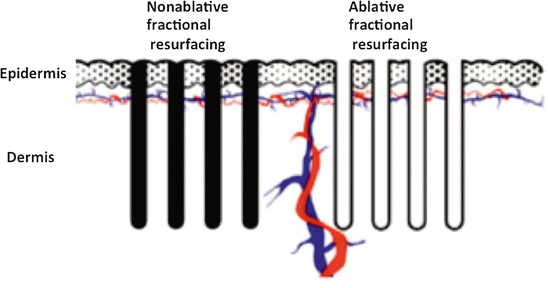
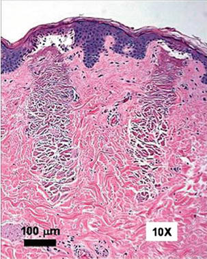
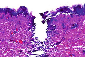
There are basically two types of fractional laser devices: non-ablative and ablative, classified according to the type of MTZs that they produce (Fig. 2). Non-ablative fractional lasers (NAFLs) simply cause thermal injury to the dermis while sparing the epidermis, whereas the ablative fractional lasers (AFLs) vaporize micro-columns of epidermis and dermis (Alexiades-Armenakas et al. 2008). The histologic changes due to non-ablative fractional photothermolysis (NAFP) and ablative fractional photothermolysis (AFP) are shown in Figs. 3 and 4.
The repair of treated skin begins by the extrusion of columns of necrotic skin, known as microscopic epidermal necrotic debris (MENDs), into the stratum corneum (Manstein et al. 2004). Manstein and colleagues (2004) showed that MENDs are button-shaped structures (40–80 μm diameter) that contain melanin and form beneath the stratum corneum above each dermal wound. The production of MENDs indicates the participation of intact keratinocytes, from uninjured skin, in the process of wound repair (Manstein et al. 2004). MENDs seen in histology correspond to the brown spots observed via epiluminescence microscopy, and these are shed from the epidermis within 7 days of FP (Manstein et al. 2004) (Fig. 5).
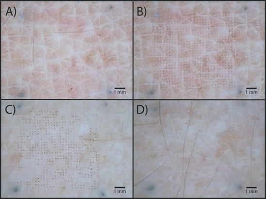
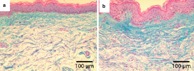
This extrusion process of cellular debris was confirmed by Hantash et al. (2006) who used anti-human elastin antibody to demonstrate the transdermal elimination of MENDs. Manstein and colleagues (2004) showed clear evidence of epidermal repair within the papillary and superficial reticular dermis, as demonstrated by increased mucin content as well as an increased undulation in the rete ridge pattern of treated skin at 3 months posttreatment (Fig. 6) (Laubach et al. 2006).
Types of Non-ablative Fractional Lasers
All of the non-ablative fractional lasers emit wavelengths in the mid-infrared range that correspond to peaks on the water absorption curve. By virtue of the mechanism of action of the fractional device, whereby only small fractions of the skin are resurfaced in each treatment session, typically 4–6 treatment sessions are required to attain a positive clinical outcome in the treatment of photodamaged skin. The treatment is relatively painful, requiring a combination of topical anesthetic creams and cryoanesthesia. Posttreatment edema and erythema are noticeable for 48 h, followed by skin desquamation over a few days.
1550-nm (Solta Fraxel Re:store, Solta Fraxel Re:store Dual) and 1540-nm (Palomar Starlux, Artisan, Icon) Erbium-Glass Lasers
The first NAFL device to be developed in 2004 was the 1550-nm erbium-glass laser that targets water as a chromophore (Geronemus 2006). The device is tunable such that the density of the MTZs and the energy can be adjusted. Most of the studies on NAFLs were performed with this device (Tierney et al. 2009).
The initial clinical studies by Manstein et al. (2004) demonstrated significant improvement in periorbital lines and wrinkles. Many other investigators consistently showed evidence of collagen remodeling as improvements in skin texture and color. The initial results continued to improve over 6–9 months posttreatment (Rahman et al. 2006; Wanner et al. 2007). Non-facial areas are also effectively treated with this NAFL device. A study by Jih et al. (2008) on the hands of ten patients showed 51–75% improvement in pigmentation and 25–50% improvement in skin texture at 3 months. DeAngelis et al. reported good efficacy of the erbium-glass laser in the treatment of striae rubra and alba ranging in maturation age from 1 to 40 years (de Angelis et al. 2011).
Wanner et al. showed fractional photothermolysis for the treatment of facial and non-facial cutaneous photodamage using a 1550-nm Er-glass fiber laser to be a useful NAFL treatment (Wanner et al. 2007).
The upgraded version of this laser, the Fraxel Dual, has a dual wavelength of 1550-nm erbium with a 1927-nm thulium laser on one platform, allowing the operator to target different treatment depths. After a single treatment with the thulium laser, a mean of 69% of subjects at 1 month demonstrated a very significant improvement in lentigines and ephelides (Brauer et al. 2014). Hence, superficial treatment of pigmented lesions can be achieved with the 1927-nm wavelength, and collagen remodeling is achieved with the deeper penetrating 1550-nm wavelength.
1440-nm Nd:YAG Laser (Cynosure Affirm) and 1440-nm Diode (Solta Clear + Brilliant)
Fractional devices that deliver 1440-nm wavelengths include the Nd:YAG and diode laser. The Cynosure device allows for uniform distribution of the energy across the skin surface, at depths of 300 μm, via combined apex pulse (CAP) technology.
Clinical studies with both devices have shown them to be only moderately effective in the remodeling of scars and treatment of photoaged skin (Marmon et al. 2014; Geraghty and Biesman 2009).
Hence, manufacturers have upgraded the original systems to include dual wavelengths. The Cynosure system offers the multiplex technology that combines a 1320-nm wavelength with a 1440-nm laser, allowing for penetration to depths of 1000–3000 μm. The 1320-/1440-nm multiplex system resulted in greater skin tightening at 6 months than the 1440-nm laser alone (Geraghty and Biesman 2009).
A new version of the Clear + Brilliant, known as the Clear + Brilliant Permea, includes both diode and thulium media, hence offering both 1440-nm and 1927-nm wavelengths.
1565-nm Erbium-Doped Laser (Lumenis ResurFx)
This wavelength has a slightly lower absorption coefficient for water than that of 1550 nm, which results in greater skin penetration. The system uses a sequential scanning system that allows for a variety of shapes, densities, and patterns of distribution of energy. It is also equipped with contact cooling for greater patient comfort. To date, there are no published studies demonstrating the relative advantage of this laser system over the established fractional near-infrared devices (Sadick 2014).
1940-nm Alexandrite Fractional Laser (Syneron-Candela)
This is a relatively novel thulium rod pumped by a pulsed alexandrite laser emitting a wavelength of 1940 nm, which corresponds to one of the water absorption peaks in the mid-infrared range. However, the absorption of this wavelength is much stronger than the other mid-infrared wavelengths (1400–1550) and weaker than the ablative wavelengths (Er:YAG and CO2). A recent study demonstrated the skin rejuvenating effects of the 1940-nm fractional device. Patients (n = 11) received a total of three facial treatments, 4–6 weeks apart, and outcome assessments were made 3 months after the final treatment. Several parameters were evaluated and the results showed a 21% reduction in pigmentation, 14.3% in rhytids, 8.9% in skin laxity, and 22.3% in solar elastosis (Miller et al. 2014).
1064-nm Fractional QS Nd:YAG Laser (Alma ClearLift on Harmony XL Pro)
This is a relatively new addition to the arsenal of NAFR lasers that has gained popularity. The 1064-nm laser beam is fractionated by a passive optical element into a 5 × 5 matrix of 25 microscopic perforations, each having a diameter of 200 μm. Each pixel receives an energy density in the range of 6–13 J/cm2. A series of five different focusing tips allow for penetration of the QS nanosecond pulses to tunable depths, selected according to the depth of the pathology being treated. The rapid laser pulses spare the epidermis, and, furthermore, since the 1064-nm wavelength is poorly absorbed by melanin and hemoglobin, the pixels penetrate up to 3 mm into the papillary and reticular dermis. Paasch, at the 2014 American Society of Lasers in Medicine and Surgery meeting, presented histological evidence of microthermal injury deep into the reticular dermis (Paasch 2014). This is advantageous, since we now know that to achieve skin firmness, penetration to deeper levels is required.
Recently, a high-speed roller has been released, known as the iPixel Scan, which allows for efficient and fast treatment of off-the-face areas such as photodamaged hands, arms, décolletage, and legs.
A pilot study by Luebberding and Armenakas (2012) showed an 11% improvement in superficial rhytids on the face and neck following a series of three treatment sessions at 2–4 week intervals. Gold et al. (2014) investigated the effect of the fractional QS 1064-nm laser on several skin parameters. Patients received a total of four treatment sessions at 2–4 week intervals with a follow-up at 3 months post last treatment. They reported an improvement of 70% in hyperpigmentation, 80% in telangiectasias, 80% in skin laxity, and 60% each in tactile roughness and actinic keratoses. A further advantage of the treatment was found to be the fact that the treatment is relatively pain-free, at a level of 0–2 on a ten-point scale, and has absolutely no downtime.
General Clinical and Treatment Considerations in Non-ablative Resurfacing
Patient Selection
Non-ablative resurfacing is contraindicated during pregnancy, lactation, and in patients who have a history of keloid formation or those that have an active infection. Patients who are on isotretinoin can only be treated 6 months after the completion of a course of treatment.
Ideal patients for non-ablative rejuvenation are those with early signs of photodamage. These are generally younger patients ranging from 35 to 50 years of age. Older patients with severe laxity and deep wrinkles will not benefit from non-ablative laser treatment and are candidates for ablative resurfacing (Fig. 7).
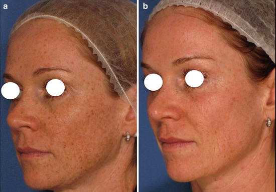
It is important to ensure that patients have realistic expectations and understand that skin rejuvenation with the non-ablative systems requires several treatment sessions.
Non-ablative resurfacing may be performed in patients of all skin types, but particular caution should be exercised when treating darker skin types. In these patients, visible light spectrum lasers that specifically target pigment must be used at conservative settings to avoid side effects such as hyper- or hypopigmentation, blisters, and scars. Although lasers in the near- and mid-infrared wavelengths are safer for darker-skinned individuals, caution must be exercised with respect to the settings. High laser fluences as well as cryogen cooling with some systems can lead to postinflammatory hyperpigmentation. This is usually transient and can be treated with bleaching agents.
Treatment Considerations
Pretreatment, the skin should be cleansed thoroughly to remove oil, makeup, and any debris that may impede the passage of the laser light. Topical anesthesia is generally required with most of the non-ablative nonfractional and fractional lasers, the exception being the fractional Q-switched Nd:YAG device. Various formulations and strengths of topical anesthetic creams have been used, from 5% up to 30% lidocaine. Fractional resurfacing with the mid-infrared laser treatments requires the more potent anesthetic formulations. There is some controversy regarding the application of topical anesthesia as, with some devices, patient feedback in terms of excessive pain is necessary to avoid epidermal injury.
The patient and all present in the laser room should wear protective eyewear as most of the wavelengths in the visible and near-infrared range pose a risk to the retina, whereas the wavelengths in the mid-infrared range can damage the cornea.
Prophylactic treatment with oral antiviral medication is advocated to avoid a herpetic breakout, specifically in patients undergoing non-ablative fractional resurfacing.
Skin cooling during and posttreatment with the use of a cool air blower or ice application is required for patient comfort and to minimize posttreatment edema. Typically with the fractional non-ablative systems, edema and erythema resolve within 3 days. Patients are required to avoid the sun and apply sunscreen diligently to avoid the possibility of postinflammatory hyperpigmentation. Additionally, in darker-skinned patients, bleaching creams are necessary posttreatment to minimize the possibility of complications.
Conclusion
Non-ablative laser treatments are ideal for the improvement of skin color, tone, and texture in relatively younger patients with skin textural imperfections associated with photoaging. These procedures have a unique role in treating skin dyschromias including vascular lesions in patients of all ages. Evidence is mounting that non-ablative lasers may also result in skin tightening; however, further studies are necessary to reinforce this theory (Gold et al. 2014; Kauvar 2014).
Non-ablative systems are often used as maintenance procedures following more aggressive ablative treatments. Furthermore, they play an important role in the prophylaxis of skin aging as they can be performed regularly with little or no downtime. Non-ablative lasers have been mainly used on facial skin; however, interest is rising in the role of these techniques on the neck, hands, arms, and other areas of the body.
Non-ablative skin resurfacing procedures produce excellent clinical outcomes with minimal risk and morbidity; hence, they are of particular importance in the treatment of photodamaged skin.
Take Home Messages
- The visible light spectrum non-ablative lasers, and in particular the Q-switched devices, are the lasers of choice for the treatment of pigmented lesions.
- The pulsed dye laser offers a unique modality for the elimination of vascular lesions such as telangiectasias and generalized erythema associated with photodamage.
- Non-ablative nonfractional skin rejuvenation offers only mild skin remodeling effects and is only suitable for patients with early signs of skin aging.
- Non-ablative fractional lasers produce impressive clinical outcomes in skin rejuvenation with respect to improvement in skin laxity and reduction in wrinkles.
- Non-ablative fractional lasers are associated with swift recovery, minimal complications, and little or no downtime.
References
Alexiades-Armenakas MR, Dover JS, Arndt KA. The spectrum of laser skin resurfacing: non-ablative, fractional, and ablative laser resurfacing. J Am Acad Dermatol. 2008;58:719–37.
Alexiades-Armenakas MR, Dover JS, Arndt KA. Fractional laser skin resurfacing. J Drugs Dermatol. 2012;11:1274–87.
Alster TS. Cutaneous resurfacing with CO2 and erbium:YAG lasers: preoperative, intraoperative, and postoperative considerations. Plast Reconstr Surg. 1999;103:619–32.
Alster TS, West TB. Resurfacing of atrophic facial acne scars with a high-energy pulsed carbon dioxide laser. Dermatol Surg. 1996;22:151–4.
Alster TS, Tanzi EL, Lazarus M. The use of fractional laser photothermolysis for the treatment of atrophic scars. Dermatol Surg. 2007;33:295–9.
Anderson RR, Parrish JA. Selective photothermolysis – precise microsurgery by selective absorption of pulsed radiation. Science. 1983;220:524–7.
Ara G, Anderson RR, Mandel KG, Otteson M, Oseroff AR. Irradiation of pigmented melanoma cells with high intensity pulsed radiation generates acoustic waves and kills cells. Lasers Surg Med. 1990;10:52–9.
Bjerring P, Clement M, Heickendorff L, Lybecker H, Kiernan M. Dermal collagen production following irradiation by dye laser and broadband light source. J Cosmet Laser Ther. 2002;4(2):39–43.
Brauer JA, McDaniel DH, Bloom BS, Reddy KK, Bernstein LJ, Geronemus RG. Non-ablative 1927 nm fractional resurfacing for the treatment of facial photopigmentation. J Drugs Dermatol. 2014;13(11):1317–22.
de Angelis F, Kolesnikova L, Renato F, Liguori G. Fractional non-ablative 1540-nm laser treatment of striae distensae in Fitzpatrick skin types II to IV: clinical and histological results. Aesthet Surg J. 2011;31(4):411–9.
DeHoratius DM, Dover JS. Non-ablative tissue remodeling and photorejuvenation. Clin Dermatol. 2007;25(5):474–9.
Dover J, Margolis RJ, Polla LL, Watanabe S, Hruza GJ, Parrish JA, Anderson RR. Pigmented guinea pig skin irradiated with Q-switched ruby laser pulses: morphologic and histologic findings. Arch Dermatol. 1989a;125:42–9.
Dover JS, Margolis RJ, Polla LL, Watanabe S, Hruza GJ, Parrish JA, Anderson RR. Pigmented guinea pig skin irradiated with Q-switched ruby laser pulses. Morphologic and histologic findings. Arch Dermatol. 1989b;125:43–9.
El-Domyati M, El-Ammawi TS, Medhat W, Moawad O, Mahoney MG, Uitto J. Effects of the Nd:YAG 1320-nm laser on skin rejuvenation: clinical and histological correlations. J Cosmet Laser Ther. 2011;13(3):98–106.
Fournier N, Dahan S, Barneon G, Rouvrais C, Diridollou S, Lagarde JM, Mordon S. Non-ablative remodeling: a 14-month clinical ultrasound imaging and profilometric evaluation of a 1540 nm Er:Glass laser. Dermatol Surg. 2002;28:926–31.
Geraghty LN, Biesman B. Clinical evaluation of single wavelength fractional laser and a novel multi-wavelength fractional laser in the treatment of photodamaged skin. Lasers Surg Med. 2009;41(6):408–16.
Geronemus RG. Fractional photothermolysis: current and future applications. Lasers Surg Med. 2006;38:169–76.
Gilchrest BA. Skin aging and photoaging. Dermatol Nursing. 1990;2(2):79–82.
Gold M. Update on fractional laser technology. J Clin Aesthet Dermatol. 2010;3:42–50.
Gold MH, Sensing W, Biron J. Fractional Q-switched 1,064-nm laser for the treatment of photoaged-photodamaged skin. J Cosmet Laser Ther. 2014;16(2):69–76.
Hantash BM, Bedi VP, Sudireddy V, Struck SK, Herron GS, Chan KF. Laser-induced transepidermal elimination of dermal content by fractional photothermolysis. J Biomed Opt. 2006;11:41–115.
Hong JS, Park SY, Seo KK, Bl G, Hwang EJ, Park GY, Eun HC. Long pulsed 1064 nm Nd:YAG laser treatment for wrinkle reduction and skin laxity: evaluation of new parameters. Int J Dermatol. 2015;54(9):e345–50.
Hsu TS, Zelickson B, Dover JS, Kilmer S, Burns J, Hruza G, Brown DB, Bernstein EF. Multicenter study of the safety and efficacy of 585 nm pulsed-dye laser for the non-ablative treatment of facial rhytids. Dermatol Surg. 2005;31(1):1–9.
Jih MH, Goldberg LH, Kimyai-Asadi A. Fractional photothermolysis for photoaging of hands. Dermatol Surg. 2008;34(1):73–8.
Jun HJ, Kim SM, Choi WJ, Cho SH, Lee JD, Kim HS. A split-face, evaluator-blind randomized study on the early effects of Q-switched Nd:YAG laser versus Er:YAG micropeel in light solar lentigines in Asians. J Cosmet Laser Ther. 2014;16(2):83–8.
Kauvar AN. Fractional non-ablative laser resurfacing: is there a skin tightening effect? Dermatol Surg. 2014;40(suppl 12):S157–63.
Kishi K, Okabe K, Ninomiya R, Konno E, Hattori N, Kasube K, Imanishi N, Nakajima H, Nakajima T. Early serial Q-switched ruby laser therapy for medium-sized to giant congenital melanocytic naevi. Br J Dermatol. 2009;161(2):345–52.
Kligman LH, Kligman AM. Photoaging. In: Fitzpatrick TB, editor. Dermatology in general medicine. New York: McGraw-Hill Co; 1986. p. 1470–5.
Krutman J, Gilchrest B. Skin aging. Berlin: Springer; 2006. p. 15–24.
Krutmann J. Ultraviolet A radiation-induced biological effects in human skin: relevance for photoaging and photodermatosis. J Derm Sci. 2000;23:S22–6.
Laubach HJ, Tannous Z, Anderson RR, Manstein D. Skin responses to fractional photothermolysis. Lasers Surg Med. 2006;38:142–9.
Lee MC, Hu S, Chen MC, Shih YC, Huang YL, Lee SH. Skin rejuvenation with 1,064-nm Q-switched Nd:YAG laser in Asian patients. Dermatol Surg. 2009;35(6):929–32.
Luebberding S, Alexiades-Armenakas MR. Fractional, non-ablative Q-switched 1,064 neodymium YAG laser to rejuvenate photoaged skin: a pilot case series. J Drugs Dermatol. 2012;11:1300–4.
Lupton JR, Williams CM, Alster TS. Laser skin resurfacing using a 1540 nm erbium glass laser: a clinical and histological analysis. Dermatol Surg. 2002;28(9):833–5.
Manstein D, Herron GS, Sink RK, Tanner H, Anderson RR. Fractional photothermolysis: a new concept for cutaneous remodeling using microscopic patterns of thermal injury. Lasers Surg Med. 2004;34(5):426–38.
Marmon S, Shek SY, Yeung CK, Chan NP, Chan JC. Evaluation of the safety and efficacy of the 1440-nm laser in the treatment of photodamage in Asian skin. Lasers Surg Med. 2014;46(5):375–9.
Miller L, Mishra V, Alsaad S, Winstanley D, Blalock T, Tingey C, Qui J, Romine S, Ross EV. Clinical evaluation of a non-ablative 1940 nm fractional laser. J Drugs Dermatol. 2014;13(11):1324–9.
Orringer JS, Voorhees JJ, Hamilton T, Hammerberg C, Kang S, Johnson TM, Karimipour DJ, Fischer G. Dermal matrix remodelling after non-ablative laser therapy. J Am Acad Dermatol. 2005;53(5):775–82.
Paasch U. Histological epidermal and dermal changes of 1064-nm Q-switched and fractionated pulses. Abstract ASLMS 2014, Phoenix, April 6, 2014.
Paul M, Blugerman G, Kreindel M, Mulholland RS. Three-dimensional radiofrequency tissue tightening: a proposed mechanism and applications for body contouring. Aesthet Plast Surg. 2011;35(1):87–95.
Polla LL, Margolis RJ, Dover JS, Whitaker D, Murphy GF, Jacques SL, Anderson RR. Melanosomes are a primary target of Q-switched ruby laser irradiation in guinea pig skin. J Invest Dermatol. 1987;89(3):281–6.
Rabe JH, Mamelak AJ, McElgunn PJ, WL M, Saunder DN. Photoaging: mechanisms and repair. J Am Acad Dermatol. 2006;55(1):1–19.
Rahman Z, Alam M, Dover JS. Fractional laser treatment for pigmentation and texture improvement. Skin Therapy Lett. 2006;11(9):7–11.
Rijken F, Bruijnzeel PLB. The pathogenesis of photoaging: the role of neutrophils and neutrophil-derived enzymes. J Invest Dermatol. 2009;14:62–72.
Rinaldi F. Laser: a review. Clin Dermatol. 2008;26(6):590–601.
Rivera AE. Acne scarring. J Am Acad Dermatol. 2008;59:659–76.
Sadick N. Advances in cosmetic dermatology, an issue of dermatologic clinics. Nils Kreuger. Amsterdam, Netherlands; 2014.
Saedi N, Jalian HR, Petelin A, Zachary C. Fractionation: past, present, future. Semin Cutan Med Surg. 2012;31(2):105–9.
Santiagos AJ, Dover JS, Arndt KA. Laser treatment of pigmented lesions – 2000: how far have we gone. Arch Dermatol. 2000;136:915–21.
Somyas K, Somsak K, Suntharee K, Pannadda L, Sansanee W. An effective treatment of dark lip by frequency-doubled Q-switched Nd:YAG laser. Dermatol Surg. 2001;27:37–40.
Stratigos AJ, Dover JS. Overview of lasers and their properties. Dermatol Ther. 2000;13:2–16.
Tanzi EL, Alster TS. Comparison of a 1450-nm diode laser and a 1320-nm Nd:YAG laser in the treatment of atrophic facial scars: a prospective clinical and histologic study. Dermatol Surg. 2004;30:152–7.
Tierney EP, Kouba DJ, Hanke CW. Review of fractional photothermolysis: treatment indications and efficacy. Dermatol Surg. 2009;35(10):1445–61.
Wanner M, Tanzi EL, Alster TS. Fractional photothermolysis: treatment of facial and nonfacial cutaneous photodamage with a 1550 nm erbium-doped fiber laser. Dermatol Surg. 2007;33:23–8.
Weiss RA, Weiss MA, Beasley KL, Munavalli G. Our approach to non-ablative treatment of photoaging. Lasers Surg Med. 2005;37(1):2–8.
Weng Y, Dang Y, Ye X, Liu N, Zhang Z, Ren Q. Investigation of irradiation by different non-ablative lasers on primary cultured skin fibroblasts. Clin Exp Dermatol. 2011;36(6):655–60.
Wu DC, Fletcher L, Guiha I, Goldman MP. Evaluation of the safety and efficacy of the picosecond alexandrite laser with specialized lens array for the treatment of photoaging décolletage. Lasers Surg Med. 48(2):188–92 (Epub ahead of print).
Yaar M, Eller MS, Gilchrest BA. Fifty years of skin aging. J Investig Dermatol Symp Proc. 2002;7(1):51–8.
Zelickson BD, Kilmer SJ, Bernstein E, Chotzen VA, Dock J, Mehregan D, Coles C. Pulsed dye laser therapy for sun damaged skin. Lasers Surg Med. 1999;25:229–36.
Zhenxiao Z, Aie X, Yuzhi J, Xiaodong W, Xiangiang J, Jing S, Han Z, Junhui Z, Xiaojun Z, Yanjun Z. Exploring the role of a non-ablative laser (1320 nm cooltouch laser) in skin photorejuvenation. Skin Res Technol. 2011;17(4):505–9.







