
Light-Emitting Diode for Acne, Scars, and Photodamaged Skin
Light-Emitting Diode for Acne, Scars, and Photodamaged Skin
Luiza Pitassi Division of Dermatology, Department of Medicine, State University of Campinas (UNICAMP), Campinas, SP, Brazil
Abstract
Light-emitting diode therapy was discovered in the late 1960s but only recently has it been widely applied in dermatology to treat a wide range of skin diseases including photoaging, scars, and acne. Since the introduction of photobiostimulation into medicine, the effectiveness and applicability of a variety of light sources have thoroughly been investigated. Light-emitting diode photomodulation is a nonthermal technology used to modulate cellular activity with light, and the photons are absorbed by mitochondrial chromophores in skin cells. Various beneficial effects of light-emitting diode at relatively low intensities have been reported, especially in indications where stimulation of healing, reduction of pain and inflammation, restoration of function, and skin rejuvenation are required. The light-emitting diode therapy is safe, nontoxic, and noninvasive with no side effects reported in the published literature.
Keywords Light-emitting diodesPhotobiostimulationPhotomodulationLow-level light therapyNonthermal technologyCellular activityMitochondrial chromophoresInflammationRestoration of functionAcneScarsSkin rejuvenationPhotodamaged skin
Introduction
Light therapy has been used in many cultures for thousands of years as a therapeutic modality to treat various health conditions (Barolet 2008). Photobiomodulation, also known as low-level light therapy (LLLT), is a medical technique in which exposures to low-level laser light or light-emitting diodes (LEDs) stimulate cellular function leading to beneficial clinical effects (Dincer 2000).
The light-emitting diode (LED) is a semiconductor device available at wavelengths ranging from ultraviolet (UV) to visible to near-infrared (NIR) bandwidth that converts electrical current into a non-coherent narrow bandwidth of the electromagnetic spectrum. LED appears to have a wide range of applications of use in dermatology. Because of the combination of high degree of penetration in the skin and absorption by respiratory chain components, light in the spectral range from 600 to 1300 nm is useful for promoting wound healing, reduction of inflammation, relief of pain, and skin rejuvenation (Anderson and Parrish 1981; Barolet et al. 2009).
LED is not an ablative or thermal mechanism but rather a photochemical effect comparable to photosynthesis in plants, whereby the light is absorbed and exerts a chemical change. Phototherapy is characterized by its ability to induce photobiological processes in cells. The effective tissue penetration of light and the specific wavelength of light absorbed by photoacceptors are two of the major parameters to be considered in light therapy (Huang et al. 2011).
LED has an additional advantage over lasers as they possess the possibility of combining wavelengths with an array of various sizes, thereby stimulating a broader range of tissue types. LED disperses over a greater surface area than most lasers and can be used where large areas are targeted, resulting in reduced treatment times (Barolet 2008).
The LED was invented in the late 1960s but only recently has it been widely applied in dermatology. Within the past 15 years, a better understanding of photobiology and an increased demand for minimally invasive yet effective dermatologic treatments have led to a growing interest in LED devices (Opel et al. 2015).
LED photomodulation can be used both alone and in combination with a variety of rejuvenation procedures. In dermatology, LED has beneficial effects on wrinkles, acne scars, hypertrophic scars, and healing of burns and can reduce UV damage both as a treatment and as a prophylactic measure (Vecchio et al. 2013).
LED photobiomodulation is the newest category of nonthermal light therapies and an effective alternative for a variety of medical treatments, with no side effects reported in the published literature when used correctly.
History
Sunlight benefits in treating skin diseases have been used as the earliest form of light therapy in ancient Egypt, India, and China (Barolet 2008). In 1904 Niels Finsen won the Nobel Prize for Physiology and Medicine for his work on using arc lamps to treat cutaneous tuberculosis and red light to prevent scarring from smallpox (Hamblin et al. 2015).
Low-level laser (light) therapy (LLLT), also known as photobiomodulation, was discovered in the late 1960s by the surgeon Endre Mester in Budapest, Hungary.
Mester began a series of laser experiments to assess the carcinogenic potential of lasers by using a low-energy ruby red laser (694 nm) on mice. To Mester’s surprise, the laser did not cause cancer but instead improved hair growth around the shaved region on the animal’s back. This was the first demonstration of photobiostimulation (Mester et al. 1968; Barolet 2008; Avci et al. 2013).
In the 1990s, the National Aeronautics and Space Administration (NASA) developed LEDs that produced a very narrow spectrum of light that in turn allowed for their first clinical applications (Calderhead 2007). The application of light therapy with the use of NASA LEDs will significantly improve the medical care that is available to astronauts on long-term space missions. NASA LEDs stimulate the basic energy processes in the mitochondria (energy compartments) of each cell, particularly when near-infrared light is used to activate the color-sensitive chemicals (chromophores, cytochrome systems) inside. NASA LED arrays have already flown on Space Shuttle missions for studies of plant growth.
The US Food and Drug Administration (FDA) has approved human trials. The use of light therapy with LEDs is an approach to help increase the rate of wound healing in the microgravity environment, reducing the risk of treatable injuries becoming mission catastrophes (Whelan et al. 2000).
Whelan et al. have used the LED originally developed for NASA plant growth experiments in space to assess the effects of near-infrared light treatment on wounds in a genetically diabetic mouse model and have found that certain tissue-regenerating genes are significantly upregulated upon LED treatment.
NASA-developed LEDs offer an effective alternative to lasers. These diodes can be configured to produce multiple wavelengths; can be arranged in large, flat arrays (allowing treatment of large wounds); and produce no heat. It is also important to note that LED light therapy has been deemed as a nonsignificant risk treatment by the FDA (Whelan et al. 2003).
In the period of almost 50 years since Mester’s discovery, the number of diseases and conditions that can be effectively treated by LLLT has grown exponentially. No longer confined to wound healing, pain, and inflammation, many major organ systems of the body such as the heart, brain, eyes, spinal cord, digestive tract, and respiratory tract can be benefited by light therapy provided the correct light source, parameters, and delivery methods that are used (Hamblin et al. 2015).
Mechanisms of Action
LEDs are complex semiconductors that convert electrical current into incoherent narrow spectrum light. Phototherapy is based on the direct transfer of incident photon energy between the incoming photons and the cellular energy pool resulting in a viable clinical reaction, but without heat or damage (Calderhead et al. 2015). The basic mechanism for phototherapy involves absorption of the incident photon energy (photoreception), transduction, and amplification of the signal within the target followed by a photoresponse (Karu 1999).
Photobiomodulation is considered to stimulate fibroblast proliferation, collagen synthesis, growth factors, and extracellular matrix production and enhances cutaneous microcirculation through activating the mitochondrial respiratory system of the cells (Lee et al. 2007a).
LED photomodulation has an effect on the human skin that is nonthermal and most likely mediated by mitochondrial cytochrome light absorption, in particular cytochrome c oxidase (CCO), which is contained in the respiratory chain. This complex enzyme has two different heme centers and two different copper centers, each of which can be reduced or oxidized which affects the absorption spectrum. Consequently, a cascade of events occurs in the mitochondria, leading to biostimulation of various processes (Mahmoud et al. 2008; Avci et al. 2013; Weiss et al. 2005).
The mechanism of LLLT is the absorption of red and near-infrared light by chromophores present in the protein components of the respiratory chain located in mitochondria (Mahmoud et al. 2008). The main therapeutic target for the visible light wavelengths is the cytochrome c oxidase enzyme in the mitochondrial respiratory chain in the target cells, resulting in a photochemically induced cascade leading to the production of adenosine triphosphate (ATP) and eventual cellular photoactivation (Karu 2007).
Respiratory chain activation is the central point and can occur by an alteration in redox properties, acceleration of electron transfer, generation of reactive oxygen species, as well as induction of local transient heating of absorbing chromophores. This leads to increased cellular metabolic activity by targeted cells, such as increased ATP, modulation of reactive oxygen species, the induction of transcription factors, alteration of collagen synthesis, and stimulation of angiogenesis (Barolet 2008; Weiss et al. 2005; Opel et al. 2015).
Activation of respiratory chain components stimulates the expression of genes related to cellular migration and proliferation, changes in the cellular homeostasis, alterations in ATP or cAMP levels, modulation of DNA and RNA synthesis, membrane permeability alterations, alkalization of cytoplasm, and cell membrane depolarization. It also alters the production of growth factors and cytokines (Mahmoud et al. 2008).
LEDs appear to affect cellular metabolism by triggering intracellular photobiochemical reactions. Observed effects include increased ATP, modulation of reactive oxygen species, the induction of transcription factors, alteration of collagen synthesis, stimulation of angiogenesis, and increased blood flow (Barolet 2008; Opel et al. 2015). LLLT stimulates the expression of genes related to cellular migration and proliferation; it also alters the production of growth factors and cytokines (Mahmoud et al. 2008; Evans and Abrahamse 2008).
Important cell types for skin and tissue regeneration are fibroblasts, keratinocytes, and immune cells (mast cells, neutrophils, and macrophages), which can be stimulated using specific wavelengths with significant tissue penetration properties (Huang et al. 2011).
LEDs are small, robust devices that emit a narrow band of electromagnetic radiation ranging from ultraviolet to visible and infrared wavelengths. Emitted light is available at wavelengths ranging from ultraviolet (UV) to visible to near-infrared (NIR) bandwidth (247–1300 nm). LED is different from other types of laser by having a low intensity, which causes low-temperature changes (Weiss et al. 2005). A significant difference between lasers and LEDs is the way the light energy is delivered. The peak power output of LEDs is measured in milliwatts, whereas that of lasers is measured in watts. LEDs provide a much gentler delivery of the same wavelengths of light compared to lasers and at a substantially lower-energy output (Barolet 2008).
While laser procedures require intensive posttreatment care and prolonged downtime and may lead to complications, the LED therapy is an effective, safe, and non-painful treatment (Wunsch and Matuschka 2014).
Other advantages over lasers include the possibility to combine wavelengths with an array of various sizes. LED disperses over a greater surface area than lasers and can be used where large areas are targeted, resulting in a faster treatment time (Barolet 2008).
The absorption of red and near-infrared light by photoacceptor molecules within the respiratory chains can cause alteration in the redox status of the cells and activate the nucleic acid synthesis to accelerate cell proliferation (Karu 1987, 1999).
Different wavelengths have different chromophores and can have various effects on tissue. Wavelengths are often referred to using their associated color and include blue (400–470 nm), green (470–550 nm), red (630–700 nm), and NIR (700–1200) lights. Depth of tissue penetration is primarily dependent upon the wavelength of the light (Barolet 2008). Wavelengths of 630–900 nm can penetrate and be absorbed through the entire papillary dermis.
For best effects, the wavelength used should allow for optimal penetration of light in the targeted cells or tissue. Because cytochrome c oxidase is the most likely chromophore in LLLT, two absorption peaks are considered in the red (~660 nm) and NIR (~850 nm) spectra (Karu et al. 2005; Barolet 2008).
Analysis of the gene expression profiles in human fibroblasts revealed an influence of low-intensity red light with a 628 nm wavelength on 111 different genes that are involved in cellular functions, such as cell proliferation; apoptosis; stress response; protein, lipid, and carbohydrate metabolism; mitochondrial energy metabolism; DNA synthesis and repair; antioxidant-related functions; and cytoskeleton- and cell-cell interaction-related functions (Zhang et al. 2003; Wunsch and Matuschka 2014).
Red light (wavelength range from 620 to 770 nm), which is part of the visible light spectrum, is able to activate fibroblast growth factor, increase type 1 procollagen, increase matrix metalloproteinase-9 (MMP-9), and decrease MMP-1 of the skin dermis due to its capability to deeply penetrate the skin to about 6 mm; thus, it is favored in photodynamic therapy (PDT). An increase in fibroblast number and a mild inflammatory infiltrate following exposure have been demonstrated histologically (Barolet et al. 2009; Issa et al. 2009; Opel et al. 2015).
Red LEDs have the deepest tissue penetration of the visible wavelengths and demonstrate significant reduction of the epidermis thickness, elastotic material, and the dermal inflammatory infiltrate as well as an increase of collagen and procollagen type I and III in the upper dermis. TGF-β is a growth factor responsible for inducing collagen synthesis from fibroblasts and is significantly increased after PDT (Karrer et al. 2013).
Because of the deeper light penetration into the skin, red light is widely preferred in PDT and may also exert anti-inflammatory effects via regulating the release of inflammatory factors. Important cell types for skin and tissue regeneration are fibroblasts, keratinocytes, and immune cells (mast cells, neutrophils, and macrophages), which can be stimulated using specific wavelengths with significant tissue penetration properties (Calderhead et al. 2015).
PDT typically involves the application of a topical photosensitizer such as 5-aminolevulinic acid (ALA) or its methyl ester (MAL), which is activated by exposure to a visible light source. Following activation of a photosensitizer with light of the appropriate wavelength, ROS, in particular singlet oxygen, are generated. As a consequence of the combination of light, photosensitizer, and tissue oxygen, cytotoxic ROS are formed in diseased tissues, inducing necrosis and apoptosis of the malignant and premalignant cells (Hamblin and Demidova 2006). The oncologic indications, actinic keratoses, nodular or superficial BCCs, and Bowen’s disease are approved indications for PDT with ALA/MAL (Morton et al. 2002).
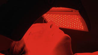
PDT in the treatment of acne is based on the fact that Propionibacterium acnes contain endogenous porphyrins, in particular coproporphyrin III (Babilas et al. 2005). The red light module is used in addition to blue light phototherapy to promote new tissue growth, enhance healing, and stimulate collagen, thereby reducing lines and wrinkles (Fig. 1).
The beneficial effect of red light on wound healing can be explained by considering several basic biological mechanisms including the induction of expression of cytokines and growth factors such as VEGF responsible for the neovascularization necessary for wound healing (Hamblin and Demidova 2006).
Near-infrared light (IR), also known as monochromatic infrared energy, is believed to stimulate circulation by inducing the release of guanylate cyclase and nitric oxide, which, in turn, promotes vasodilation and growth factor production as well as angiogenesis, leading to subsequent wound healing (Karu 1987; Opel et al. 2015) (Fig. 2).
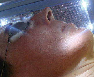
IR LED constitutes the wave band longer than 760 nm and can penetrate the skin between 5 and 10 mm. Near-infrared light 830 nm has been used to treat wounds, pain, ulcers, recalcitrant lesions, skin rejuvenation, and acne vulgaris (Opel et al. 2015).
A single treatment of the normal skin with 830 nm LED-LLLT compared with unirradiated controls demonstrated rapid photomediated degranulation of mast cells at 48 h, accompanied by an ultrastructurally demonstrated inflammatory response with the appearance of significantly greater numbers of mast cells, macrophages, and neutrophils (Calderhead et al. 2008).
LED irradiation with a combination of wavelengths, such as 630 or 660 nm combined with 830 or 850 nm, increased cell number and type I collagen expression more than treatment with each wavelength alone and decreased MMP-1 expression (Tian et al. 2012).
Lee et al. compared the effects among LED treatment with 633 and 830 nm on their own and the combination of 830 and 633 nm with a control group. Although all the LED-treated groups in this study showed statistically significant gross and histological results compared with the controls, the 830 nm group proved superior to the other LED groups in all aspects including collagenesis, skin elasticity, expression of tissue inhibitors of matrix metalloproteinase 1, and subjective patient satisfaction (Lee et al. 2007a).
Blue light (wavelength range from 400 to 480 nm) is UV-free irradiation and appears to exert its effect on acne via its influence on Propionibacterium acnes and its anti-inflammatory properties (Fig. 3).
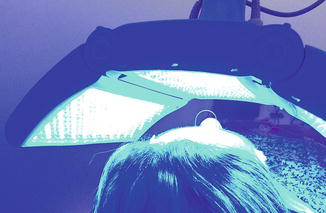
P. acnes contains naturally occurring porphyrins, mainly coproporphyrin and protoporphyrin IX. Absorption of blue light by these molecules is believed to induce a natural PDT effect with destruction of the bacteria via the formation of oxygen free radicals (Dai et al. 2012; Opel et al. 2015).
One mechanism of action of phototherapy for acne is through the absorption of light (specifically blue light) by porphyrins that have been produced by P. acnes as a part of its normal metabolism and that act as endogenous photosensitizers (Lee et al. 2007a; Avci et al. 2013). Because of the effectiveness in reducing cell proliferation, blue light is also propitious to treat hyperplastic diseases and chronic skin inflammation.
The use of a dual-wavelength (red and blue) LED light source enhances PDT results for acne and other sebaceous disorders. Red wavelength (630 nm) can reach the sebaceous glands, and blue (405 nm) light photobleaches any residual protoporphyrin IX (PpIX) in the epidermis, thereby reducing posttreatment photosensitivity (Barolet 2008).
Topical PDT is also a treatment for nonmelanoma skin cancer (NMSC) and the improvement of photoaging. PDT typically involves the application of a topical photosensitizer such as 5-aminolevulinic acid (ALA) or its methyl ester (MAL), which is activated by exposure to a visible light source. As a consequence of the combination of light, photosensitizer, and tissue oxygen, cytotoxic ROS are formed in diseased tissues, inducing necrosis and apoptosis of the malignant and premalignant cells (Gold et al. 2006; Szeimies et al. 2012).
A number of studies have indicated that exposing patients to a combination of LED wavelengths is more effective than monotherapy. This synergistic effect has been investigated on a variety of skin disorders (Russell et al. 2005; Goldberg et al. 2006; Goldberg and Russel 2006; Lee et al. 2007a; Park et al. 2014).
Indications
Review of the literature revealed that differing wavelengths of light-emitting diode devices used in dermatology have many beneficial effects to treat a wide range of skin diseases (Table 1), including acne, scars, and photodamaged skin, and side effects were either mild or not reported. It remains prudent to screen individuals with photosensitive dermatoses, or those taking photosensitizing medications, as these are contraindications to treatment. Caution must be emphasized especially for epileptic and photophobic patients, especially if LEDs are pulsed (Barolet 2008; Opel et al. 2015).
| Wound healing |
| Photorejuvenation |
| Hypertrophic scars and keloids |
| Acne |
| Post-inflammatory hyperpigmentation |
| Erythema |
| Edema |
| Analgesia |
| Burn |
| Alopecia |
| Photodynamic therapy treatment (actinic keratosis, Bowen’s disease, and basal cell carcinoma) |
| Vitiligo |
| Psoriasis |
| Rosacea |
| Herpes simplex |
Wavelengths in the 600–700 nm range are chosen for treating superficial tissue, and wavelengths between 780 and 950 nm are chosen for deeper-seated tissues due to longer optical penetration distances through tissue. Because of the possible existence of a biphasic dose-response curve referred to above, choosing the correct dosage of light (in terms of energy density) for any specific medical condition is difficult (Hamblin and Demidova 2006) (Table 2).
| Color | Wavelength (nm) | Skin treatment | Treatment time |
|---|---|---|---|
| Red | 610–760 | Skin rejuvenation, fine lines, roughness, skin tone, texture, pore size, depigmentation, wound healing, hypertrophic scars, keloids, edema, and erythema | 20–30 min per session; two sessions per week |
| Blue | 450–500 | Acne, hyperpigmentation, keloids, fibrotic skin diseases | 20–30 min per session; two sessions per week |
| Infrared | 850–940 | Skin rejuvenation, hypertrophic scars, keloids | 20–30 min per session; two sessions per week |
LED for Acne, Scars and Photodamaged Skin
Acne
Phototherapy with visible light (mainly blue light, red light, or a combination of both) has been proposed as an alternative therapeutic modality to treat acne vulgaris. One mechanism of action of phototherapy for acne is through the absorption of light (specifically blue light) by porphyrins that have been produced by P. acnes as a part of its normal metabolism and that act as endogenous photosensitizers (Avci et al. 2013).
Blue light therapy (415 nm) is effective at activating coproporphyrin III and protoporphyrin IX, subsequently destroying the P. acnes bacteria (Fig. 4). It has been shown to significantly reduce acne lesions in studies on mild-to-moderate, inflammatory, and pustular acne when irradiating over eight to ten treatments (Goldberg and Russel 2006).
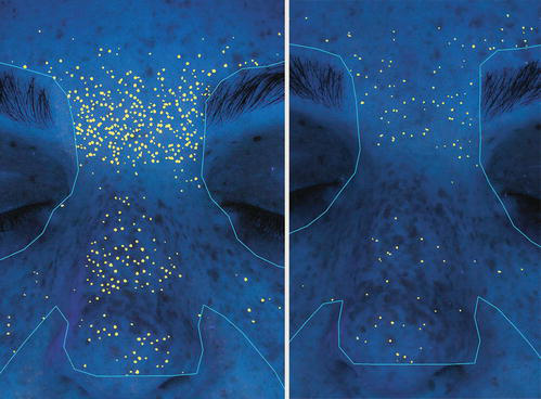
Blue light treatment also appears to have anti-inflammatory effects on keratinocytes by decreasing the cytokine-induced production of IL-1 alpha and ICAM-1 markers (Shnitkind et al. 2006). Blue LED light (400–470 nm) has a maximal penetration of up to 1 mm. It is best suited for the treatment of more superficial conditions, such as to target P. acnes in acne vulgaris (Opel et al. 2015). Blue light applied to P. acnes colonies induces photoexcitation of bacterial porphyrins, stimulates the production of singlet oxygen, and, finally, results in the endogenous photodynamic destruction of P. acnes (Lee et al. 2007b; Joo et al. 2012).
Shalita et al. demonstrated the use of blue light (405–420 nm) for acne treatment, using 10-min light exposures twice weekly. A total of 35 subjects with lesions on the face and back were treated over a 4-week period; 80% demonstrated a significant improvement of noninflammatory, inflammatory, and total facial lesions, with a 70% mean decrease in inflammatory lesion count 2 weeks after the last treatment (Shalita et al. 2001).
Morton et al. treated 30 patients with mild-to-moderate acne with 8-, 10-, or 20-min blue LED (415 nm) treatments over a period of 4 weeks. The inflammatory lesion counts decreased, with minimal effect on noninflammatory lesions (Morton et al. 2005).
Tremblay et al. gave patients with mild-to-moderate inflammatory acne two 20-min treatments of blue LED (415 nm) per week for 4–8 weeks. Ninety percent of patients were satisfied with the result. Objectively, patients had a 50% reduction in lesion counts (Tremblay et al. 2006).
The blue light treatment is associated with significant reductions in lesion size and number, severity, and redness of flare-ups and improvements in the skin’s overall appearance, as well as in clarity, radiance, tone, texture, and smoothness (Ash et al. 2015; Opel et al. 2015).
Red light (633 nm) is less effective at activating coproporphyrin III than blue light but is a potent activator of protoporphyrin IX, also found in P. acnes bacteria. Since red light penetrates deeper into the tissue than blue, it is possible that red light actively destroys P. acnes bacteria residing in the lower regions of the sebaceous gland (Goldberg and Russel 2006).
Red light is believed to stimulate cytokine release from various cells including macrophages and reduce inflammation. The effect of visible red light on the local vasculature is also well recognized. The red light brings more oxygen and nutrients into the area, further helping to reduce inflammation and enhance the wound repair process (Goldberg and Russel 2006; Sadick 2008; Avci et al. 2013; Sawhney and Hamblin 2014).
The combination of LED for the treatment of acne is also promising. Phototherapy with mixed blue–red light, probably by combining antibacterial and anti-inflammatory action, is a safe, effective, and non-painful treatment for mild-to-moderate acne vulgaris, with no significant short-term adverse effects (Avci et al. 2013; Opel et al. 2015).
Karu demonstrated that when Propionibacterium acnes was exposed to blue and red light simultaneously in vivo, there was a marked inhibition of cell activity compared to that seen when red and blue lights were delivered independently (Karu 1999).
Lee et al. treated patients with moderate acne with a combination of blue (415 nm) and red (640 nm) LED devices. A 34% improvement in comedone count and a 78% improvement in the number of inflammatory lesions were observed. Most studies revealed that improvement in inflammatory lesions was higher than the improvement in comedones (Lee et al. 2007b). Kwon et al. demonstrated a decrease of both inflammatory and noninflammatory acne lesions by 77% and 54%, respectively, following home-use combined blue and red LED (Kwon et al. 2013).
Although blue light has been tried in conjunction with ALA in the treatment of acne in 20 patients, patients experienced greater side effects, and the results were not clinically significant when compared with blue LED alone (Weinstabl et al. 2011; Opel et al. 2015).
Red light can prevent or treat acne post-inflammatory hyperpigmentation (PIH). On the basis of photographic analysis and melanin content measurements, most patients can achieve substantial reduction or absence of PIH lesions in the LED-treated areas (Barolet 2008). Red light therapy is generally performed using wavelengths from the visible spectrum, 670 nm, which is a wavelength absorbed by melanin. The amount of light energy being absorbed by pigments in the skin, such as melanin, should be taken into account when designing a light therapy treatment regimen for a patient. In order to achieve the same energy delivery to structures below the epidermis, patients with skin types in the range of 5 or 6 on the Fitzpatrick scale will require a higher dose of light (fluence) than patients with skin types in the range of 1 or 2 (Brondon et al. 2007).
Various LED wavelengths can affect melanogenesis in normal human melanocytes. LED irradiation at 830, 850, and 940 nm reduced melanogenesis through decreased tyrosinase expression without exerting any cytotoxic effects. The LED wavelength at 830 nm also reduces melanin synthesis and might be a helpful therapeutic tool for treating patients with hyperpigmentation (Kim et al. 2012).
Papageorgiou et al. investigated the effects of a combination blue and red light treatment in a randomized study of 107 patients with mild-to-moderate acne. Results displayed a 76% reduction in inflammatory lesions in the combination group. This result was significantly superior to that achieved by blue light alone (Papageorgiou et al. 2000).
Blue and red light combination LED phototherapy is an effective, safe, and non-painful treatment for mild-to-moderately severe acne vulgaris, particularly for papulopustular acne lesions (Lee et al. 2007b).
Scars
Scars, hypertrophic scars, and keloids are cosmetically and psychosocially disfiguring and may result in patients seeking a variety of treatments to address the aesthetic and functional concerns attributed to scarring. Hypertrophic scars and keloids can form after surgery and trauma and are characterized by fibroblastic proliferation and excess collagen deposition (Uitto and Kouba 2000; Lev-Tov et al. 2012).
Light-emitting diode devices have been shown to stimulate fibroblast activity and hasten wound healing. Studies have revealed the possible benefit of red and infrared low-level light therapies to improve scars. It has been reported that collagen synthesis is reduced, and interstitial matrix metalloproteinases (MMP-1), the collagenase involved in normal turnover of skin collagen, are upregulated in aged skin (Brondon et al. 2007).
It has been proposed that interleukin (IL)-6 signaling pathways play a central role in this process and thus that IL-6 pathway inhibition could be a promising therapeutic target for scar prevention. As LED therapy has been shown to decrease IL-6 mRNA levels, it may potentially be preventing aberrant healing (Ghazizadeh et al. 2007; Uitto 2007; Barolet 2008).
LED phototherapy using near-infrared 805 nm light is a method to prevent or attenuate the development of hypertrophic scars or keloids in patients that underwent surgical excision or CO2 laser ablation of keloids or hypertrophic scars (Mamalis et al. 2014).
In vitro studies demonstrate that LED phototherapy, at both red and near-infrared wavelengths, can suppress fibroblast proliferation and may provide a mechanistic foundation for future treatment of keloids (Barolet and Boucher 2010) (Fig. 5).
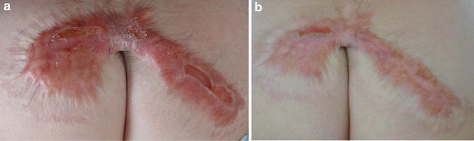
Scars treated for 15 min daily for 30 days with an infrared LED device (805 nm at 30 mW/cm2) showed significant improvement with no associated side effects, as evidenced by improvements in VSS score, measurement of scar height by quantitative skin topography, and blinded clinical assessment of photographs (Mamalis et al. 2014).
A recent study has demonstrated that light-emitting diode blue can inhibit adult human skin dermal fibroblast proliferation and migration speed and is associated with increased reactive oxygen species generation in a dose-dependent manner without altering viability. LED blue has the potential to contribute to the treatment of keloids and other fibrotic skin diseases (Mamalis et al. 2015).
The potential of PDT on scar tissue has been previously investigated. In vitro studies have shown that ALA-PDT induced collagen-degrading matrix metalloproteinase (MMP)-1 and MMP-3 in dermal fibroblasts while reducing collagen type 1 mRNA expression (Karrer et al. 2003). A retrospective blinded study by Sakamoto et al. found aminolevulinic acid (ALA) or methyl ester aminolevulinic acid (MAL) combined with red LED to statistically improve scar appearance after two or more treatments (Sakamoto et al. 2012).
Nie et al. reported a positive effect of PDT in a patient with a persistent keloid which had not responded to a number of routine therapies. Following five MAL-PDT sessions, scar color had improved, and the keloid had reduced in size, become flatter, with reduced erythema in the surrounding margin (Nie et al. 2010).
PDT can reduce scar formation in keloids, as evidenced by lack of recurrence and improvement in signs and symptoms as well as by decreased blood flow, increased pliability, and decreased collagen and hemoglobin levels (Ud-Din et al. 2013).
Photodamaged Skin
The use of LEDs for skin rejuvenation is unique in that it does not produce any thermal damage. It is proposed that specific LED light wavelengths are absorbed in the skin and used to modulate cell function, proliferation, and repair in sun-damaged tissue, in a process termed photobiomodulation (Baez and Reilly 2007).
Photobiomodulation is considered to stimulate growth factor production (i.e., fibroblast growth factor, TGF, PDGF), extracellular matrix production, and increased synthesis of collagen and procollagen and enhances cutaneous microcirculation through activating the mitochondrial respiratory system of the cells (Lee et al. 2007a).
A number of clinical studies provide evidence of the effectiveness of LED therapy in photorejuvenation using a variety of LED light sources. LEDs have been shown to improve UV-damaged skin conditions, including photoaging, suggesting that the mechanism of action between LEDs and UV radiation is different (Kim et al. 2012).
Trelles has suggested that LED therapy represents a potential approach in antiaging prevention. The prevention can be achieved via irradiating low-level photoenergy with specific wavelengths that, based on the photobiological findings, can stimulate both epidermal and dermal cells (Trelles 2006).
Recent studies suggest that LLLT (specifically red or NIR radiation) may provide effective protection against UV-induced photodamage. This is believed to be due to the fact that, earlier or during the day (morning time), red/NIR wavelengths of the solar spectrum predominate and prepare the skin for the potentially harmful UV radiation that predominates later on in the day (noon/afternoon) (Barolet 2008; Sawhney and Hamblin 2014).
Irradiation with red light increased fibroblastic growth factor synthesis from photoactivated macrophages and accelerated mast cell degeneration (Lam et al. 1986).
The induction of collagen synthesis by LED in the red spectrum has been shown to occur largely in the papillary dermis. Barolet et al. showed that LED therapy reversed collagen downregulation and MMP-1 upregulation. This could explain the improvements in skin appearance observed in LED-treated individuals. These findings suggest that LED at 660 nm is a safe and effective collagen-enhancement strategy (Barolet et al. 2009).
The red light is used to increase new tissue growth, enhance healing, and stimulate collagen, thereby reducing lines and wrinkles. It also has been used to improve skin roughness, depth of rhytids, skin tone, texture, pore size, dyspigmentation, edema, and erythema (Sauder 2010).
A split-face study of red LED (633 nm) in patients who had undergone blepharoplasty and periocular resurfacing demonstrated a statistically significant improvement of edema, erythema, bruising, and pain on the treated side of the face (Opel et al. 2015).
Light at 830 nm (near-infrared) wavelength is absorbed in the cellular membrane rather than in cellular organelles, which remain the target when using light in the visible spectrum. Irradiation at 830 nm has accelerated fibroblast–myofibroblast transformation and mast cell degranulation. In addition, chemotaxis and phagocytic activity of leucocytes and macrophages are enhanced on cellular stimulation by this wavelength (Russell et al. 2005).
Infrared wavelength phototherapy with an LED at 830 nm has anti-inflammatory effects and is useful for the regeneration of damaged skin. The wavelength of 830 nm is well known to dramatically increase the action potentials of wound-healing cells, particularly those in the inflammatory and remodeling stages, and would therefore cause a considerably faster resolution of post-laser adverse effects such as erythema and pain (Russell et al. 2005; Trelles 2006).
Previous work has been published on the combination use of 633 and 830 nm LED therapy in the treatment of photoaged skin. Red (633 nm) light has been shown to increase fibroblast growth factor and collagen synthesis in the skin (Baez and Reilly 2007).
The 830 nm wavelength is well associated with photobiomodulation of the wound-healing cells: the mast cells, neutrophils, and macrophages. The subsequent doses of 633 nm concentrate on the fibroblasts but maintain the reaction level in the other cells. Both wavelengths are well associated with increases in local blood flow rate and volume, and 830 nm also stimulates the transitional remodeling phase (Trelles 2006).
The synergy of 633 and 830 nm wavelength lights combines these effects to enhance fibroblast proliferation and thus increase collagen synthesis, as well as stimulating inflammatory cell lines such as mast cells and macrophages (Calderhead et al. 2015).
Goldberg et al. investigated the combination of red (633 nm) and IR (830 nm) LED treatment on photodamaged skin and reported softening of periorbital wrinkles in 80% of subjects. There was subjective improvement of softness, smoothness, and firmness. Histologic examination demonstrated increased number and thickness of collagen fibrils (Goldberg et al. 2006).
An improvement in skin appearance in aged/photoaged individuals has been documented after full-face or split-face serial treatments with red (630, 633 nm) or red in combination with infrared (830 nm) light (Barolet et al. 2009).
Previous findings were able to correlate fibroblast activity and dermal matrix remodeling processes, with an increase in intradermal collagen density and reduced signs of aging (Lee et al. 2007a). The proposed underlying mechanisms include the photostimulation of terminal molecules in the electron transport chain and the subsequent adenosine triphosphate (ATP) concentration increase, along with the selective light-driven activation of water molecules, thereby enhancing metabolic exchange and influencing the ion transporter systems found in cellular membranes (Wunsch and Matuschka 2014).
A prospective, placebo-controlled, double-blind, split-face trial by Lee et al. randomized patients with facial rhytids to receive red LED (640 nm), IR (830 nm), both, or sham treatments. Patients demonstrated a statistically significant reduction in wrinkle severity across all treatment groups: 26%, 33%, and 36%, respectively. Skin elasticity also improved. Tissue assays were notable for an increase in collagen and elastic fibers adjacent to highly active fibroblasts (Lee et al. 2007a).
A study performed with a light device combining narrowband blue light (420 nm) and near infrared (850–890 nm), using 20 min exposure and a total dose of 60 J/cm2, has demonstrated a dramatic improvement in skin photorejuvenation when associated with glycolic acid peels and topical vitamin C (Fournier et al. 2006). LED treatments work well after any procedure that causes erythema and irritation, including chemical peels or ablative laser systems, with extremely high satisfaction levels.
The combination of LED light wavelengths was shown in in vitro results to improve cell shape homogenization, cell proliferation, and the level of major proteins involved in the healing process (Chabert et al. 2015). The LED system is known for its healing and anti-inflammatory properties and also for enhancing the results of facial cosmetic procedures.
Conclusion
LED phototherapy is a non-ablative, nonthermal, and nontraumatic treatment, which stimulates cell activities and functions through a photobiomodulative effect.
Light therapy can be a safe and effective alternative for treating acne, scars, and photodamaged skin and a variety of medical treatments, with no adverse effects or downtime reported during or after LED treatment.
Take Home Messages
- LED is non-ablative and nonthermal, making it safe for all skin types.
- The treatment is hands-free with no need for operator presence.
- The infrared spectrum is invisible to the eye.
- Eye protection is mandatory, both for the physician and the patient.
- LED can be used as an adjunctive treatment to all laser procedures.
- LED therapy helps wound healing after surgery.
- The results are better when LED is used in combination.
- LED therapy is an effective, non-painful, and no-downtime treatment modality.
References
Anderson RR, Parrish JA. The optics of human skin. J Invest Dermatol. 1981;77:13–9.
Ash C, Harrison A, Drew S, Whittall R. A randomized controlled study for the treatment of acne vulgaris using high-intensity 414 nm solid state diode arrays. J Cosmet Laser Ther. 2015;17(4):170–6.
Avci P, Gupta GK, Clark J, Wikonkal N, Hamblin MR. Low-level laser (light) therapy (LLLT) for treatment of hair loss. Lasers Surg Med. 2013;9999:1.
Babilas P, Karrer S, Sidoroff A, Landthaler M, Szeimies RM. Photodynamic therapy in dermatology – an update. Photodermatol Photoimmunol Photomed. 2005;21(3):142–9.
Baez F, Reilly LR. The use of light emitting diode therapy in the treatment of photoaged skin. J Cosmet Dermatol. 2007;6(3):189–94.
Barolet D. Light-emitting diodes (LEDs) in dermatology. Semin Cutan Med Surg. 2008;27:227–38.
Barolet D, Boucher A. Prophylactic low-level light therapy for the treatment of hypertrophic scars and keloids: a case series. Lasers Surg Med. 2010;42(6):597–601.
Barolet D, Roberge CJ, Auger FA, Boucher A, Germain L. Regulation of skin collagen metabolism in vitro using a pulsed 660 nm LED light source: clinical correlation with a single-blinded study. J Invest Dermatol. 2009;129:2751–9.
Brondon P, Stadler I, Lanzafame RJ. Melanin density affects photobiomodulation outcomes in cell culture. Photomed Laser Surg. 2007;25(3):144–9.
Calderhead RG. The photobiological basics behind light-emitting diode (LED) phototherapy. Laser Ther. 2007;16:97–108.
Calderhead RG, Kubota J, Trelles MA, Ohshiro T. One mechanism behind LED phototherapy for wound healing and skin rejuvenation: key role of the mast cell. Laser Ther. 2008;17:141–8.
Calderhead RG, Kim WS, Ohshiro T, Trelles MA, Vasily D. Adjunctive 830 nm light-emitting diode therapy can improve the results following aesthetic procedures. Laser Ther. 2015;24(4):277–89.
Chabert R, Fouque L, Pinacolo S, Garcia Gimenez N, Bonnans M, Cucumel K, Domloge N. Evaluation of light-emitting diodes (LED) effect on skin biology (in vitro study). Skin Res Technol. 2015;21(4):426–36.
Dai T, Gupta A, Murray CK, Vrahas MS, Tegos GP, Hamblin MR. Blue light for infectious diseases: Propionibacterium acnes, Helicobacter pylori, and beyond? Drug Resist Updat. 2012;15(4):223–36.
Dincer I. Renewable energy and sustainable development: a crucial review. Renew Sust Energ Rev. 2000;4(2):157–75.
Evans DH, Abrahamse H. Efficacy of three different laser wavelengths for in vitro wound healing. Photodermatol Photoimmunol Photomed. 2008;24(4):199–210.
Fournier N, Fritz K, Mordon S. Use of nonthermal blue (405- to 420-nm) and near-infrared light (850- to 900-nm) dual-wavelength system in combination with glycolic acid peels and topical vitamin C for skin photorejuvenation. Dermatol Surg. 2006;32(9):1140–6.
Ghazizadeh M, Tosa M, Shimizu H, et al. Functional implications of the IL-6 signaling pathway in keloid pathogenesis. J Invest Dermatol. 2007;127:98–110.
Gold MH, Bradshaw VL, Boring MM, et al. Split face comparison of photodynamic therapy with 5-aminolevulinic acid and intense pulsed light versus intense pulsed light alone for photodamage. Dermatol Surg. 2006;32:795–803.
Goldberg DJ, Russel BA. Combination blue (415 nm) and red (633 nm) LED phototherapy in the treatment of mild to severe acne vulgaris. J Cosmet Laser Ther. 2006;8:71–5.
Goldberg DJ, Amin S, Russell BA, Phelps R, et al. Combined 633-nm and 830-nm LED treatment of photoaging skin. J Drugs Dermatol. 2006;5:748–53.
Hamblin MR, Demidova TN. Mechanisms for Low-Light Therapy. In: Hamblin MR, Waynant R, Anders J (eds), Proceedings of the SPIE. 2006;6140:1–12.
Hamblin MR, Pires de Sousa MV, Arany PR, Carroll JD, Patthoff D. Low-level laser (light) therapy and photobiomodulation: the path forward. Proc SPIE. 2015;9309:930902.
Huang YY, Sharma SK, Carroll J, Hamblin MR. Biphasic dose response in low level light therapy – an update. Dose-Response. 2011;9:602–18.
Issa MCA, Piñeiro-Maceira J, et al. Immunohistochemical expression of matrix metalloproteinases in photodamaged skin by photodynamic therapy. Br J Dermatol. 2009;161:647–53.
Joo Y, Kang H, Choi EH, Nelson JS, Jung B. Characterization of a new acne vulgaris treatment device combining light and thermal treatment methods. Skin Res Technol. 2012;18(1):15–21.
Karrer S, Bosserhoff A, Weiderer P, Landthaler M, Szeimies RM. Influence of 5-aminolevulinic acid and red light on collagen metabolism of human dermal fibroblasts. J Invest Dermatol. 2003;120:325–31.
Karrer S, Kohl E, Feise K, et al. Photodynamic therapy for skin rejuvenation: review and summary of the literature – results of a consensus conference of an expert group for aesthetic photodynamic therapy. J Dtsch Dermatol Ges. 2013;11(2):137–48.
Karu TI. Photobiological fundamentals of low-power laser therapy. J Quantum Electron. 1987;23:1703–17.
Karu T. Primary and secondary mechanisms of action of visible to near-IR radiation on cells. J Photochem Photobiol B. 1999;49:1–17.
Karu T. Identification of the photoreceptors. In: Ten lectures on basic science of laser phototherapy. Grangesberg: Prima Books AB; 2007.
Karu TI, Pyatibrat LV, Afanasyeva NI. Cellular effects of low power laser therapy can be mediated by nitric oxide. Lasers Surg Med. 2005;36:307–14.
Kim JM, Kim NH, Tian YS, Lee AY. Light-emitting diodes at 830 and 850 nm inhibit melanin synthesis in vitro. Acta Derm Venereol. 2012;92(6):674–9.
Kwon HH, Lee JB, Yoon JY, et al. The clinical and histological effect of home-use, combination blue-red LED phototherapy for mild-to-moderate acne vulgaris in Korean patients: a double-blind, randomized controlled trial. Br J Dermatol. 2013;168:1088–94.
Lam TS, Abergel RP, Meeker CA, Castel JC, Dwyer RM, Uitto J. Laser stimulation of collagen synthesis in human skin fibroblast cultures. Lasers Life Sci. 1986;1(1):61–77.
Lee SY, Park KH, Choi JW, Kwon JK, Lee DR, Shin MS, et al. A prospective, randomized, placebo-controlled, double-blinded, and split-face clinical study on LED phototherapy for skin rejuvenation: clinical, profilometric, histologic, ultrastructural, and biochemical evaluations and comparison of three different treatment settings. J Photochem Photobiol B Biol. 2007a;88(1):51–67.
Lee SY, You CE, Park MY. Blue and red light combination LED phototherapy for acne vulgaris in patients with skin phototype IV. Lasers Surg Med. 2007b;39:180–8.
Lev-Tov H, Brody N, Siegel D, Jagdeo J. Inhibition of fibroblast proliferation in vitro using low-level infrared light-emitting diodes. Dermatol Surg. 2012;39:422–5.
Mahmoud BH, Hexsel CL, Hamzavi IH, Lim HW. Effects of visible light on the skin. Photochem Photobiol. 2008;84(2):450–62.
Mamalis AD, Lev-Tov H, Nguyen DH, Jagdeo JR. Laser and light-based treatment of keloids – a review. J Eur Acad Dermatol Venereol. 2014;28(6):689–99.
Mamalis A, Garcha M, Jagdeo J. Light emitting diode-generated blue light modulates fibrosis characteristics: fibroblast proliferation, migration speed, and reactive oxygen species generation. Lasers Surg Med. 2015;47:210–5.
Mester E, Ludany G, Sellyei M, Szende B, Gyenes G, Tota GJ. Studies on the inhibiting and activating effects of laser beams. Langenbecks Arch Chir. 1968;322:1022–7.
Morton CA, Brown SB, Collins S, et al. Guidelines for topical photodynamic therapy: report of a workshop of the British photodermatology group. Br J Dermatol. 2002;146:552–67.
Morton CA, Scholefield RD, Whitehurst C, Birch J. An open study to determine the efficacy of blue light in the treatment of mild to moderate acne. J Dermatol Treat. 2005;16:219–23.
Nie Z, Bayat A, Behzad F, Rhodes L. Positive response of a recurrent keloid scar to topical methyl aminolevulinate-photodynamic therapy. Photodermatol Photoimmunol Photomed. 2010;26:330–2.
Opel DR, Hagstrom E, Pace AK, Sisto K, Hirano-Ali SA, Desai S, Swan J. Light-emitting diodes: a brief review and clinical experience. J Clin Aesthet Dermatol. 2015;8(6):36.
Papageorgiou P, Katsambas A, Chu A. Phototherapy with blue (415 nm) and red (660 nm) light in the treatment of acne vulgaris. Br J Dermatol. 2000;142:973–8.
Park KY, Choi SY, Mun SK, Kim BJ, Kim MN. Combined treatment with 578/511 nm copper bromide laser and light-emitting diodes for postlaser pigmentation: a report of two cases. Dermatol Ther. 2014;27(2):121–5.
Russell BA, Kellett N, Reilly LR. A study to determine the efficacy of combination LED light therapy (633 nm and 830 nm) in facial skin rejuvenation. J Cosmet Laser Ther. 2005;7(3–4):196–200.
Sadick NS. Handheld LED array device in the treatment of acne vulgaris. J Drugs Dermatol. 2008;7:347–50.
Sakamoto FH, Izikson L, Tannous Z, et al. Surgical scar remodeling after photodynamic therapy using aminolevulinic acid or its methyl ester: a retrospective, blinded study of patients with field cancerization. Br J Dermatol. 2012;166:413–6.
Sauder DN. Light-emitting diodes: their role in skin rejuvenation. Int J Dermatol. 2010;49(1):12–6.
Sawhney MK, Hamblin MR. Low-level light therapy (LLLT) for cosmetics and dermatology. Proc SPIE 8932, Mechanisms for Low-Light Therapy IX, 89320X (February 18, 2014).
Shalita AR, Harth Y, Elman M, Slatkine M, Talpalariu G, Rosenberg Y, Korman A, Klein A. Acne phototherapy using UV-free high intensity narrow band blue light: a three-center clinical study. Proc SPIE. 2001;4244:61–73.
Shnitkind E, Yaping E, Geen S, Shalita AR, Lee WL. Antiinflammatory properties of narrow-band blue light. J Drugs Dermatol. 2006;5:605–10.
Szeimies RM, Torezan L, Niwa A, Valente N, Unger P, Kohl E, et al. Clinical, histopathological and immunohistochemical assessment of human skin field cancerization before and after photodynamic therapy. Br J Dermatol. 2012;167(1):150–9.
Tian YS, Kim NH, Lee AY. Antiphotoaging effects of light-emitting diode irradiation on narrow-band ultraviolet B–exposed cultured human skin cells. Dermatol Surg. 2012;38(10):1695–703.
Trelles MA. Phototherapy in anti-aging and its photobiologic basics: a new approach to skin rejuvenation. J Cosmet Dermatol. 2006;5:87–91.
Tremblay JF, Sire DJ, Lowe NJ, Moy RL. Light-emitting diode 415 nm in the treatment of inflammatory acne: an open-label, multicentric, pilot investigation. J Cosmet Laser Ther. 2006;8:31–3.
Ud-Din S, Thomas G, Morris J, Bayat A. Photodynamic therapy: an innovative approach to the treatment of keloid disease evaluated using subjective and objective non-invasive tools. Arch Dermatol Res. 2013;305(3):205–14.
Uitto J. IL-6 signaling pathway in keloids: a target for pharmacologic intervention? J Invest Dermatol. 2007;127:6–8.
Uitto J, Kouba D. Cytokine modulation of extracellular matrix gene expression: relevance to fibrotic skin diseases. J Dermatol Sci. 2000;24(suppl):S60–9.
Vecchio D, Pam Z, Pam N, Hamblin MR. Low-level laser (light) therapy (LLLT) in skin: stimulating, healing, restoring. Semin Cutan Med Surg. 2013;32:41–52.
Weinstabl A, Hoff-Lesch S, Merk HF, von Felbert V. Prospective randomized study on the efficacy of blue light in the treatment of psoriasis vulgaris. Dermatology. 2011;223:251–9.
Weiss RA, McDaniel DH, Geronemus RG, et al. Clinical experience with light-emitting diode (LED) photomodulation. Dermatol Surg. 2005;31:1199–205.
Whelan HT, Houle JM, Whelan NT, et al. The NASA light-emitting diode medical program: progress in space flight and terrestrial applications. Space Technol Appl Intell Forum. 2000;504:37–43.
Whelan HT, Buchmann EV, Dhokalia A, Kane MP, Whelan NT, Wong-Riley MTT, et al. Effect of NASA light-emitting diode irradiation on molecular changes for wound healing in diabetic mice. J Clin Laser Med Surg. 2003;21(April):67–74.
Wunsch A, Matuschka KA. Controlled trial to determine the efficacy of red and near-infrared light treatment in patient satisfaction, reduction of fine lines, wrinkles, skin roughness, and intradermal collagen density increase. Photomed Laser Surg. 2014;32(2):93–100.
Zhang Y, Song S, Fong CC, et al. cDNA microarray analysis of gene expression profiles in human fibroblast cells irradiated with red light. J Invest Dermatol. 2003;120:849–57.







