
Laser Treatment of Vascular Lesions
Laser Treatment of Vascular Lesions
Andréia S. Fogaça Dermatology/Medicine, University Santo Amaro (UNISA), São Paulo, São Paulo, Brazil
Abstract
Laser surgery has become the treatment of choice for many vascular lesions. The most common indications are vascular anomalies including port-wine stain and hemangiomas, as well as facial erythema and telangiectasias. In this chapter, we are going to approach different types of lasers and their indications, results, and side effects.
Keywords Vascular lesionsOxyhemoglobinDeoxyhemoglobinMethemoglobinPulsed dye laserKTP laserNear-infrared radiation
Introduction
One of the first indications of lasers in dermatology was the removal of vascular lesions, using the theory of selective photothermolysis, introduced by Anderson and Parrish in 1983 (Anderson and Parrish 1983). Three components are necessary for selective photothermolysis: (1) a laser wavelength with preferential absorption of the target chromophore, (2) appropriate pulse duration matched to the target size, and (3) a fluence that both treats the target and minimizes nonspecific thermal-related injury. The classic target chromophore for vascular lesions has been oxyhemoglobin, which has the greatest absorption peaks at 418, 542, and 577 nm. The laser light is absorbed by oxyhemoglobin and converted to heat, which is transferred to the vessel wall causing coagulation and vessel closure. Other hemoglobin species have more recently been recognized as appropriate targets, depending on the vascular lesion. They are deoxyhemoglobin and methemoglobin. In the case of deoxyhemoglobin, the greatest absorption peak is 755 nm and it has been used for refractory or hypertrophic PWS (port-wine stain), a venocapillary malformation.
The first lasers to treat vascular lesions were CO2 and argon lasers. The argon laser at 488 and 515 nm enjoyed a high absorption coefficient for hemoglobin (HgB), but the pulse durations (continuous wave) were longer than the thermal relaxation time of the targeted blood vessels, and in the absence of surface/epidermal cooling, the high absorption by epidermal melanin resulted in a high risk of hypopigmentation and scarring.
In 1968, Dr. Leon Goldman and colleagues demonstrated the histopathology of the laser treatment of port-wine lesions (Solomon et al. 1968).
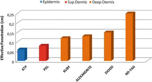
Although Goldman accurately predicted that vascular lesions could be selectively heated, the pulsed dye laser (PDL) was the first laser, in 1986, to show that this selectivity was effective. Initially it was developed at 577 nm to target the yellow absorption peak of oxyhemoglobin. Currently, PDL lasers have shifted to 585 nm and 595 nm, allowing a depth of penetration of approximately 1.16 mm.
In general, vascular laser technologies can be divided into three spectral ranges (Graphic 1):
- Green-yellow (GY) light sources, such as PDL (pulsed dye laser/585, 595 nm) and KTP (potassium titanyl phosphate laser 532 nm);
- the diode laser (800 nm) and alexandrite (755 nm) lasers;
- near-infrared radiation (NIR), lasers with a smaller ratio of melanin to HgB absorption and deeper penetration (940, 980, and 1064 nm).
In summary, smaller lesions in lighter skin are treated with GY light sources, and larger lesions in darker skin are treated with NIR lasers.
Intense pulsed light (IPL) devices emit polychromatic noncoherent broadband light from 420 to 1400 nm with varying pulse durations, can cover all of the HgB peaks, and cannot be “framed” into one of the three aforementioned categories.
There are alternatives to treat vascular lesions, such as ablative lasers (CO2) or photodynamic therapy (PDT).
The microanatomy of the vascular lesion should always be considered.
Laser Parameters
Wavelength
In general, the first parameter to be considered is wavelength, and it should be selected to achieve three goals: (1) to absorb the vascular target (O2 saturation of cutaneous blood ranges from 50% to 80%); the peaks of oxy-HgB absorption are 418, 542, and 577 nm with a smaller peak at 940 nm (Anderson and Parrish 1981a); (2) to avoid the melanin target in darker-skinned patients, it is possible to use the Nd:YAG laser at 1064 nm (melanin absorption is much less than with GY light); (3) to achieve the desirable depth of penetration.
The lasers used to treat vascular lesions have wavelengths that are well absorbed by hemoglobin. However, we should be aware of concurrent absorption by melanin, which is maximal at short wavelengths (Anderson and Parrish 1981b). This may result in unwanted clinical effects such as hypopigmentation, especially when treating persons with dark skin types. As melanin in normally pigmented skin is situated in the epidermis, overheating of the melanosomes may even induce epidermal necrosis, eliciting clinical blistering or in the long term even scarring.
In addition, penetration depth is also mainly determined by wavelength. As a rule, longer wavelengths have less scattering and a larger penetration depth. Theoretically, longer wavelengths may therefore be preferred for deeper and larger vascular lesions such as reticular veins, whereas shorter wavelengths may be suited for superficial vascular lesions such as telangiectasias.
Pulse Duration
The ideal pulse duration should be equal to or only slightly larger than the thermal relaxation time (the time required for the heated tissue to lose about half of its heat). Very short pulses confine heat not only to the vessels but also to the erythrocytes. Accordingly, thermal confinement is excessive, and localized vessel rupture and intravascular thrombosis are observed in the treated area. With longer pulses (6–40 ms), intravascular thrombosis and spot-sized purpura are mitigated, as gentle heating results in vessel wall stenosis and thrombosis of the larger vessels but not of the microvessels that produce widespread purpura (Table 1).
| Diameters (μm) | Tr (ms) |
|---|---|
| 10 | 0.048 |
| 20 | 0.19 |
| 50 | 1.2 |
| 100 | 4.8 |
| 200 | 19.0 |
| 300 | 42.6 |
Spot Size
Larger spots increase the number of photons that penetrate deeper into the dermis, compared with smaller spots. However, with the same fluence, a larger spot will produce more epidermal damage and pain. The rule is that larger spots should be accompanied by lower fluences than their small-spot counterparts.
Surface Cooling
Epidermal cooling was introduced in the 1990s to protect the epidermis, minimizing pigmentary changes and epidermal necrosis. In the treatment of all vascular lesions, cooling of the skin surface is crucial to minimize epidermal damage, to allow high radiant exposure, and to minimize the discomfort associated with treatment (Nelson et al. 1995). In summary, there are three types of surface cooling:
- Cryogen spray (nitrogen or tetrafluoroethane): a significant reduction in pain score can be achieved when adequate dynamic cooling is used (on the order of tens of milliseconds). Disadvantages of the spray are the possibility of pigmentation changes (excessive cooling) and the need for purchasing cryogen canisters.
- Contact cooling (sapphire windows or copper plate) that is placed on the skin during laser treatment. The main caution is to avoid high compression of the vascular lesion. Risks of contact cooling are fogging and poor contact.
- Refrigerated air cooling (cooling devices) works well but requires a second hand to hold it near the surface or an accessory that allows one-hand operation. Air cooling has the advantage that the device can be used for different procedures and for large areas of the skin. However, caution is mandatory, especially in patients with darker skin types. Post-inflammatory hyperpigmentation due to air cooling has been widely reported (Manuskiatti et al. 2007).
Physiological Influences
Superficial Vessels
Superficial vessels located over the deep vessels work as a shield, reducing the efficacy of the treatment. Therefore, these vessels should be treated first for better results.
Skin Temperature, Vascular Dilatation, and Blood Flow
Low temperature of the skin over the area to be treated requires a slightly higher fluence to reach a good effect (purpuric response). It is also described that high temperature with increased blood flow in the target can improve the treatment effect. On the other hand, vascular dilatation, suction, pressure, or erythema induced by UVB seems to have no influence on clinical response (Paul et al. 1983; Aguilar et al. 2012).
Vascular Lasers and Light Sources
Pulsed Dye Laser (PDL: 577, 585, and 595 nm)
Pulsed dye lasers use a rhodamine dye dissolved in a solvent and pumped by a flashlamp. The PDL was the initial “test” for selective photothermolysis (SPT). The first PDLs were slow (0.5 Hz or less), equipped with only a small-diameter spot size (3–5 mm), lacked cooling, and used a 577 nm wavelength near one of the peaks of oxy-HgB absorption. Over the years from 1981 to 1990, the wavelength was changed to 585 nm. Later, cooling devices were added and the laser wavelength was again increased to 595 nm to further enhance epidermal/vascular penetration.
Pulsed dye lasers have been proven safe and effective in the treatment of a variety of vascular lesions, including port-wine stains (Faurschou et al. 2011). Dierickx et al. identified the ideal pulse duration for PWS treatment to be 1–10 ms. In practice, treatment often begins at 1.5 ms, though this may be adjusted down to 0.45 ms and up to 6 ms. Parameters to consider include a 7–10 mm spot size, pulse duration of 0.45–6 ms, and fluence of 5.5–9.5 J/cm2 with appropriate epidermal cooling. Lower energies are used for larger spot sizes with shorter pulse durations. Longer pulse durations are advisable in darker skin types. Treatment should start at lower energies, and these can be increased if treatment is tolerated well. The fluence is adjusted to achieve the desired end point. For the PDL, the desired end point is immediate purpura. Parameters vary by device.
Proper eye protection is essential, and surgical lubricant may be placed on eyebrows and eyelashes to avoid singeing. Although hair often regrows, permanent hair loss can occur with PDL treatment, given the close proximity of the follicles to the surface.
Potassium Titanyl Phosphate Laser (KTP: 532 nm)
KTP lasers are Nd:YAG lasers with the frequency doubled to 532 nm. This wavelength (532 nm) has high absorption by oxy-HgB and melanin, and therefore should be avoided in darker skin types because of the high risk of hypopigmentation (Fournier et al. 2002). KTP laser penetration is approximately 1 mm, and it is more appropriately used to treat telangiectasias. The KTP laser may be used to treat individual vessels, with the advantage of no purpura.
There are many devices with this wavelength, all equipped with sapphire contact cooling.
Alexandrite Laser (755 nm)
The alexandrite is a long-pulsed near-infrared laser. It was initially used for laser hair removal. There is strong absorption of deoxy-HgB, and overall blood absorption at 755 nm is 2× that of the 1064 nm Nd:YAG laser. However, melanin is highly absorbed at 755 nm; for this reason, it is better indicated in lighter skin types and darker vessels.
There are many alexandrite lasers with long-pulsed capability. Cryogen spray, contact cooling, and refrigerated air are integrated into these devices. The alexandrite laser is typically used for PDL-resistant lesions, though it may be implemented as a first-line treatment for hypertrophic violaceous lesions in adults. The end point is a subtle gray-blue discoloration followed by deeper purpura. Care must be taken not to overlap pulses, as scarring can occur.
Diode Laser (800–983 nm)
Multiple diode lasers are now available, and different systems can emit infrared light at a variety of wavelengths, including 800, 810, 940, and 983 nm. Above 900 nm, absorption by melanin is lower than absorption by oxyhemoglobin. This makes diode lasers a safer treatment option for patients with darker skin types than the alexandrite laser. Like the alexandrite laser, the 810 nm laser is better indicated for deeper vascular lesions in relatively fair-skinned patients (Wall et al. 2007).
Nd:YAG Laser (1064 nm)
The Nd:YAG laser has relatively poor HgB absorption; therefore, this device should be used at higher fluences. These higher fluences necessitate epidermal cooling to achieve epidermal protection. Absorption by melanin at this wavelength is lower than for any other laser type used for vascular procedures. Nd:YAG laser treatment is considered to be especially effective and safe in darker skin types. At 1,064 nm, penetration depth is at its peak, at more than 4 mm. This makes the Nd:YAG laser a suitable treatment modality for deeply located veins (Meesters et al. 2013a).
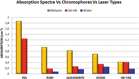
Although the depth of penetration can be increased, there is a narrow therapeutic window with these devices, and caution is advised owing to the risk of scarring. It is recommended that only experienced laser surgeons use these devices.
Intense Pulsed Light (IPL: 500–1200 nm)
IPLs rely on a xenon flashlamp that emits high-intensity, noncoherent, polychromatic broad-spectrum light (500–1200 nm), with varying pulse durations. By appropriate filtering, one can customize the spectrum for specific disorders. For most vascular applications, shorter wavelength ranges are applied. IPL devices are commonly used with the 550 and 570 nm filters to deliver primarily yellow and red light, with a minor component of near-infrared light. The use of IPL for vascular lesions has increased because improvements in power supplies, cooling, and filtering are now available, and IPL devices are safer and more efficient (Ross 2006; Goldman et al. 2005; Goldman and Eckhouse 1996) (Graphic 2).
Vascular Lesions
Facial Telangiectasia and Spider Telangiectasia
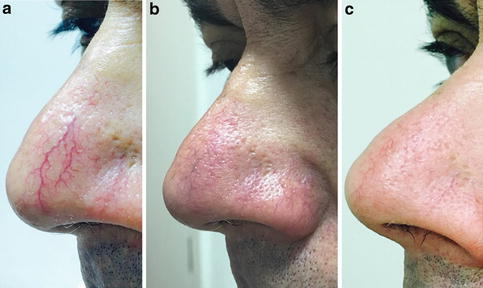
Telangiectasias are small superficial vessels 0.1–1.5 mm in diameter that are commonly associated with sun damage, aging, and/or genetics (Goldman and Bennett 1987). Spider telangiectasia represents telangiectasia with a central feeding arteriole. Both can be treated with PDL, KTP, and IPL. Near-infrared lasers, specifically diode and Nd:YAG, have been used to treat deeper- or larger-caliber vessels (Fig. 1a–c). Laser treatment is efficient and is the gold standard for treating these lesions. The KTP laser produces better results than the PDL laser when used to treat individual vessels, with the advantage of no purpura. There is relatively stronger absorption of hemoglobin at 532 nm, and care must be taken in patients with darker skin. Rule of thumb: for discrete, smaller (0.1–0.6 mm) telangiectasias, PDL, KTP laser, or IPL can be applied; for larger vessels (1 mm or more), the Nd:YAG laser or alexandrite laser is preferred, but care must be taken to apply the smallest fluence and smallest spot sufficient for vessel closure. The end point for treating vessels is vessel clearance, a transient blue coagulum, or purpura (Table 2).
| Infantile hemangiomas | Incidence |
|---|---|
| Superficial hemangiomas | 50% |
| Deep hemangiomas | 15% |
| Combined hemangiomas | 35% |
Vessels around the nasal ala carry more risk of scarring; therefore, in this area cooling is essential and overlapping pulses should be avoided. We use epidermal cooling devices before and after shooting. The advantages of IPL are the relatively large spot sizes, which can minimize the polka-dot effect common to PDL and KTP lasers.
Rosacea
Rosacea is a cutaneous vascular disorder associated with follicular inflammation. Patients with rosacea often have associated background facial erythema. Lasers and IPL are the first-choice treatment for telangiectasias and facial erythema (Fig. 2). A comprehensive review of the literature finds that of the 18 histologic studies on rosacea, 14 showed an increase in Demodex mites (Schmidt and Gans 2004). It is hypothesized that these mites may play a role in the inflammation of rosacea. Studies have demonstrated thermal destruction of these mites after IPL therapy, which may contribute to the therapeutic effects of IPL (Prieto et al. 2002). Usually two to three sessions demonstrate good results; a certain percentage of patients (20%) do not respond to IPL and need to be treated with PDL. PDL provides, like IPL, effective and relatively risk-free results.
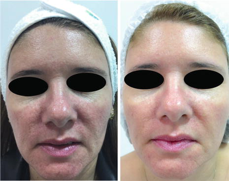
In our experience, two treatments per year is the best method to control rosacea. Aminolevulinic acid photodynamic therapy (ALA-PDT) or methyl aminolevulinate photodynamic therapy (MAL-PDT) has also been reported for resistant rosacea (Baglieri and Scuderi 2011).
Poikiloderma of Civatte
Poikiloderma of Civatte presents in chronically sun-exposed areas, most commonly on the neck, chest, and lateral cheeks, with red-brown discoloration and associated telangiectasias. In our experience, the best results are achieved with IPL. Multiple sessions are necessary, and some cases are resistant to treatment. Several studies demonstrated that patients presenting typical changes of poikiloderma on the neck were treated with IPL at various settings every 4 weeks until the desired improvement occurred. A 50–75% improvement in the extent of telangiectasia and hyperpigmentation comprising poikiloderma was observed after an average of 2.8 treatments. The incidence of hypopigmentation was 5%. Approximately 75% improvement occurs after one treatment. Side effects include transitory erythema lasting from 24 to 72 h. Purpura occurs only 10% of the time, and resolution occurs within 3–5 days (Weiss et al. 2000; Goldman and Weiss 2001). We advise our patients that “footprints” can be seen; they represent the shape of the contact crystal, and this is a normal transitory response. PDL has also been used to treat the vascular component of poikiloderma of Civatte with good results. Larger spot sizes and lower fluences are advised for PDL to limit potential side effects such as hypopigmentation or “polka dot.” Fractional lasers have been studied to treat poikiloderma and have been shown to reduce the redness and hyperpigmentation (Behroozan et al. 2006; Tierney and Hanke 2009).
Venous Lakes
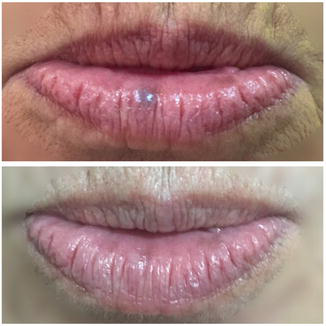
Venous lakes are red-blue nodules, usually seen as a single lesion, that typically occur on the lip and can be treated successfully by PDL, KTP, or Nd:YAG lasers and/or IPLs. For superficial lesions, KTP and PDL are helpful. For deeper lesions, diode, alexandrite, and Nd:YAG lasers are more effective. Often one treatment session will reduce the lesion by 80% in volume, and occasionally the lesion will disappear after one treatment (Fig. 3). Surface cooling is necessary to avoid epidermal injury. Any device with contact cooling should be held against the skin surface with only gentle pressure to avoid vessel collapse.
Infantile Hemangiomas
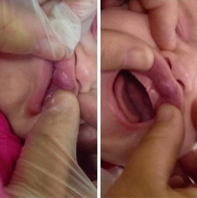
Infantile hemangiomas constitute the most common vascular tumors of early infancy. The estimated incidence of IHs ranges from 1% to 2.6% in healthy infants in the immediate newborn period to about 4–10% of infants by 1 year of age. A female predominance has been described, ranging from 2:1 to 9:1 (Esterly 1996). Histopathologically, these lesions are composed of benign proliferations of plump endothelial cells with a unique vascular phenotype. Glutaminase transferase 1 staining of these lesions is positive and demonstrates small, scanty capillaries, a feature that persists throughout the natural progression of IH and can help confirm the diagnosis (North et al. 2000). Studies of the natural history of IHs reveal that complete resolution occurs in 50% of children by age 5 years and 70% by age 7 years, with continued improvement in the remaining children until 10–12 years of age (Bivings 1954; Garzon and Frieden 2000). IHs tend to be classified based on the depth of tissue involvement (Table 2) (Chiller et al. 2002). Many studies have demonstrated that PDL lasers are more effective and safer than KTP lasers and Nd:YAG (Leonard-Bee et al. n.d.; Raulin and Greve 2001). According to our experience, the best results are achieved with the following parameters: fluence 5–9 J/cm2, spot size 7–10 mm, and pulse duration 0.45–1.5 ms. The alexandrite laser or Nd:YAG can be used to complement the results in the treatment of deeper lesions. Ablative fractional lasers are also being explored for their role in treating fibro-fatty residual tissue from involuted hemangiomas and scars secondary to previous surgical excision. Alternatives to treat IHs are imiquimod 5% cream or topical timolol maleate 0.5% gel (Jiang et al. 2011; Pope and Chakkittakandiyil 2010) (Fig. 4).
Port-Wine Stain Birthmarks
Port-wine stains are congenital, though in rare cases they may be acquired. PWS are found in approximately 0.3% of newborns. They tend to occur on the head and neck, although they may appear anywhere on the body. PWS persist throughout life and many thicken with time. Early treatment may improve responsiveness, decrease the number of treatments, and reduce the likelihood of permanent adverse sequelae (Reyes and Geronemus 1990; Chapas et al. 2007; Chapas and Geronemus 2005). PDL remains the gold standard of treatment for most PWS, although newer-generation IPL (narrow-band IPL) and KTP lasers have evolved as reasonable options. Anesthesia is an important concern when performing laser surgery in the pediatric population. While older children may tolerate the laser procedure using topical anesthetics only, infants and young children may require general anesthesia. Cooling the skin is crucial to minimize damage to surrounding tissue and to reduce the risk of postoperative complications such as swelling, scarring, and post-inflammatory pigmentary changes (especially in darker-skinned patients).
The PDL remains the most studied device in PWS treatment. Over the years, different parameters have been used to treat PWS, with significantly good results. The fluence is adjusted to achieve the desired end point. For the PDL, the desired end point is immediate purpura. A confluent gray color signifies that the fluence is too high. The most commonly applied pulse duration is 1.5 ms. However, for lighter PWS, 0.45 ms has been advocated. Treatment intervals are normally about 3–6 weeks apart. A recent study showed that more frequent treatments might be preferable (Minkis et al. 2009; Chapas and Geronemus 2009). In general, improvement and clearance are gradual and require five to ten treatments.
The alexandrite laser is typically used for PDL-resistant lesions, though it may be implemented as a first-line treatment for hypertrophic violaceous lesions in adults. Care must be taken not to overlap pulses, as scarring can occur. Note that the range of appropriate fluences for alexandrite laser use is quite broad. The Nd:YAG laser and IPL can also be used for PWS. Photodynamic therapy (PDT) has been used successfully to treat PWS, primarily in China. The use of systemically administered hematoporphyrin photosensitizers results in prolonged photosensitivity (weeks), which limits its use. Alternative photosensitizers, such as benzoporphyrin derivative monoacid ring A and mono-L-aspartyl chlorin e6 (Npe6), have shorter periods of photosensitivity and may be a good option (Eppley and Sadove 1994).
Laser treatment according to the vascular lesion is summarized in Table 3, parts 1 and 2.
| Diagnostic | Clinical characteristics | Laser | Parameters | End point | Interval of treatment | Treatment expectations | Comments |
|---|---|---|---|---|---|---|---|
| Facial telangiectasia | 0.1–1 mm diameter; red-purple; macules or linear papules | KTP PDL IPL Nd:YAG |
7–16 J/cm2, 3–7 mm, 10–30 ms 5–8 J/cm2, 10–12 mm, 0.45–6 ms 24–40 J/cm2, 10–30 ms (550–570 nm) 90–115 J/cm2, 3–5 mm, 20–30 ms |
Vessel blanching or closure | 4–6 weeks | 1–2 treatments are indicated; vessels around the nasal ala are more resistant and cannot disappear; new telangiectasias can appear | Hereditary hemorrhagic telangiectasia, lupus erythematosus, systemic sclerosis (CREST syndrome), rosacea, hyperestrogenic state, basal cell carcinoma |
| Spider telangiectasia | Red-purple papules | PDL Nd:YAG |
5–8 J/cm2, 7–10 mm, 1–5 ms 90–100 J/cm2, 3–5 mm, 20–30 ms |
Vessel blanching or closure | 4–6 weeks | 1–2 treatments are indicated | Hyperestrogenic state, pregnancy |
| Rosacea | Facial erythema; telangiectasias | IPL Nd:YAG (Genesis) |
24–45 J/cm2, 10–20 ms (550–570 nm) 13–15 J/cm2; 6,000–10,000 shots |
Homogeneous erythema | 4 weeks | After 3–6 treatments, a clearance of 50–80% is usually achieved; retreatment once or twice a year | Hyperestrogenic state, tabagism, dietary habits, sun exposure, stress |
| Venous lake | 2–10 mm diameter; blue-purple papules | KTP Nd:YAG Nd:YAG |
7 J/cm2, 4 mm, 14 ms 90–110 J/cm2, 5–7 mm, 20–30 ms 100–150 J/cm2, 3–7 mm, 20–30 ms |
Vessel blanching or purpura | 4–6 weeks | 1–2 treatments are indicated | Associated with photodamage |
| Poikiloderma of Civatte | Telangiectasias; hyperpigmentation; hypopigmentation; skin atrophy | IPL PDL |
17–24 J/cm2, 20–30 ms (550–570 nm) 5–7 J/cm2, 7–10 mm, 0.45–2 ms |
Homogeneous erythema; purpura | 4–6 weeks | After 3–6 treatments, a clearance of 50–80% is usually achieved | Associated with photodamage |
| Port-wine stain | Pink-red-purple-blue macules or papules; present at birth | PDL | 6–8 J/cm2, 10 mm, 1.5 ms | Purpura | 4–12 weeks | Gradual lesion discoloration (10% per treatment) | Glaucoma; Sturge–Weber syndrome; Klippel–Trenaunay; GLUT-1 negative |
| Infantile hemangioma | Pink-red macules; first few weeks of life | PDL | 6–8 J/cm2, 7–10 mm, 0.45–1.5 ms | Purpura | 2–8 weeks | Slow growth rate | Care must be taken when the lesion is near the ocular globe; GLUT-1 positive |
| Pyogenic granuloma | 0.5–2 cm diameter; red-purple papules | PDL Nd:YAG |
6.5–9 J/cm2, 7–12 mm, 0.45–3 ms 100–150 J/cm2, 3–7 mm, 20–30 ms |
Subtle gray-blue discoloration followed by deeper purpura | 3–4 weeks | 1–3 treatments are indicated | Electrosurgery and excision are the most indicated treatments |
Pretreatment
Clean the skin gently. Topical anesthesia should be avoided (blanching of the skin caused by anesthesia can decrease the results of the treatment). In some cases it may be necessary, taking into account the patient’s age, the extent of the lesions, and preference.
- Intralesional lidocaine or nerve blocks may be required to treat deeper lesions such as venous malformations.
- Systemic anesthesia should be considered for children when treating hemangioma.
- Proper eye protection is essential.
Posttreatment
Surface cooling is very important to stop the heating and minimize side effects (scarring, pigmentary changes).
Physical sunscreen is recommended after treatment.
My Experience
In my experience, the success of vascular lesion treatment, with safety and efficacy, is related to surface cooling just before and after each session. Proper cooling is essential to protect the epidermis and minimize side effects (scarring).
I am therefore summing up the key points:
- Do not treat tanned skin.
- Use longer pulse durations and/or lower fluences for darker skin.
- To treat port-wine stains, my first choice is the pulsed dye laser.
- For hypertrophic lesions or PDL-resistant PWS, we can use the alexandrite laser.
- For venous lakes, Nd:YAG is a good choice. One to two sessions will reduce 80–100% of the lesion.
- The use of the 1064 nm long-pulse Nd:YAG laser requires fluences over ten times those used with the 532 and 595 nm lasers, since the absorption of Hb and HbO2 at 1,064 nm is ten times less.
- My best results for treatment of rosacea include two to three IPL sessions per year.
- To avoid the polka-dot effect when treating vessels around the nasal ala, my best choice is IPL.
Conclusion
Laser treatment of vascular lesions was the first to use selective photothermolysis, and nowadays it is one of the most important treatments using this theory. The treatment is safe and efficient for most vascular lesions. The selection of parameters guarantees treatment success (wavelength, spot size, pulse duration, surface cooling). New possibilities arise with technological advances and the growth of medical knowledge.
Take Home Messages
- Pulsed dye laser remains the gold-standard treatment for port-wine stains.
- Early laser treatment improves port-wine stain response.
- The end point for treating vessels is vessel clearance, a transient blue coagulum, or purpura.
- Lasers and IPL are the first-choice treatment for telangiectasias and facial erythema.
- Multiple sessions are necessary to treat poikiloderma of Civatte, and some cases are resistant to treatment.
- The alexandrite laser or Nd:YAG can be used to complement the results in the treatment of deeper lesions.
- It is indicated to use longer pulse durations and lower fluences for darker skin.
- Do not treat tanned skin.
References
Aguilar G, Choi B, Broekgaarden M, et al. An overview of three promising mechanical, optical, and biochemical engineering approaches to improve selective photothermolysis of refractory port wine stains. Ann Biomed Eng. 2012;40(2):486–506.
Amir J, Metzker A, Krikler R, Reisner SH. Strawberry hemangioma in preterm infants. Pediatr Dermatol. 1986;3:331–2.
Anderson RR, Parrish JA. Microvasculature can be selectively damaged using dye lasers: a basic theory and experimental evidence in human skin. Lasers Surg Med. 1981a;1:263–76.
Anderson RR, Parrish JA. The optics of human skin. J Invest Dermatol. 1981b;77:13–9.
Anderson RR, Parrish JA. Selective photothermolysis: precise microsurgery by selective absorption of pulsed radiation. Science. 1983;220(4596):524–7.
Baglieri F, Scuderi G. Treatment of recalcitrant granulomatous rosacea with ALA-PDT: report of a case. Indian J Dermatol Venereol Leprol. 2011;77:536.
Behroozan DS, Goldberg LH, Glaich AS, Dai T, Friedman PM. Fractional photothermolysis for treatment of poikiloderma of Civatte. Dermatol Surg. 2006;32:298–301.
Bivings L. Spontaneous regression of angiomas in children; twenty-two years’ observation covering 236 cases. J Pediatr. 1954;45:643–7.
Boscolo E, Bischoff J. Vasculogenesis in infantile hemangioma. Angiogenesis. 2009;12:197–207.
Chapas AM, Geronemus RG. Our approach to pediatric dermatologic laser surgery. Lasers Surg Med. 2005;37:255–63.
Chapas AM, Geronemus RG. Physiologic changes in vascular birthmarks during early infancy: mechanisms and clinical implications. J Am Acad Dermatol. 2009;61:1081–2.
Chapas AM, Eickhorst K, Geronemus RG. Efficacy of early treatment of facial port-wine stains in newborns: a review of 49 cases. Lasers Surg Med. 2007;39:563–8.
Chiller KG, Passaro D, Frieden IJ. Hemangiomas of infancy: clinical characteristics, morphologic subtypes, and their relationship to race, ethnicity, and sex. Arch Dermatol. 2002;138:1567–76.
Comi AM, Bellamkonda S, Ferenc LM, Cohen BA, Germain-Lee EL. Central hypothyroidism and Sturge–Weber syndrome. Pediatr Neurol. 2008;39:58–62.
Davidson AJ. Anesthesia and neurotoxicity to the developing brain: the clinical relevance. Paediatr Anaesth. 2011;21:716–21.
Eppley BL, Sadove AM. Systemic effects of photothermolysis of large port-wine stains in infants and children. Plast Reconstr Surg. 1994;93:1150–3.
Esterly NB. Cutaneous hemangiomas, vascular stains and malformations, and associated syndromes. Curr Probl Pediatr. 1996;26:3–39.
Faurschou A, Olesen AB, Leonardi-Bee J, et al. Lasers or light sources for treating port-wine stains. Cochrane Database Syst Rev. 2011;CD007152.
Fournier N, Brisot D, Mordon S. Treatment of leg telangiectasias with a 532 nm KTP laser in multipulse mode. Dermatol Surg. 2002;28:564–71.
Garden JM, Bakus AD, Paller AS. Treatment of cutaneous hemangiomas by the flashlamp-pumped pulsed dye laser: prospective analysis. J Pediatr. 1992;120:555–60.
Garzon MC, Frieden IJ. Hemangiomas: when to worry. Pediatr Ann. 2000;29:58–67.
Geronemus RG. Long-pulsed neodymium:yttrium-aluminum-garnet laser treatment for port-wine stains. J Am Acad Dermatol. 2006;54:923.
Goldman MP, Bennett RG. Treatment of telangiectasia: a review. J Am Acad Dermatol. 1987;17(2 pt 1):167–82.
Goldman MP, Eckhouse S. Photothermal sclerosis of leg veins. ESC Medical Systems, LTD Photoderm VL Cooperative Study Group. Dermatol Surg. 1996;22:323–30.
Goldman MP, Weiss RA. Treatment of poikiloderma of Civatte on the neck with an intense pulsed light source. Plast Reconstr Surg. 2001;107:1376–81.
Goldman MP, Weiss RA, Weiss MA. Intense pulsed light as a non-ablative approach to photoaging. Dermatol Surg. 2005;31:1179–87; discussion 1187.
Gonzalez S, Vibhagool C, Falo LD Jr, et al. Treatment of pyogenic granulomas with the 585 nm pulsed dye laser. J Am Acad Dermatol. 1996;35:428–31.
Hammes S, Kaiser K, Pohl L, et al. Pyogenic granuloma: treatment with the 1064 nm long-pulsed neodymium-doped yttrium aluminum garnet laser in 20 patients. Dermatol Surg. 2012;38:918–23.
Izikson L, Nelson JL, Anderson RR. Treatment of hypertrophic and resistant port-wine stains with a 755 nm laser: a case series of 20 patients. Lasers Surg Med. 2009;41:427–32.
Jiang C, Hu X, Ma G, et al. A prospective self-controlled phase II study of imiquimod 5% cream in the treatment of infantile hemangioma. Pediatr Dermatol. 2011;28:259–66.
Leonard-Bee J, Batta K, O’Brien C, Bath-Hextall FJ. Interventions for infantile hemangiomas (strawberry birthmarks) of the skin. Cochrane Database Syst Rev. n.d.;CD006545.
Li W, Yamada I, Masumoto K, Ueda Y, Hashimoto K. Photodynamic therapy with intradermal administration of 5-aminolevulinic acid for port-wine stains. J Dermatolog Treat. 2010;21:232–9.
Manuskiatti W, Eimpunth S, Wanitphakdeedecha R. Effect of cold air cooling on the incidence of postinflammatory hyperpigmentation after Q-switched Nd:YAG laser treatment of acquired bilateral nevus of Ota-like macules. Arch Dermatol. 2007;143:1139–43.
Meesters AA, Pitassi LHU, Campos V, Wolkerstorfer A, Dierickx CC. Transcutaneous laser treatment of leg veins. Lasers Med Sci. 2013a;Original Article.
Meesters AA, Pitassi LHU, Campos V, Wolkerstorfer A, Dierickx CC. Transcutaneous laser treatment of leg veins. Lasers Med Sci. 2013b.
Minkis K, Geronemus RG, Hale EK. Port-wine stain progression: a potential consequence of delayed and inadequate treatment? Lasers Surg Med. 2009;41:423–6.
Moehrle M, Léauté-Labrèze C, Schmidt V, et al. Topical timolol for small hemangiomas of infancy. Pediatr Dermatol. 2012.
Nelson JS, Milner TE, Anvari B, et al. Dynamic epidermal cooling during pulsed laser treatment of port-wine stain: a new methodology with preliminary clinical evaluation. Arch Dermatol. 1995;131:695–700.
North PE, Waner M, Mizeracki A, Mihm MC Jr. Glut1: a newly discovered immunohistochemical marker for juvenile hemangiomas. Hum Pathol. 2000;31:11–22.
Paul BS, Anderson RR, Jarve J, Parrish JA. The effect of temperature and other factors on selective microvascular damage caused by dye laser. J Invest Dermatol. 1983;81(4):333–6.
Pope E, Chakkittakandiyil A. Topical timolol gel for infantile hemangiomas: a pilot study. Arch Dermatol. 2010;146:564–5.
Prieto VG, Sadick NS, Lloreta J, Nicholson J, Shea CR. Effects of intense pulsed light on sun-damaged human skin, routine, and ultrastructural analysis. Lasers Surg Med. 2002;30:82–5.
Raulin C, Greve B. Retrospective clinical comparison of hemangioma treatment by flashlamp-pumped (585 nm) and frequency-doubled Nd:YAG (532 nm) lasers. Lasers Surg Med. 2001;28:40–3.
Reyes BA, Geronemus R. Treatment of port-wine stains during childhood with the flashlamp-pumped pulsed dye laser. J Am Acad Dermatol. 1990;23:1142–8.
Ross EV. Laser versus intense pulsed light: competing technologies in dermatology. Lasers Surg Med. 2006;38:261–72.
Schmidt N, Gans E. Demodex and rosacea I: the prevalence and numbers of Demodex mites in rosacea. Cosmet Dermatol. 2004;17:497–502.
Solomon H, Goldman L, Henderson B, Richfield D, Franzen M. Histopathology of the laser treatment of port-wine lesions: biopsy studies of treated areas observed up to three years after impacts. J Invest Dermatol. 1968;50(2):141–6.
Tan OT, Sherwood K, Gilchrest BA. Treatment of children with port-wine stains using the flashlamp pulsed tunable dye laser. N Engl J Med. 1989;320:416.
Taner JL, Dechert MP, Frieden IJ. Growing up with a facial hemangioma: parent and child coping and adaptation. Pediatrics. 1998;101:446–52.
Tierney EP, Hanke CW. Treatment of poikiloderma of Civatte with ablative fractional laser resurfacing: prospective study and review of the literature. J Drugs Dermatol. 2009;8:527–34.
Wall TL, Grassi AM, Avram MM. Clearance of multiple venous lakes with an 800 nm diode laser: a novel approach. Dermatol Surg. 2007;33:100–3.
Weiss RA, Goldman MP, Weiss MA. Treatment of poikiloderma of Civatte with an intense pulsed light source. Dermatol Surg. 2000;26:823–7; discussion 828.
Xiao Q, Li Q, Yuan KH, Cheng B. Photodynamic therapy of port-wine stains: long-term efficacy and complications in Chinese patients. J Dermatol. 2011;38:1146–52.
Yohn JJ, Huff JC, Aeling JL, Walsh P, Morelli JG. Lesion size is a factor for determining the rate of port-wine stain clearing following pulsed dye laser treatment in adults. Cutis. 1997;59:267–70.
Zhao Y, Zhou Z, Zhou G, et al. Efficacy and safety of hemoporfin in photodynamic therapy for port-wine stain: a multicenter and open-label phase IIa study. Photodermatol Photoimmunol Photomed. 2011;27:17–23.







