
Laser for Onychomycosis
Laser for Onychomycosis
Claudia Maria Duarte de Sá Guimarães, Taissa Vieira Machado Vila and Sergio Bittencourt-Sampaio Av. Nossa Sra. de Copacabana, 435, sala 903, Rio de Janeiro, Brazil Laboratory of Cell Biology of Fungi, Carlos Chagas Filho Institute of Biophysics, Federal University of Rio de Janeiro, Rio de Janeiro, Brazil Department of Histology and Embryology, UFRJ, Souza Marques School of Medicine, Av. Nossa Senhora de Copacabana 605/907, CEP 22050-902 Rio de Janeiro, RJ, Brazil
Abstract
Onychomycosis is a common fungal infection that affects the nail plate or the nail bed and is responsible for approximately 50% of the pathologies affecting the nails (Ghannoum Microbiology 157:3232–42, 2011). Its treatment remains a challenge, even though progress has occurred with the introduction of new antifungal drugs in the 1990s, since many cases linger for decades without a clinical cure (Sigurgeirsson J Europ Acad Dermatol Venerol 24:679–684, 2010). Most cases are caused by dermatophyte fungi; however, in recent years there has been a progressive increase of records of onychomycosis caused by nondermatophyte fungi (yeast and filamentous fungi) that do not respond to antifungal agents (Ranawaka et al. Dermatol Online Journal 18(1):7, (2012); Hwang et al. Ann. Dermatol 24(2):175–180, 2012). Photobiomodulation and photoinactivation studies indicate that lasers in the range of the infrared electromagnetic spectrum (870, 930, 1,064 nm), when applied with biochemical energy, are able to improve microcirculation, to stimulate the metabolism of cells, and to inhibit fungal and bacterial multiplication through action on the wall of the microorganisms, by altering the electric charges and favoring the formation of ROS (singlet oxygen radicals, free radicals). This technique can be associated with the application to the nails of the fractional CO2 laser so that, in a drug delivery system, it can be combined with antifungal and/or antibacterial agents in order to act synergistically and thus reduce the number of sessions of sub-millisecond 1,064 nm laser.
Keywords OnychomycosisAntifungalDermatophyteNondermatophyteBiofilmPhotobiomodulationNd:YAG laserCO2 laserDrug delivery
Introduction
Onychomycosis is a common fungal infection that affects the nail plate or the nail bed and is responsible for approximately 50% of the pathologies affecting the nails (Ghannoum et al. 2000). Its treatment remains a challenge, even though progress has occurred with the introduction of new antifungal drugs in the 1990s, as many cases linger for decades without clinical cure (Sigurgeirsson 2010). It is estimated that the rate of clinical cure with oral medications reaches slightly over 50% of the cases, in the most optimistic statistics, while topical treatments do not reach 20% cure rates (Scher et al. 2007).
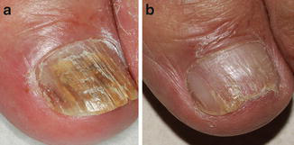
Onychomycosis can complicate other foot problems in elderly persons, affect social interactions, impair employment in which there is direct contact with the public, and serve as a reservoir for potentially aggressive fungi in immunosuppressed individuals. Some factors are considered predisposing factors, such as advanced age, diabetes, hyperhidrosis, immunocompromised individuals (tumors of hematologic origin, HIV/AIDS), alteration of peripheral circulation, and nail trauma (tight shoes, athletes, etc.), tinea pedis, and trauma from manicure or pedicure, in addition to family history (probably because of transmission) and nail psoriasis (Fig. 1) (Pariser et al. 2013).
Basic Concepts
The nails are keratinized plates of hardened consistency located on the distal ends of the fingers and toes. The first signs of the structure that will lead to the nails appear near the end of the first trimester of fetal life. The epidermis of the extremities of the fingers begins to proliferate and grow in the shape of a curved line toward the dermis. Next, the proliferative plate is divided into segments forming the nail groove. Cells in the deepest part of this groove give rise to the nail matrix, where the upper cells are keratinized and form the plate or body of the nail. As the mitoses in the matrix continue, the nail plate is displaced along the dorsal surface of the finger. This process in the hands precedes the one in the feet. Thus, approximately at the 32nd week, the fingernails reach the ends of the fingers, while, in the feet, this only happens at approximately the 36th week of intrauterine life.
The proximal part of the nail is called the root, where the nail matrix is located, which continues in the exposed part named body. Matrix cells become keratinized and, unlike the other parts of the body, do not come off; on the contrary, they become compact and move in the linear path of the nail plate, where they remain included. In the matrix, Merkel cells and melanocytes are also found. The dermis of that proximal part presents short papillae. The matrix is firmly attached to the dermis, so that, when the nail is extracted, the entire matrix cannot be removed. The root is covered with a small skinfold (proximal nail fold), which covers a small portion of the body of the nail, thus constituting the eponychium, formed by soft keratin.
Underlying the body of the nail, the epidermis differs from other anatomical regions by presenting only the Malpighian layer without cells containing keratohyalin granules, existing in the skin throughout the body. In this case, the nail plate performs the function of the cornea layer. The proximal portion of the nail bed has an epidermis that is very thick, generating an opalescent image known as the lunula, most evidenced in the thumb and which may appear in other fingers (Montagna and Parakkal 1974). The cells responsible for the production and growth of the nail (nail matrix) are located there. On a layer of prismatic (basal) cells, 6–10 layers of polyhedral cells and 3–12 layers of flat cells can be found. It is assumed that the white discoloration is derived from this thick condition, which does not allow the indirect viewing of the blood inside the capillaries (Bloom 1977; Weiss 1977; Montagna and Parakkal 1974; Snell 1985); however, the lunula does not always coincide with the location of the matrix (Montagna and Parakkal 1974).
The nail plate contains a type of keratin that is very high in sulfurous amino acids (hard keratin), which explains its structural stability and chemical resistance. In this region, the cells have a thick cell membrane and are firmly attached to each other, with a large amount of thick and birefringent tonofibrils, measuring from 60 to 80 Å, in their interior, and surrounded by dense amorphous material (Weiss 1977). On the surface of the nail plate, there are very evident longitudinal grooves in the elderly, but they are rarely present in the young population (Montagna and Parakkal 1974). Despite being thick and compact, the nail, as well as hair, is more permeable to water than the stratum corneum of the skin in general.
Underlying the body of the nail (nail bed), the dermis rests on the periosteum of the phalanx. In this segment, the dermal papillae form longitudinal and parallel ridges, which accompany the longitudinal axis of the nail. The vascularization is intense, which gives the pink opaque color observed through the nail plate. The side edges are involved by skinfolds, the lateral nail folds, separated from the bed where the body of the nail rests. The location that establishes the continuity of the epidermis of the finger with the distal end of the nail bed is named the hyponychium, from where the free edge of the nail emerges.
Fingernails grow from 0.5 to 1.2 mm per week, while toenails show a slower evolution (Snell 1985). Several conditions can interfere in this growth: circadian rhythms (the growth is greater during the day than during the night and greater in summer than in winter), age group (greatly reduced growth after the seventh decade), hormonal factors (which speed up during pregnancy, slow down with hypothyroidism, etc.), nutritional conditions, and traumatic conditions.
Etiology and Epidemiology
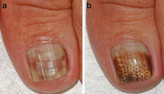
Most cases of onychomycosis are caused by dermatophyte fungi, which are microorganisms found in soil (geophilic), animals (zoophilic), or humans (anthropophilic). The anthropophilic species are Trichophyton, Microsporum, and Epidermophyton. Dermatophytes are able to invade keratinized tissues (cornea layer, hair, and nails) and therefore are called keratinophilic microorganisms. Nondermatophyte fungi are not keratolytic, which is why they are found in the intercellular cement or fixed in the keratin previously destroyed by the dermatophytes, by trauma, or by other nail disease (Pariser et al. 2013; Hwang et al. 2012). Filamentous nondermatophyte fungi are most common in tropical regions (e.g., Fusarium and Aspergillus), as well as yeasts (especially Candida spp.). Nondermatophyte fungi have presented increasing incidence and are considered more difficult to treat, because they often do not respond to antifungal agents (Fig. 2) (Ranawaka et al. 2012; Hwang et al. 2012).
A large-scale American study isolated dermatophyte fungi in 59% of the cases and nondermatophyte fungi and yeasts in approximately 20% of the cases. Trichophyton rubrum and Trichophyton mentagrophytes are among the most frequently encountered dermatophytes, while Trichophyton tonsurans, Microsporum canis, and Epidermophyton floccosum represented 0.8% of the dermatophytes. Among the isolated nondermatophytes, Acremonium, Fusarium, and Scopulariopsis spp. deserve to be highlighted, with frequencies of 29.5%, 34.1%, and 20%, respectively. Among the yeasts, Candida parapsilosis represented 66.7% and Candida albicans 16.7% of the cultures (Ghannoum et al. 2000).
A study carried out in Rio de Janeiro in 2001 evaluated 2,271 patients and diagnosed 400 patients, these diagnoses being in 264 fingernails and 136 toenails, through direct mycological examination and culture; this study indicated the presence of dermatophyte fungi in 46.5% of the cases of onychomycosis in toenails, Candida in 49% of the fingernails of women, and 4.5% of emerging fungi (nondermatophytes and other microorganisms). Among the nondermatophyte fungi capable of causing onychomycosis, Scopulariopsis brevicaulis, Fusarium spp., Acremonium spp., Aspergillus spp., Scytalidium spp., and Onychocola canadensis were identified (Araujo et al. 2003). A study performed in Sri Lanka recorded onychomycosis caused by nondermatophyte fungi in 45.8%, Candida spp. in 34.1%, and dermatophytes in 20% of the cases. This variation of pathogens was attributed to contact with soil, the habit of walking barefoot, the frequent immersion of the hands in water, and the moist and warm climate. The most frequently encountered nondermatophyte fungi were Aspergillus niger, Aspergillus flavus, and Fusarium spp. Onychomycosis was accompanied by paronychia in 76% of the cases. Nondermatophyte fungi accounted for 22% of the onychomycoses in India, 35.5% in Malaysia, 51.6% in Thailand, and 68% in Pakistan (Ranawaka et al. 2012).
The Fusarium spp. have been highlighted as plant pathogens and can occasionally infect animals and humans. They can be found in soil, underground, and aerial parts of plants, organic substrates, and water, where they are part of the structure of biofilms. Among humans, they cause superficial infections (such as keratitis and onychomycosis), local invasive disease, or disseminated infections, the latter affecting severely immunocompromised patients (with deep and prolonged neutropenia and/or severe T cell immunodeficiency) and patients with hematologic diseases, mainly patients with acute leukemia. Furthermore, they can cause allergic sinusitis in immunocompetent individuals and mycotoxicosis in humans and animals from the intake of food contaminated by the toxin produced by these fungi. Among the 50 known species, 12 are capable of causing infection, and preexisting onychomycosis can be the source of disseminated fusariosis (Nucci et al. 2007).
Biofilms
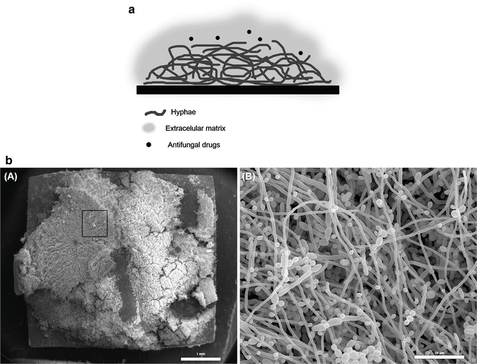
Since the seventeenth century, biofilms have been described in multiple systems. Most bacteria preferentially grow as biofilms in all self-sustaining aquatic ecosystems, and these sessile bacterial cells differ deeply from their planktonic counterparts (cells in suspension) (Costerton et al. 1995). The definitions of biofilm have evolved over the years, in parallel to the advances in biology and research studies on the subject. The definition used today was proposed by Donlan and Costerton (2002), and it describes a biofilm as a microbial community in which the cells are connected to a substrate, or to each other, embedded in a matrix of extracellular polymeric substances (produced by themselves) and exhibiting an altered phenotype regarding the rate of growth and transcription of genes (Fig. 3) (Donlan and Costerton 2002).
In fungi, the ability to colonize surfaces and to form biofilms was initially demonstrated for Candida albicans and Saccharomyces cerevisiae, in the 1990s and early 2000s (Hawser and Douglas 1994; Reynolds and Fink 2001). However, the growing awareness of the importance of fungal biofilms can be seen in the increase in the number of publications describing the formation of biofilms by other species of Candida spp. (Bizerra et al. 2008; Lattif et al. 2010; Silva et al. 2011) as well as other yeasts that cause opportunistic infections and pneumonia in humans, such as Malassezia pachydermatis (Cannizzo et al. 2007), Rhodotorula sp. (Nunes et al. 2013), Trichosporon asahii (Di Bonaventura et al. 2006), Blastoschizomyces (D’Antonio et al. 2004), Pneumocystis spp. (Cushion et al. 2009), and Cryptococcus neoformans (Martinez and Casadevall 2007). Moreover, the ability to form biofilms has also been demonstrated in several filamentous fungi, including Aspergillus fumigatus (Mowat et al. 2009) and Fusarium spp. (Imamura et al. 2008); in fungi that cause endemic mycoses, such as Histoplasma capsulatum (Pitangui et al. 2012), Paracoccidioides brasiliensis (Sardi et al. 2014), and Coccidioides immitis (Davis et al. 2002); and in zygomycetes, such as Mucorales (Singh et al. 2011).
Biofilms in Dermatology
Despite the participation of biofilms in nail infections still being an open discussion, their involvement in other dermatological infections, including acne, miliaria, atopic dermatitis, and wounds, is well accepted, especially in relation to bacterial biofilms (Vlassova et al. 2011).
Biofilms in Onychomycosis
During the course of the nail infection, the formation of a thick biomass can be observed, with fungal elements embedded in an extracellular matrix (Burkhart et al. 2002). Several factors, including the firm adhesion of the fungi to the nail plate, the presence of persister cells, and the difficulty of eradicating the infection, suggest that biofilms are an important factor in the pathogenesis of onychomycoses (Nusbaum et al. 2012). Complementing this hypothesis, the ability to form biofilms was demonstrated in vitro for the primary causative agent of onychomycosis, the dermatophyte Trichophyton sp. (Costa-Orlandi et al. 2014). A model for biofilm formation in human nail fragments has recently been established using Candida albicans and Fusarium oxysporum (Vila et al. 2014) and may help to elucidate the involvement of biofilms in the pathogenesis of onychomycoses and to validate the antibiofilm activity of new molecules and new treatments (Fig. 4).
Antifungal Resistance Associated with Biofilms
According to the definition of a biofilm, the cells that make up its structure have an altered phenotype and differ from planktonic cells in the expression of genes, in the rate of growth, and, mainly, in susceptibility to antifungal agents. The increase in antifungal resistance in cells of Candida spp. grown as biofilms, in relation to their planktonic forms, is the most medically relevant behavioral change (Ramage et al. 2002). Multiple mechanisms have been suggested to explain the increased antifungal resistance of the biofilm, including cell density, alteration of drug targets, expression of drug efflux pumps, the extracellular matrix, and the presence of persistent cells (Kuhn et al. 2002a; Lattif et al. 2011; Mukherjee and Chandra 2004; Perumal et al. 2007; Ramage et al. 2002, 2012).
Role of the Extracellular Matrix of the Biofilm in Resistance
In most biofilms, the population of microorganisms corresponds to 10% of the total mass, and the extracellular matrix (ECM) corresponds to 90%. The matrix consists of a cluster of different biopolymers responsible for keeping the cells adhered to the surface and maintaining the cohesion of the biofilm, for restraining the cells, and for keeping them close, thereby allowing cell–cell communication and diffusion of signaling molecules (Flemming and Wingender 2010).
The ECM, by definition, provides protection to cells against environmental factors, such as host immunity and antifungal agents (Seneviratne et al. 2008). One of the described key components of the ECM of C. albicans is a β-1,3-glucan responsible for sequestering azoles, echinocandins, pyrimidines, and polyenes (Nett et al. 2010a, b; Vediyappan et al. 2010), behaving as a “drug sponge” and contributing to the increased resistance of biofilms of C. albicans (Nett et al. 2010a, b). The latest study published on the subject suggests that, in addition to β-1,3-glucan, a polysaccharide complex formed by mannan-glucan (which is, in fact, the polysaccharide in greatest abundance in the ECM of biofilms of C. albicans) is also capable of binding to fluconazole (and possibly to other drugs) and contributes to the resistance (Zarnowski et al. 2014). This work emphasizes that most polysaccharides of the ECM probably act as drug sequestrants and contribute to the resistance of the biofilm to antifungal agents.
In addition to the polysaccharides, the extracellular DNA present in the ECM of biofilms of C. albicans also appears to have a role in resistance to non-azole agents, as the addition of DNase increases the antibiofilm activity of polyene agents and echinocandins, but not the activity of azoles (Martins et al. 2009, 2012).
Even though the resistance mechanisms associated with the lower susceptibility of biofilms to the available antifungals are not fully elucidated, several studies show that biofilms of C. albicans, C. parapsilosis, and C. tropicalis are resistant to treatments with different commercially available antifungals, among them fluconazole, nystatin, terbinafine, amphotericin B, voriconazole, and ravuconazole (Ferreira et al. 2009; Kuhn et al. 2002b). Moreover, in vitro studies with biofilms of Fusarium spp. also highlight their lower susceptibility to the commercial antifungal agents amphotericin B, voriconazole, itraconazole, and fluconazole (Mukherjee et al. 2012; Zhang et al. 2012).
Biofilms of Candida spp.
The in vitro development of biofilms of Candida spp. (on abiotic surfaces) can be didactically described in four sequential steps: (1) adherence, the initial phase, in which yeasts in suspension and those circulating (planktonic cells) adhere to the surface; (2) an intermediate phase, concerning development of the biofilm; (3) a maturation phase, in which the polymer matrix completely soaks all layers of cells adhered to the surface in a three-dimensional structure; (4) dispersion, in which the most superficial cells leave the biofilm and colonize areas surrounding the surface (Chandra et al. 2001; Ramage et al. 2005; Seneviratne et al. 2008). At the end of development, the biofilm consists of a dense network of cells in the form of yeasts, hyphae, and pseudohyphae soaked by a polymeric extracellular matrix and with water channels between the cells, which facilitate the diffusion of nutrients from the environment through the biomass to the lower layers and which also allow the elimination of waste (Chandra et al. 2001; Ramage et al. 2001, 2005).
Biofilms of Candida spp. formed in in vivo models seem to follow the same sequence of formation (Andes et al. 2004); however, maturation occurs more rapidly and the final thickness is greater in these biofilms than in those grown in in vitro systems.
The final architecture of the biofilm is variable and depends, in part, on the substrate on which it is formed and the growing conditions, such as the culture medium used (in vitro), concentration and types of sugars, presence of serum proteins, pH, and temperature (Kumamoto 2002; Seneviratne et al. 2008).
Biofilms of Fusarium spp.
Species of the genus Fusarium are soil saprophytes and important pathogens of plants and humans. The clinical form of fusariosis depends on the immune status of the host. In immunocompetent individuals, keratitis and onychomycoses are the most common infections. In immunocompromised individuals, disseminated fusariosis is the second most common filamentous fungal infection, and it particularly affects patients treated with high-dose corticosteroids and patients with severe neutropenia, with a mortality rate of up to 100% (Nucci and Anaissie 2007). Fusarium spp. is an important causative agent of microbial keratitis, and biofilm formation has been suggested as a contributing factor in recent outbreaks, mainly associated with the use of contact lenses (Mukherjee et al. 2012). Fusarium spp. is also commonly isolated as a causative agent in onychomycoses (de Araújo et al. 2003; Morales-Cardona et al. 2014). In nails, fungal cells form thick fungal biomasses, with fungal elements embedded in an extracellular matrix (Burkhart et al. 2002); this behavior suggests the participation of biofilms in the pathogenesis of onychomycosis (Nusbaum et al. 2012). In the northern hemisphere, onychomycosis caused by dermatophytes, especially T. mentagrophytes and T. rubrum, is more prevalent (Ghannoum et al. 2000). However, in warmer and more humid countries such as Brazil, the incidence of nondermatophytic filamentous fungi (such as Fusarium spp.) and yeasts (such as Candida spp.) as causative agents of these infections is very significant (de Araújo et al. 2003; Morales-Cardona et al. 2014). These data are extremely relevant since, while most dermatophytes are sensitive to the terbinafine and azoles commonly used in the treatment of onychomycoses (ketoconazole, clotrimazole, and fluconazole), Fusarium spp. is often resistant to the available antifungals (Bueno et al. 2010; Ortoneda et al. 2004).
Diagnosis
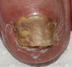
The diagnosis of onychomycosis involves researching the involvement of the nail unit (nail plate, nail bed, and periungual tissues). Dermatophytes affect toenails more than fingernails, where infections by Candida spp. are more frequent. The physical examination should be careful in order to record all the nail units involved and to observe the clinical signs of onychomycosis: onycholysis, material under the nail plate, subungual hyperkeratosis, change of color (white or yellow, brown), and the intensity of destruction of the nail plate (Fig. 5) (Pariser et al. 2013).
Clinical Evaluation
The clinical evaluation begins by gathering information such as age, sex, presence of vascular diseases, diabetes, hypertension, number of infected nails, duration of infection, history of previous treatment, type of onychomycosis, percentage of nail involvement, thickness of the nail, presence of dermatophytoma, involvement of the matrix, and exclusively lateral involvement (Ghannoum et al. 2000; Scher et al. 2007). In Brazil, there is also the need to include the family history of onychomycosis and the habit of removing the cuticles during manicures, which facilitates involvement of the nails with mixed infections (bacterial and fungal). Examination of areas of intertrigo assists in the identification and treatment of fungal reservoirs. Individuals with a family history of diabetes, metabolic syndrome, peripheral circulatory problems, or immunodeficiency should be treated by a multidisciplinary team. Young individuals from 20 to 30 years old with chronic infection that is difficult to treat should be investigated for family onychomycosis, because of contamination of the nails in infancy, the use of bathrooms with contaminated relatives and/or locker rooms, and fungi resistant to traditional treatment.
Identification of the clinical presentation is important for the choice of an appropriate therapeutic approach for each case (Scher et al. 2007). The white-yellow or orange-brown spots may be caused by fungi that do not respond to the medication used, as in the case of nondermatophyte fungi. Brown spots are common in the fifth toe and should be differentiated from nail melanoma (although these two pathologies may occur at the same time). Patients with chronic onychomycosis should be examined for signs that indicate active infection, even if the culture is negative:
- Change of more than 10% of the nail plate compatible with infection caused by dermatophytes by dermatoscopy
- Presence of white, yellow, orange, or brown spots of plates on the nail
- Lateral onycholysis with debris
- Lateral hyperkeratosis on the nail plate or bed (Fig. 6) (Ortiz et al. 2014)
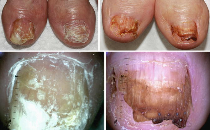
The following are some fungi that can cause melanonychia: Scytalidium, Scopulariopsis, Aspergillus, Fusarium, Trichophyton rubrum, Trichophyton mentagrophytes, Trichophyton soudanense, Candida albicans, Candida tropicalis, Curvularia, etc. (Finch et al. 2012).
Differential Diagnosis
- Irritant contact — enamels, acrylics, or artificial nails
- Nail trauma — athletes, contusions
- Psoriasis — secondary infection by fungus
- Lichen planus
- Neoplasms — subungual
- Bacterial infection — Gram-positive bacteria
Clinical Classification
- Distal and lateral subungual onychomycosis — mixed infection by Candida and bacteria, dermatophytes
- Proximal subungual onychomycosis — affects the base of the nail and can be caused by fungi of the genus Fusarium spp.
- White superficial onychomycosis — T. mentagrophytes
- Endonyx onychomycosis — dermatophytes
- Total onychomycosis — dermatophytes and mixed infections with nondermatophyte fungi (Pariser et al. 2013; Hwang et al. 2012; Carney et al. 2011)
Prognostic Factors
Onychomycoses are under the influence of factors that make the prognosis grim. They act to worsen the nail infection. Their characteristics are:
Host — impaired peripheral circulation, diabetes, and immunosuppression (Figs. 7 and 8)
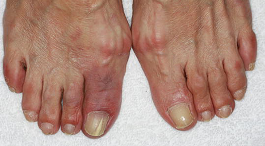
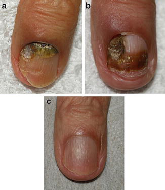
Nails — hyperkeratosis greater than 2 mm, lateral disease, dermatophytoma (biofilm), involvement of more than 50% of the nail plate, slow growth of the nails, total dystrophic onychomycosis, involvement of the matrix, and severe onycholysis
Microorganism — filamentous nondermatophyte fungi, yeasts, and mixed infections (Carney et al. 2011)
Clinically, it is not possible to identify or differentiate onychomycosis caused by dermatophytes, filamentous nondermatophytes, or yeasts (Ranawaka et al. 2012). The collection of samples is carried out with sterile instruments, with curettage after cutting part of the free edge of the nail, with curettage of the nail plate when white superficial onychomycosis is suspected, or with samples of the nail and nail bed when the involvement is closer to the matrix. The material is collected after cleaning the nail with alcohol, then stored and transported in a sterile collector. Part of this material will be used for direct examination and another part for culture in Sabouraud agar medium. The direct examination, although nonspecific, aids in differential diagnosis from other diseases, such as psoriasis or lichen planus, although secondary bacterial and/or fungal infections may occur in relation to the other inflammatory pathologies. It is important to inform the laboratory when there is a need to research a nondermatophytic fungus and/or bacteria so that the material can be grown in suitable medium.
Culture is a method considered the gold standard for the diagnosis of onychomycosis in 3–4 weeks, although it can show a false negative in 15% of the cases (Pariser et al. 2013; Liu et al. 2000). The isolation of a nondermatophyte fungus or yeast can be considered environmental contamination or contamination from the local microbiota. The recommendation for diagnosis of these pathogens is based on the lack of growth of the dermatophyte fungus in culture and the growth of five colonies of the same microorganism in two consecutive samples (Araujo et al. 2003; Ranawaka et al. 2012).
Examination using nucleic acid amplification (PCR) technology has offered the opportunity to improve the quality and speed of diagnosis of dermatophytes, by up to 4 h. It identifies species and subspecies of dermatophytes and sets them apart from nondermatophytes and Candida spp., and it does not require viable microorganisms for the test (Liu et al. 2000).
Treatment
Topical and Systemic Treatment
The treatment of onychomycoses faces several difficulties, among them the need for long-term monitoring, the side effects of systemic medications, and the difficulty of delivering the medication to the target area to be treated (Vural et al. 2008). The traditional therapeutics of onychomycoses consists of palliative care, chemical or mechanical debridement (Chiacchio et al. 2004), and use of systemic or topical medications. The definition of “complete healing” by the US regulatory agency, the Food and Drug Administration (FDA), for evaluation of clinical results, is negative results of direct examination and culture, as well as a completely normal appearance of the nail (Pariser et al. 2013).
The choice of treatment depends on the clinical presentation, the severity of the disease, and the cost and duration of treatment. The systemic drugs used – terbinafine, itraconazole, and fluconazole – are associated with significant adverse effects, which hinder prescription for patients with hepatic and/or renal problems. Topical medications are safer but ineffective. In clinical practice, patients with long-term infections or chronic onychomycosis with serious changes of the nail plate, nail bed, and matrix can be observed. The study by Pariser et al. (2013) indicates that the cure rate with topical ciclopirox 8% for 48 weeks is 5.5–8.5%. Therefore it is not recommended as monotherapy. Cure rates with therapeutic treatment with systemic medications range from 14% to 54%, according to the medication used, the dose, and the duration of administration.
Treatment with Light and Laser
The application of lights to tissues and organisms can have both stimulatory and inhibitory responses according to the parameters used. Some terms are used to describe these effects, such as biostimulation, low-level laser (or light) therapy (LLLT), low-intensity laser therapy, low-power laser therapy, cold laser, soft laser, photobiostimulation, and photobiomodulation. The most used term is low-level laser therapy (LLLT), a term often mentioned in the Medical Subject Headings (MeSH), the controlled-vocabulary thesaurus of the National Library of Medicine. The conference of the North American Association for Light Therapy and the World Association for Laser Therapy in September 2014 determined by consensus that the term that best designates it is photobiomodulation, proposed for inclusion in the MeSH Section at the National Library of Medicine (Anders et al. 2015).
Photodamage by ultraviolet light in bacteria and fungi without the use of a photosensitizing agent was demonstrated at the beginning of the 1900s with lamps that radiated wavelengths between 226 and 328 nm (UVC + UVB). Germicidal activity against prokaryotic and eukaryotic pathogens was observed in 2002; however, its use was abandoned because of the photocarcinogenic effect in human cells (Bornstein et al. 2009a). Since then, other wavelengths in the near-infrared (NIR) range of the electromagnetic spectrum have been studied in order to achieve photoinactivation of pathogens, both with the use of light and a photosensitizing agent and with the use of laser (Vural et al. 2008; Kosarev et al. 2010).
Studies of light and lasers with antimicrobial objectives have followed parallel paths in areas such as physical chemistry, microbiology, and clinical practice. Although studies across the electromagnetic spectrum are unraveling the behavior of certain wavelengths and some tests using laser on cultures of T. rubrum and bacteria indicate photoinhibition of certain microorganisms, there is still a lack of in vitro studies that mimic clinical conditions and of clinical studies with broader sampling. One of the difficulties is the wide variety of clinical presentations of onychomycoses and the combinations of mixed infections that form biofilms (Meral et al. 2003; Vural et al. 2008; Knappe et al. 2004; Pasquini 2003; Landsman et al. 2010).
Interaction of the Laser with the Skin
The extent and intensity of the action of the laser on the skin depend on the structure of the tissue, determined by its water content and blood circulation, which influence absorption, scattering, reflection, thermal conductivity, heat capacity, and density, which in turn are influenced by the parameters of the laser beam (energy intensity and wavelength). Depending on the duration of laser irradiation in the tissues, different energy densities lead to three types of interaction: photochemical effects (10 s–1000 s; 10−3–1 W/cm2), photothermal effects (1 ms–100 s; 1–106 W/cm2), and photoionization and photomechanical effects (10 ps–100 ns; 108–1012 W/cm2). Photothermal and photochemical effects are obtained using less energy and with longer pulses than those used for photoionization and photomechanical effects. These physical parameters are important for classifying the type of laser equipment available and its purposes. In this way, the same wavelength may be indicated for different clinical conditions according to the energy of the equipment and its ability to generate short or long pulses (Knappe et al. 2004).
The increase in temperature and its distribution in the area exposed to laser radiation depend on the energy absorbed by the tissue and its thermal properties. According to the temperature achieved, different effects occur. When the temperature reaches 45 °C, there is no irreversible tissue damage. Temperatures between 45 and 50 °C generate alterations of enzymes and edema. Temperatures above 60 °C for a few seconds generate denaturing coagulation of the tissue proteins, and temperatures between 90 and 100 °C cause vaporization of the plasma cell (as with the CO2 laser) (Knappe et al. 2004).
Currently, there are studies on the effects of lasers whose wavelength lies in the near-infrared (NIR) range of the electromagnetic spectrum. Lasers in the range between 700 and 1,400 nm have in common the ability to penetrate deeply into the dermis with virtually no dispersion of the light beams. A study performed with a diode laser system at 870 nm and 930 nm demonstrated in vitro photoinactivation at physiological temperatures of Staphylococcus aureus, Escherichia coli, Candida albicans, and Trichophyton rubrum by reducing membrane potential and increasing the generation of reactive oxygen species (ROS), without harmful effects to human cells.
A clinical study with the 870 nm and 930 nm laser diode performed by Landsman et al. (2010), based on previous studies by Bornstein, obtained improvement in cases of onychomycosis in 63% of the cases followed for 180 days, through 3 mm of growth of treated nails and periodic acid–Schiff-negative cultures in 30% of the cases (Landsman et al. 2010; Bornstein 2009; Bornstein et al. 2009). Exposures were performed as two applications of 2 min duration and accompanied by temperature measurement with an infrared thermometer.
The 1,064 nm Nd:YAG Laser
The 1,064 nm Nd:YAG laser has been used for remodeling of collagen (Dang et al. 2005; Koh et al. 2010; Dayan et al. 2003a, b; Schmults et al. 2004; Tan et al. 2004), treatment of pseudofolliculitis barbae (Ross et al. 2002) and pyogenic granuloma, stimulation of angiogenesis (Kipshidze et al. 2001), and as an antibacterial and antifungal agent, as noted in photobiomodulation performed with visible light and near-IR (Guffey et al. 2014).
A new in vitro model was created to study the effectiveness of the Nd:YAG laser through irradiation of nails previously sterilized and subsequently contaminated with biofilms of Candida albicans and Fusarium oxysporum. Evaluation of the colonies by electron microscopy showed 50% inhibition of the biofilm of Candida albicans and 100% inhibition of the biofilm of Fusarium oxysporum, with the presence of holes in the wall of the latter microorganism and emptying of its contents, indicating injury to the fungal wall. The tests indicated reduced viability of the colonies of Candida albicans and Fusarium oxysporum studied (Vila et al. 2014).
The Nd:YAG laser is used in cases in which systemic medication is contraindicated, such as when nail growth is slow (because of circulatory problems, diabetes, trauma, etc.). The 1,064 nm Nd:YAG laser belongs to a range of the electromagnetic spectrum characterized by the ability to penetrate deeply into the skin with little dissipation and dispersion, and it has hemoglobin and water as known target chromophores and little affinity for melanin. In this way, it can be used safely in high phototypes (IV, V, VI).
Dayan et al. (2003b) indicate that the Nd:YAG laser stimulates the formation of collagen fibers and improves microcirculation (Bornstein 2009; Dayan et al. 2003a, b). The 1,064 nm Nd:YAG laser facilitates revascularization of the extremities through a nonthermal effect. Clinically, there is stimulation of nail growth and improvement of its structure and vascularization, which is why it is used in cases of slow nail growth or onychoschizia.
When dermatophytomas are present (adherent fungal abscesses protected by biofilms and refractory to oral antifungal therapy), chemical and/or surgical debridement is recommended. It can be done with urea at 40% or by trimming the nail with a CO2 laser.
The sub-millisecond Nd:YAG laser can be used in treatment of the nails. Depending on the equipment, it can be used with a 5 or 6 mm spot, which allows good dispersion of the laser in the treated area with a reduced Gaussian curve (which concentrates the energy in the center of the spot), forming a top-hat profile, with fewer joules and deeper penetration than smaller spots.
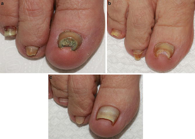
Although clinical studies use small samples, treatment with the Nd:YAG laser can be an alternative used alone or in combination with other treatments (Sá Guimarães 2014). Currently, some neodymium-doped yttrium aluminum garnet (Nd:YAG) laser devices have been approved for temporary whitening of the nails: Pinpointe Footlaser (Nuvolase), GenesisPlus (Cutera), Q-Clear (Light Age), CoolTouch VARIA (CoolTouch), and Joule ClearSense (Sciton) (Ortiz et al. 2014; Kimura et al. 2012; Hochman et al. 2011). The Joule ClearSense tip (Sciton equipment) emits a 6 mm spot, with proficiency ranging from 5 to 6 J/cm2, a pulse duration of 0.3 ms, 4.0 Hz, and real-time temperature control measured through the infrared thermometer built into the handpiece. Application is performed in concentric circles repeated until a temperature between 42 and 44 °C is reached. Three initial sessions are performed at weekly intervals, followed by monitoring until complete growth of the treated nails, with intervals ranging from 1 to 3 months according to the intensity of nail involvement and clinical improvement (Fig. 9).
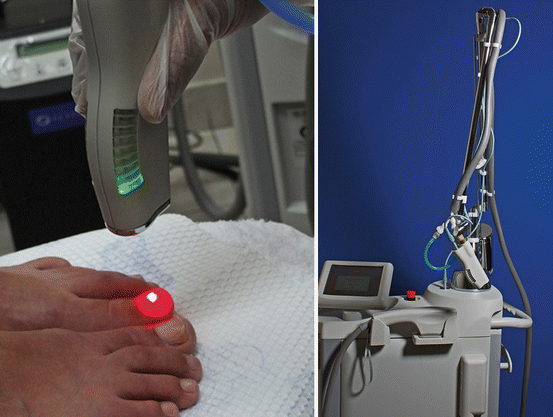
Prevention of recurrences can be achieved with long-term application of antifungal powder with miconazole 2% to the feet and shoes (Warshaw et al. 2005). This measure treats and prevents moccasin-type tinea pedis, usually caused by T. rubrum or T. mentagrophytes (interdigitalis).
The 10,600 nm CO2 Laser
The CO2 laser has water as its main chromophore. In continuous mode, it is used at parallel points (3 or 4 W, in continuous mode, with control of the cut perpendicular to the pedal) in order to cut the plate while preserving the nail bed. Cutting the hyperkeratotic nail facilitates collection of material for culture and surgical debridement.
The experimental use of the fractional CO2 laser as a means of drug delivery was described by Haedersdal et al. (2010). In this experiment, methyl 5-aminolevulinate (MAL), a porphyrin precursor, was used as the test drug. Significant absorption deep into the skin was observed, with creation of intradermal channels by the 3 mm laser and increased concentration of porphyrin in the hair follicles due to diffusion of MAL in the tested area (Haedersdal et al. 2010).
The fractional CO2 laser can be used in the treatment of onychomycosis caused by Fusarium spp., with application (12 W, 1,000 μm spacing, 700 μs pulse duration, and five stacks; Smartxide, Deka laser) along the entire length of the nail plate and eponychium, followed by application of a cream with tetracycline and topical amphotericin B (occlusive dressing at night) (Lurati et al. 2011), followed by morning brushing with hydrogen peroxide 10 vol (Garcez et al. 2010) until complete nail growth. Application of amphotericin B produces a brown pigmentation on the diseased part of the nail, where debris of dehydrated appearance is present. This coloration serves as an auxiliary chromophore for application of the Nd:YAG laser, causing a greater concentration of heat in these areas.
Conclusions
Anders et al. (2015) suggest that the term photobiomodulation therapy would be “a form of light therapy which uses non-ionizing forms of light sources, including lasers, LEDs, and broadband light, in visible light and infrared. It is a nonthermal process that involves endogenous chromophores that promote photophysical and photochemical events at multiple biological scales.” This therapy is applied to reduce pain, inflammation, and for immune modulation, and it promotes wound healing and tissue regeneration (Anders et al. 2015). Meral et al. (2003), Ortiz et al. (2014), Vila et al. (2014), and Vural et al. (2008) found that the lights and lasers used in photobiomodulation are able to inhibit bacterial and fungal growth. Research studies are only beginning, and they will certainly bring new solutions to infections caused by biofilms from different sources.
Take Home Messages
- The rate of clinical cure with oral medications reaches slightly over 50% of cases, while topical treatments do not reach 20% cure rates.
- Nondermatophyte fungi have shown an increasing incidence and are considered more difficult to treat.
- Biofilm formation was demonstrated in vitro for the primary causative agent of onychomycosis.
- The Nd:YAG laser stimulates the formation of collagen fibers, improves microcirculation, and acts as an antibacterial and antifungal agent.
- The fractional CO2 laser can be used for drug delivery.
References
Anders JJ, Lanzafame RJ, Arany PR. Low-level light/laser therapy versus photobiomodulation therapy. Photomed Laser Surg. 2015;33(4):183–4.
Andes DR, Nett J, Oschel P, Albrecht R, Marchillo K, Pitula A. Development and characterization of an in vivo central venous catheter Candida albicans biofilm model. Infect Immun. 2004;72:6023–31.
Araujo AJG, Souza MAJ. Onicomicoses por fungos emergentes: análise crítica, diagnóstico laboratorial e revisão. An Bras Dermatol. 2003;78(4):445–55.
Bizerra FC, Nakamura CV, De Poersch C, Estivalet Svidzinski TI, Borsato Quesada RM, Goldenberg S, Krieger MA, Yamada-Ogatta SF. Characteristics of biofilm formation by Candida tropicalis and antifungal resistance. FEMS Yeast Res. 2008;8:442–50.
Bloom W, Fawcett DW. Tratado de Histologia. Rio de Janeiro: Interamericana; 1977.
Bornstein E. A review of current research in light-based technologies for treatment of podiatric infectious disease states. JAPMA. 2009;99:348–52.
Bornstein ES, Hermann W, Gridley S. Near-infrared photoinactivation of bacteria and fungi at physiologic temperatures. Photochem Photobiol. 2009a;85:1364–74.
Bornstein E, Hermans W, Gridley S, Manni J. Near-infrared photoinactivation of bacteria and fungi at physiologic temperatures. Photochem Photobiol. 2009b;85:1364–74.
Bueno JG, Martinez C, Zapata B, Sanclemente G, Gallego M, Mesa AC. In vitro activity of fluconazole, itraconazole, voriconazole and terbinafine against fungi causing onychomycosis. Clin Exp Dermatol. 2010;35:658–63.
Burkhart CN, Burkhart CG, Gupta AK. Dermatophytoma: recalcitrance to treatment because of existence of fungal biofilm. J Am Acad Dermatol. 2002;47:629–31.
Cannizzo FT, Eraso E, Ezkurra PA, Villar-Vidal M, Bollo E, Castellá G, Cabañes FJ, Vidotto V, Quindós G. Biofilm development by clinical isolates of Malassezia pachydermatis. Med Mycol. 2007;45:357–61.
Carney C, Tosti A, Daniel R, Scher R, Rich P, DeCoster J, Elewski B. A new classification system for grading the severity of onychomycosis. Arch Dermatol. 2011;147(11):1277–82.
Chandra J, Kuhn DM, Mukherjee PK, Hoyer LL, McCormick T, Ghannoum MA. Biofilm formation by the fungal pathogen Candida albicans: development, architecture, and drug resistance. J Bacteriol. 2001;183:5385–94.
Chiacchio N, Kadunc BV, Almeida ART, Madeira CL. Nail abrasion. J Cosmet Dermatol. 2004;2:150–2.
Costa-Orlandi CB, Sardi JCO, Santos CT, Fusco-Almeida AM, Mendes-Giannini MJS. In vitro characterization of Trichophyton rubrum and T. mentagrophytes biofilms. Biofouling. 2014;30:719–27.
Costerton JW, Lewandowski Z, Caldwell DE, Korber DR, Lappin-Scott HM. Microbial biofilms. Annu Rev Microbiol. 1995;49:711–45.
Cushion MT, Collins MS, Linke MJ. Biofilm formation by Pneumocystis spp. Eukaryot Cell. 2009;8:197–206.
D’Antonio D, Parruti G, Pontieri E, Di Bonaventura G, Manzoli L, Sferra R, Vetuschi A, Piccolomini R, Romano F, Staniscia T. Slime production by clinical isolates of Blastoschizomyces capitatus from patients with hematological malignancies and catheter-related fungemia. Eur J Clin Microbiol Infect Dis. 2004;23:787–9.
Dang YY, Ren QS, Liu HX, Ma JB, Zhang JS. Comparison of histologic, biochemical and mechanical properties of murine skin treated with 1,064 nm and 1,320 nm Nd:YAG lasers. Exp Dermatol. 2005;14:876–82.
Davis LE, Cook G, Costerton JW. Biofilm on ventriculoperitoneal shunt tubing as a cause of treatment failure in coccidioidal meningitis. Emerg Infect Dis. 2002;8:376–9.
Dayan SH, Vartanian J, Mernaker G, Mobley SR, Dayan AN. Nonablative laser resurfacing using the low-pulse (1,064 nm) Nd:YAG laser. Arch Facial Plast Surg. 2003a;5:310–5.
Dayan S, Damrose JF, Bhattacharyya K, Mobley SR, Patel MK, O’Grady K, Mandrea S. Histological evaluations following 1,064 nm Nd:YAG laser resurfacing. Lasers Surg Med. 2003b;33:126–31.
de Araújo AJ, Souza MAJ, Bastos OM, de Oliveira JC. Ocorrência de onicomicose em pacientes atendidos em consultórios dermatológicos da cidade do Rio de Janeiro. An Bras Dermatol. 2003;78:299–308.
Di Bonaventura G, Pompilio A, Picciani C, Iezzi M, D’Antonio D, Piccolomini R. Biofilm formation by the emerging fungal pathogen Trichosporon asahii: development, architecture, and antifungal resistance. Antimicrob Agents Chemother. 2006;50:3269–76.
Donlan RM, Costerton JW. Biofilms: survival mechanisms of clinically relevant microorganisms. Clin Microbiol Rev. 2002;15:167–93.
Ferreira JAG, Carr JH, Starling CEF, de Resende MA, Donlan RM. Biofilm formation and effect of caspofungin on biofilm structure of Candida species bloodstream isolates. Antimicrob Agents Chemother. 2009;53:4377–84.
Finch J, Arenas R, Baran R. Fungal melanonychia. J Am Acad Dermatol. 2012;66(5):830–41.
Flemming HC, Wingender J. The biofilm matrix. Nat Rev Microbiol. 2010;8:623–33.
Garcez AS, Núñez SC, Baptista MS, Daghastanli NA, Itri R, Hamblin MR, Ribeiro MS. Antimicrobial mechanisms behind the photodynamic effect in the presence of hydrogen peroxide. Photochem Photobiol Sci. 2010;10(4):483–90.
Ghannoum MA, Hajjeh RA, Scher R, Konnikov N, Gupta AK, Summerbell R, Sullivan S, Daniel R, Krusinski P, Fleckman P, Rich P, Odom R, Aly R, Parise D, Zaiac M, Rebell G, Lesher J, Gerlach B. A large-scale North American study of fungal isolates from nails: the frequency of onychomycosis, fungal distribution, and antifungal susceptibility patterns. J Am Acad Dermatol. 2000;43(4):641–8.
Guffey JS, Motts S, Payne W. Using visible and near-IR light to facilitate photobiomodulation: a review of current research. Eur J Acad Essay. 2014;1(3):113–6.
Haedersdal M, Sakamoto FH, Farinelli WA, Doukas AG, Tam J, Anderson RR. Fractional CO2 laser-assisted drug delivery. Lasers Surg Med. 2010;42:113–22.
Hawser SP, Douglas LJ. Biofilm formation by Candida species on the surface of catheter materials in vitro. Infect Immun. 1994;62:915–21.
Hochman LG. Laser treatment of onychomycosis using a novel 0.65-millisecond pulsed Nd:YAG 1,064-nm laser. J Cosmet Laser Ther. 2011;13:2–5.
Hwang SM, Suh MK, Ha GY. Onychomycosis due to nondermatophytic molds. Ann Dermatol. 2012;24(2):175–80.
Imamura Y, Chandra J, Mukherjee PK, Lattif AA, Szczotka-Flynn LB, Pearlman E, Lass JH, O’Donnell K, Ghannoum MA. Fusarium and Candida albicans biofilms on soft contact lenses: model development, influence of lens type, and susceptibility to lens care solutions. Antimicrob Agents Chemother. 2008;52:171–82.
Kimura U, et al. Treating onychomycosis of the toenail: clinical efficacy of the sub-millisecond 1,064 nm Nd:YAG laser using a 5 mm spot diameter. J Drugs Dermatol. 2012;11(4):496–504.
Kipshidze N, Nikolaychik V, Muckerheide M, Keelan MH, Chekanov V, Maternowski M, Chawla P, Hernandez I, Iyers S, Danga G, Sahota H, Leon MB, Rowbin G, Moses JW. Effect of short-pulsed nonablative infrared laser irradiation on vascular cells in vitro and neointimal hyperplasia in a rabbit balloon injury model. Circulation. 2001;104:1850–5.
Knappe V, Frank F, Rohde E. Principles of lasers and biophotonic effects. Photomed Laser Surg. 2004;22(5):411–7.
Koh BK, Lee CK, Chae K. Photorejuvenation with submillisecond neodymium-doped yttrium aluminum garnet (1,064 nm) laser: a 24-week follow-up. Dermatol Surg. 2010;36:1–8.
Kosarev J, Vizintin Z. Novel laser therapy in treatment of onychomycosis. J LAHA. 2010;1:1–8.
Kuhn DM, George T, Chandra J, Mukherjee PK, Ghannoum MA. Antifungal susceptibility of Candida biofilms: unique efficacy of amphotericin B lipid formulations and echinocandins. Antimicrob Agents Chemother. 2002a;46:1773–80.
Kuhn DM, Chandra J, Mukherjee PK, Ghannoum MA. Mechanism of fluconazole resistance in Candida albicans biofilms: phase-specific role of efflux pumps and membrane sterols. Infect Immun. 2002b;71:878–88.
Kumamoto CA. Candida biofilms. Curr Opin Microbiol. 2002;5:608–11.
Landsman AS, Robbins AH, Angelini PF, Wu CC, Cook J, Oster M, Bornstein ES. Treatment of mild, moderate, and severe onychomycosis using 870 and 930 nm light exposure. J Am Podiatr Med Assoc. 2010;100(3):166–77.
Lattif AA, Mukherjee PK, Chandra J, Swindell K, Lockhart SR, Diekema DJ, Pfaller MA, Ghannoum MA. Characterization of biofilms formed by Candida parapsilosis, C. metapsilosis, and C. orthopsilosis. Int J Med Microbiol. 2010;300:265–70.
Lattif AA, Mukherjee PK, Chandra J, Roth MR, Welti R, Rouabhia M, Ghannoum MA. Lipidomics of Candida albicans biofilms reveals phase-dependent production of phospholipid molecular classes and a role for lipid rafts in biofilm formation. Microbiology. 2011;157:3232–42.
Liu D, Coloe S, Baird R, Pedersen J. Application of PCR to the identification of dermatophyte fungi. J Med Microbiol. 2000;49:493–7.
Lurati M, Rosselet FB, Vernez M, Spring P, Bontems O, Fratti M, Monod M. Efficacious treatment of non-dermatophyte mould onychomycosis with topical amphotericin B. Dermatology. 2011;223:289–92.
Martinez LR, Casadevall A. Cryptococcus neoformans biofilm formation depends on surface support and carbon source and reduces fungal cell susceptibility to heat, cold, and UV light. Appl Environ Microbiol. 2007;73:4592–601.
Martins M, Uppuluri P, Thomas DP, Cleary IA, Henriques M, Lopez-Ribot JL, Oliveira R. Presence of extracellular DNA in the Candida albicans biofilm matrix and its contribution to biofilms. Mycopathologia. 2009;169:323–31.
Martins M, Henriques M, Lopez-Ribot JL, Oliveira R. Addition of DNase improves the in vitro activity of antifungal drugs against Candida albicans biofilms. Mycoses. 2012;55:80–5.
Meral G, et al. Factors affecting the antibacterial effects of Nd:YAG laser in vivo. Lasers Surg Med. 2003;32:197–202.
Montagna W, Parakkal LPF. The structure and function of skin. New York: Academic; 1974.
Morales-Cardona CA, Valbuena-Mesa MC, Alvarado Z, Solorzano-Amador A. Non-dermatophyte mould onychomycosis: a clinical and epidemiological study at a dermatology referral centre in Bogota, Colombia. Mycoses. 2014;57:284–93.
Mowat E, Williams C, Jones B, McChlery S, Ramage G. The characteristics of Aspergillus fumigatus mycetoma development: is this a biofilm? Med Mycol. 2009;47(Suppl 1):S120–6.
Mukherjee PK, Chandra J. Candida biofilm resistance. Drug Resist Updat. 2004;7:301–9.
Mukherjee PK, Chandra J, Yu C, Sun Y, Pearlman E, Ghannoum MA. Characterization of Fusarium keratitis outbreak isolates: contribution of biofilms to antimicrobial resistance and pathogenesis. Invest Ophthalmol Vis Sci. 2012;53:4450–7.
Nett JE, Crawford K, Marchillo K, Andes DR. Role of Fks1p and matrix glucan in Candida albicans biofilm resistance to an echinocandin, pyrimidine, and polyene. Antimicrob Agents Chemother. 2010a;54:3505–8.
Nett JE, Sanchez H, Cain MT, Andes DR. Genetic basis of Candida biofilm resistance due to drug-sequestering matrix glucan. J Infect Dis. 2010b;202:171–5.
Nucci M, Anaissie E. Fusarium infections in immunocompromised patients. Clin Microbiol Rev. 2007;20:695–704.
Nunes JM, Bizerra FC, Ferreira RCE, Colombo AL. Molecular identification, antifungal susceptibility profile, and biofilm formation of clinical and environmental Rhodotorula species isolates. Antimicrob Agents Chemother. 2013;57:382–9.
Nusbaum AG, Kirsner RS, Charles CA. Biofilms in dermatology. Skin Therapy Lett. 2012;17:1–5.
Ortiz AE, Avram MM, Wanner MA. A review of lasers and light for the treatment of onychomycosis. Lasers Surg Med. 2014;46:117–24.
Ortoneda M, Capilla J, Pastor FJ, Pujol I, Guarro J. In vitro interactions of licensed and novel antifungal drugs against Fusarium spp. Diagn Microbiol Infect Dis. 2004;48:69–71.
Pariser D, Elewski B, Rich P, Scher RK. Update on onychomycosis: effective strategies for diagnosis and treatment. Semin Cutan Med Surg. 2013;32(2S):S1–13.
Pasquini C. Near-infrared spectroscopy: fundamentals, practical aspects and analytical applications. J Braz Chem Soc. 2003;14(2):198–219.
Perumal P, Mekala S, Chaffin WL. Role for cell density in antifungal drug resistance in Candida albicans biofilms. Antimicrob Agents Chemother. 2007;51:2454–63.
Pitangui NS, Sardi JCO, Silva JF, Benaducci T, Moraes da Silva RA, Rodríguez-Arellanes G, Taylor ML, Mendes-Giannini MJS, Fusco-Almeida AM. Adhesion of Histoplasma capsulatum to pneumocytes and biofilm formation on an abiotic surface. Biofouling. 2012;28:711–8.
Ramage G, Van de Walle K, Wickes BL, Lopez-Ribot JL. Characteristics of biofilm formation by Candida albicans. Rev Iberoam Micol. 2001;18:163–70.
Ramage G, Bachmann S, Patterson TF, Wickes BL, López-Ribot JL. Investigation of multidrug efflux pumps in relation to fluconazole resistance in Candida albicans biofilms. J Antimicrob Chemother. 2002;49:973–80.
Ramage G, Saville SP, Thomas DP, López-Ribot JL. Candida biofilms: an update. Eukaryot Cell. 2005;4:633–8.
Ramage G, Rajendran R, Sherry L, Williams C. Fungal biofilm resistance. Int J Microbiol. 2012;2012:1–14.
Ranawaka RR, Silva N, Ragunathan RW. Non-dermatophyte mold onychomycosis in Sri Lanka. Dermatol Online J. 2012;18(1):7.
Reynolds TB, Fink GR. Bakers’ yeast, a model for fungal biofilm formation. Science. 2001;291:878–81.
Ross V, Cooke LM, Timko AL, Overstreet KA, Graham BS, Barnette DJ. Treatment of pseudofolliculitis barbae in skin types IV, V, and VI with a long-pulsed neodymium:yttrium aluminum garnet laser. J Am Acad Dermatol. 2002;47:263–70.
Sá Guimarães CMD. Tratamento da onicomicose com laser Nd:YAG: resultados em 30 pacientes. Surg Cosmet Dermatol. 2014;6(2):155–60.
Sardi JCO, Pitangui NDS, Rodríguez-Arellanes G, Taylor ML, Fusco-Almeida AM, Mendes-Giannini MJS. Highlights in pathogenic fungal biofilms. Rev Iberoam Micol. 2014;31:22–9.
Scher RK, Tavakkol A, Sigurgeirsson B. Onychomycosis: diagnosis and definition of cure. J Am Acad Dermatol. 2007;56:939–44.
Schmults CD, Phelps R, Goldberg DJ. Nonablative facial remodeling. Arch Dermatol. 2004;140:1373–6.
Seneviratne C, Jin L, Samaranayake L. Biofilm lifestyle of Candida: a mini review. Oral Dis. 2008;14:582–90.
Sigurgeirsson B. Prognostic factors for cure following treatment of onychomycosis. J Eur Acad Dermatol Venerol. 2010;24:679–84.
Silva S, Negri M, Henriques M, Oliveira R, Williams DW, Azeredo J. Adherence and biofilm formation of non-Candida albicans Candida species. Trends Microbiol. 2011;19:241–7.
Singh R, Shivaprakash MR, Chakrabarti A. Biofilm formation by zygomycetes: quantification, structure and matrix composition. Microbiology. 2011;157:2611–8.
Snell RS. Histologia Clínica. Rio de Janeiro: Interamericana; 1985.
Tan M, Dover J, Hsu T, Arndt KA, Stewart B. Clinical evaluation of enhanced nonablative skin rejuvenation using a combination of 532 nm and 1,064 nm laser. Lasers Surg Med. 2004;34:349–55.
Vediyappan G, Rossignol T, d’Enfert C. Interaction of Candida albicans biofilms with antifungals: transcriptional response and binding of antifungals to beta-glucans. Antimicrob Agents Chemother. 2010;54:2096–111.
Vila TVM, Rosental S, de Sá Guimarães CMD. A new model of in vitro fungal biofilms formed on human nail fragments allows reliable testing of laser and light therapies against onychomycosis. Lasers Med Sci. 2014;30(3):1031–9.
Vlassova N, Han A, Zenilman JM, James G, Lazarus GS. New horizons for cutaneous microbiology: the role of biofilms in dermatological disease. Br J Dermatol. 2011;165:751–9.
Vural E, Winfield HL, Shingleton AW, Horn TD, Shafirstein B. The effects of laser irradiation on Trichophyton rubrum growth. Lasers Med Sci. 2008;23:349–53.
Warshaw EM, St. Clair KR. Prevention of onychomycosis reinfection for patients with complete cure of all 10 toenails: results of a double-blind, placebo-controlled pilot study of prophylactic miconazole powder 2%. J Am Acad Dermatol. 2005;53(4):717–20.
Weiss L, Greep RO. Histology. New York: McGraw-Hill; 1977.
Zarnowski R, Westler WM, Lacmbouh GA, Marita JM, Bothe JR, Bernhardt J, Lounes-Hadj AS, Fontaine J, Sanchez H, Hatfield RD, et al. Novel entries in a fungal biofilm matrix encyclopedia. MBio. 2014;5:1333–14.
Zhang X, Sun X, Wang Z, Zhang Y, Hou W. Keratitis-associated fungi form biofilms with reduced antifungal drug susceptibility. Invest Ophthalmol Vis Sci. 2012;53:7774–8.







