
Lasers for Aesthetic and Functional Vaginal Rejuvenation
Lasers for Aesthetic and Functional Vaginal Rejuvenation
André Vinícius de Assis Florentino, Thales Lage Bicalho Bretas and Maria Claudia Almeida Issa Department of Gynecology, Faculdade de Ciências Médicas de Campina Grande, Campina Grande, PB, 58411-020, Brazil Brazilian Federation of Gynecology and Obstetrics, São Paulo, SP, Brazil Universidade Federal Fluminense, Niterói, RJ, Brazil Department of Clinical Medicine – Dermatology, Fluminense Federal University, Niterói, RJ, Brazil
Abstract
Vaginal rejuvenation has popped up recently due to the increase in life expectancy and the search for a better quality of life. Nowadays, women live one-third of their lives after menopause, demanding efforts to keep their sexual life up to their body and mind’s health. Advances in laser therapy have made this possible, reversing vulvovaginal atrophy and its symptoms, known as genitourinary syndrome of menopause, including vaginal dryness, dyspareunia, decrease in sexual arousal and orgasm, stress urinary incontinence, and vaginal bleeding. Carbon dioxide laser (CO2) and the erbium laser (Er:YAG) are the most common lasers used for female rejuvenation. They promote neocollagenesis and neovascularization in the connective tissue of the vaginal wall, as well as recover the mucosal epithelium, restoring lubrication and elasticity of vaginal walls.
Keywords CO2 laserErbium laserEr:YAGGenitourinary syndrome of menopauseIntimate laserMenopauseRadiofrequencyUrinary incontinenceVaginal laxityVaginal rejuvenationVulvovaginal atrophy
Introduction
Vaginal rejuvenation is commonly defined as a combination of minimally invasive procedures that stimulate regeneration of the female lower genital tract, aiming to regain aesthetic and functional features lost with the aging process in menopausal women. It is also indicated to treat vagina relaxation in young women.
Vagina relaxation, for example, is one of the biggest responsibilities for sexual dissatisfaction. It represents the loss of the optimum structural architecture of the vagina, generally associated with natural aging and especially affected by childbirth, whether vaginal or not. Multiple pregnancies increase the alteration of these structures, making vaginal muscles relaxed with poor tone, strength, control, and support. The vaginal canal becomes wider and stretched, and sexual gratification consequently diminishes, since it has been attributed to frictional forces generated during intercourses. Therefore, many women, especially after menopause, seek vaginal treatments to regain their sexual health and well-being (Sturdee et al. 2010).
As we know, menopause is the cessation of menstruation for more than 1 year, reflecting the depletion of ovarian function. As the life expectancy of women has increased, nowadays approximately one-third of a woman’s life is postmenopausal (NAMS 2013). The progressive cessation of circulating estrogen levels leads to metabolic and tissue changes, more evident in the genital tract due to its particular sensitivity to variations in sexual hormone levels. With menopause, the vaginal epithelium starts to get thinner and vaginal walls become less elastic with loss of rugations, looking more pale, dry, and friable at the specular exam, often bleeding after minimal trauma. The entire vaginal canal becomes shorter and narrower. The vulvar area, particularly the clitoris, becomes atrophic and more vulnerable (Mehta and Bachmann 2008). These alterations lead to a constellation of symptoms grouped as genitourinary syndrome of menopause (GSM).
The GSM is a new term in substitution for vulvovaginal atrophy as agreed by the North American Menopause Society and the International Society for the Study of Women’s Sexual Health (Portman et al. 2014). This syndrome is characterized by a combination of symptoms such as vaginal pain during sexual intercourse (dyspareunia), itching, urinary incontinence, dysuria, and the main complaint of aging women: vaginal dryness, an early symptom. The GSM affects around 20–45% of women (Santoro and Komi 2009) and reflects directly on the general quality of life, mostly causing a profound negative impact in the sexual field.
Vaginal health can be objectively assessed by the vaginal health index score (VHIS). The VHIS evaluates the appearance of the vaginal mucosa (elasticity, pH, vaginal discharge, mucosal integrity, and moisture). Each parameter is graded from 1 to 5; the higher the score, the better the vaginal health. If the total score is smaller than 15, the vagina is considered atrophic (Bachmann et al. 1992) – see Table 1.
| Parameter | 1 | 2 | 3 | 4 | 5 |
|---|---|---|---|---|---|
| Elasticity | None | Poor | Fair | Good | Excellent |
| Vaginal discharge (fluid volume) | None | Scant amount, vault not entirely covered | Superficial amount, vault entirely covered | Moderate amount of dryness (small areas of dryness on cotton tip applicator) | Normal amount (fully saturates on cotton tip applicator) |
| pH | >6.0 | 5.6–6.0 | 5.1–5.5 | 4.7–5.0 | <4.7 |
| Mucosal integrity | Petechiae noted before contact | Bleeds with light contact | Bleeds with scraping | Not friable – thin epithelium | Normal |
| Moisture | None, surface inflamed | None, surface not inflamed | Minimal | Moderate | Normal |
There are several treatment options to minimize GSM symptoms, including nonhormonal products for mild cases, local vaginal hormone therapy for persistent symptoms, and systemic hormonal replacement therapy (HRT) as a broader approach for severe symptoms (Sturdee et al. 2010).
The nonhormonal therapy is mostly based on the use of vaginal lubricants and moisturizers on a regular basis, providing only temporary relief prior to intercourse. Lubricants have been demonstrated to decrease vaginal irritation during sexual activity but do not provide a long-term solution (Bygdeman and Swahn 1996).
Low-dose local estrogen therapies can be useful for women with GSM symptoms without systemic climacteric complaints, in the absence of contraindications, such as a personal history of either endometrial or breast cancer. The major disadvantage of this approach is the recurrence of symptoms once it has been suspended, generating a dependency relation among women.
The systemic HRT relieves not only the vaginal symptoms but also general complaints like hot flushes, mood lability, and sleep disturbances. It is not indicated for exclusive GSM, since it has more contraindications and side effects than the local HRT, such as the increased risk of thrombosis and breast/endometrial cancer (Santoro and Komi 2009).
In this context, vaginal laser (an acronym for “light amplification by stimulated emission of radiation”) therapy appears as a feasible option for women desiring nonhormonal therapy with longer efficacy compared to topical local therapy. The laser device is a coherent, collimated, and monochromatic source of light, which is absorbed by tissue according to its light absorbance coefficient. Considering vaginal indications, the most used kinds of lasers are the fractional carbon dioxide (CO2) and erbium-doped yttrium aluminum garnet (Er:YAG) lasers, both of them targeting the water of the tissue through different wavelengths: 10,600 nm for the CO2 and 2,940 nm for the Er:YAG laser (Vizintin et al. 2012).
History
The concept of stimulated light was conceived by Albert Einstein back in 1905, in a phenomenon called the photoelectric effect. The first laser was constructed in 1960, a chromium-ruby device. The first carbon dioxide (CO2) laser was built in 1964 by Patel and his team.
Laser is considered one of the most versatile therapeutic instruments in several medical fields. In dermatology, fractional lasers like Er:YAG and CO2 lasers have been used mainly for resurfacing, rejuvenation, and scar improvement, with significant effects in the connective tissue, stimulating tissue remodeling, with similar effects being reproducible within the vaginal epithelium (Sasaki et al. 2009).
Gynecologists have been using lasers successfully to treat vaginal and cervical pathologies over the past five decades (Kaplan et al. 1973). In this last decade, a wave of publications involving vaginal rejuvenation has caught the attention of the scientific community worldwide. The mechanism of action involves neocollagenesis and restoration of the trabecular architecture of the vaginal wall, regaining the appearance observed in premenopausal women.
The CO2 and the Er:YAG lasers are among the most studied and spoken technologies aiming at vaginal rejuvenation. More recently, radiofrequency has also gained some strength in treating this area, with prominent findings, especially using a transcutaneous temperature-controlled radiofrequency for vulvovaginal rejuvenation (Vanaman et al. 2016).
Mechanisms of Action
The medical use of laser technology is based on the interaction between the light emitted and tissue chromophores, including hemoglobin, melanin, connective tissue, and water (Alexiades-Armenakas et al. 2008), which depends on the different absorption of light in the electromagnetic spectrum.
The vaginal wall is composed of four layers: squamous epithelium, lamina propria, muscular layer, and the adventitia. Like the epidermis, the squamous epithelium has basal and suprabasal layers, and differentiates to form a cornified envelope comprised of a flattened layer of specialized cells. Electron microscopy shows lipid lamella, but they do not form an impermeable intercellular lipid envelope as they do in the epidermis, making the vaginal wall permeable to water and soluble proteins. Also unlike the epidermis, the vaginal epithelium usually is not keratinized and stores glycogen. The synthesis of glycogen is diminished by estrogen cessation after menopause, as well as the epithelial thickness already mentioned (Anderson et al. 2014).
The CO2 laser is a fractional, ablative laser that emits light at the wavelength of 10,600 nm, which is strongly absorbed by the tissue’s water (Fisher 1992), promoting microthermal ablation of the epithelium (the so-called microthermal zones – MTZ), with preservation of healthy tissue islands between the ablated areas. This ablation induces the body to repair and promote re-epithelization of the vaginal tissue, restoring the flora, the thickness, and lubrication. It also causes an important heating deep in the dermis surrounding the ablated tissue, which, in turn, promotes neocollagenesis and reorganization of the elastic and collagen fibers of the connective tissue, restoring the healthy architecture of the vagina. Differently from local therapies, the fractional CO2 laser can act in deep layers stimulating collagen synthesis (Gaspar et al. 2011).
The Er:YAG laser is a fractional, nonablative laser that emits light at the wavelength of 2,940 nm, which is also absorbed by the tissue’s water, with 15 times more affinity than the CO2, with less recovery downtime but also less collagen production, since its rays reach smaller depths. It induces photothermal heating of the vaginal wall without ablating its surface, which boosts neocollagenesis and remodeling of the connective tissue and also stimulates regrowth of the epithelium, but with comparatively less thermal injury and less mucosa swelling than the CO2 laser (Gaviria and Lanz 2012).
The Er:YAG 2,940 nm Smooth Mode is a modified erbium 2,940 nm mode in which laser energy is delivered onto the mucosa tissue in a fast sequence of low-fluence laser pulses inside an overall super-long pulse of several hundred milliseconds. The delivered laser energy thus results in an overall nonablative buildup of heat and creates a temperature increase within the mucous tissue. With this Er:YAG laser (IntimaLase™, Fotona®), human tissue can be nonablatively heated to a depth of 100 microns, which is just what is required for a depth-controlled thermal treatment of vaginal mucosa tissue (Vizintin et al. 2012).
However, through diverse ways, Er:YAG and CO2 lasers share some similarities in their mechanism of action, since both target the water, resulting in tissue heating. This mechanism involves a controlled heat-shock response that stimulates the production of a small family of proteins called the heat-shock proteins. Heat-shock proteins 43, 47, and 70 (a protein subtype acting as chaperone of collagen, which is overexpressed after laser irradiation) appear to play an important role, stimulating the production of many growth factors. Among these factors are: transforming growth factor-A (stimulates matrix protein synthesis such as collagen), basic fibroblast growth factor (stimulates angiogenic activity with endothelial cell migration and proliferation), epidermal growth factor (stimulates re-epithelization), platelet-derived growth factor (stimulates fibroblasts to produce extracellular matrix components), and vascular endothelial growth factor (regulates vasculogenesis and angiogenesis).
Therefore, the laser stimuli activate fibroblasts to produce new collagen, other components of the extracellular matrix (proteoglycans, glycosaminoglycans, etc.), and new vessels, with specific effects on epithelial tissue (Prignano et al. 2009). Such a bodily response of recovery results in a thicker epithelium, neocollagenesis, reorganization of the trabecular architecture of the collagen, neovascularization with recovery of the papillary disposition of the subepithelial connective tissue, and regained production of mucopolysaccharides by the extracellular matrix (Salvatore et al. 2015). These alterations lead to reestablishment of vaginal health, with return of lubrication, amelioration of epithelial pallor, reversal of vaginal laxity, return of vaginal pH and microbiota, improvement in elasticity of the vaginal wall, and elevation of sexual arousal and satisfaction for both the woman and her partner.
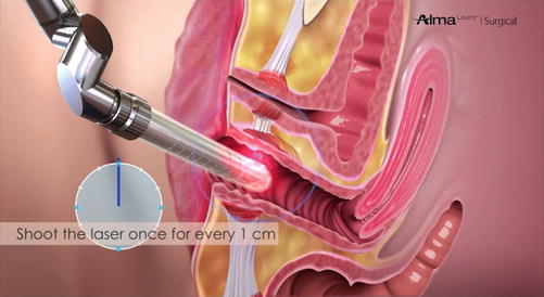
In our practice, we often use the CO2 laser Femilift™ from Alma Lasers for vaginal rejuvenation, which has a special sterile, disposable, and individual cover for each patient. This facility makes the procedure more hygienic and allows the operator to perform multiple laser sessions subsequently, without the long wait for sterilization (Fig. 1).
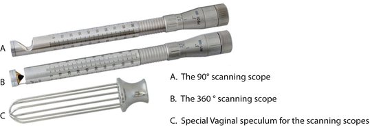
The Er:YAG laser we often use is either from Fotona (IntimaLase™ and IncontiLase™) or from LMG (Solon Femina™), and both have a laser speculum that avoids contact between the laser device and the vagina, but it needs to be sterilized before the next patient. The former, IntimaLase® and IncontiLase®, emit, respectively, a circumferential and an angular laser beam, to reach either the whole vaginal wall or selectively the anterior wall (for urinary incontinence) – Fig. 2.
Indications
The vaginal laser is indicated for women between 20 and 80 years old with one of the following symptoms (Salvatore et al. 2014; Zerbinati et al. 2015; Gambacciani and Levancini 2015; Vizintin et al. 2015; Perino et al. 2016; Gambacciani et al. 2015):
- Hypotrophy and/or vaginal atrophy
- Mild to moderate urinary incontinence
- Sexual dysfunction such as dyspareunia, dryness, low vaginal sensitivity, and vaginal wall bleeding during intercourse
- Postpartum and lactation (temporarily reduced estrogen levels)
- Vagina relaxation/laxity
- Posttreatment for gynecological cancers (breast and endometrial), to improve vaginal symptoms from the lack of estrogen
Contraindications
- Pregnancy
- Bacterial or fungal vaginal infection – laser treatment can be performed only 30 days after treatment of the vaginal infection
- HPV infection
- Active herpes viral infection – laser treatment can be performed without active infection and during prophylaxis treatment
- Gynecological oncology pathologies
- Previous surgical orthesis implant for urinary incontinence, such as transvaginal mesh/sling
- Impaired immune system or chronic corticoid therapies
- Scleroderma, lichen sclerosus, vitiligo, or psoriasis
- Uncontrolled diabetes
- Anticoagulant therapy
- Patients who have used isotretinoin in the last 12 months (Salvatore et al. 2014; Zerbinati et al. 2015; Gambacciani and Levancini 2015; Vizintin et al. 2015; Perino et al. 2016; Gambacciani et al. 2015)
Orientations Prior to Treatment
Prior to treatment, patients should fill out the pretreatment questionnaire and sign the Informed Consent Form. It is highly recommended that the physician take a detailed patient medical history, including previous treatment modalities, and examine the gynecological condition for treatment suitability with the vaginal laser (Salvatore et al. 2014; Zerbinati et al. 2015; Gambacciani and Levancini 2015; Vizintin et al. 2015; Perino et al. 2016; Gambacciani et al. 2015).
It is also important to determine why the patient is seeking treatment and to understand her expectations. It is advisable to warn the patient that there may be very mild discomfort associated with the treatment.
Patients shall perform a pregnancy test at least 1 day before the session, and show a recent cervical screening test result (best if within less than 30 days). Patients with a personal history of previous genital herpes simplex infection should undergo the prophylaxis protocol, which is valacyclovir 500 mg every 12 h for 7 days, starting 24 h prior to the laser session. Acyclovir or famciclovir can also be used (Beeson and Rachel 2002).
Procedure
- The patient should be in the lithotomy position.
- Insert a disposable or sterile vaginal speculum to look for active lesions, signs of infection, or alterations of either the vaginal fluid or the mucosa. If a normal aspect is observed, dry the pathway with sterile gauze to avoid burning from water absorption.
- After taking out the speculum, carefully introduce the laser device completely, until you feel it touching the cervix or the patient complains of pain. Some lubricant oil, like mineral oil, might be used in the introit if it is too dry, avoiding the laser window. Usually, the laser probes are marked circumferentially and have a sign that represents the vaginal depth, between 7 and 13 cm. They also have a security distance from the tip to the laser window, which prevents irradiation of the patient’s cervix.
- Some devices have a 360° laser-beam delivery system, enabling 360° irradiation of the vaginal canal, whereas others carry a limited laser window, which needs to be systematically rotated clockwise to reach the totality of the vaginal wall circumference. These latter devices are also designed to make rotation easy and patterned, so the doctor is sure to perform the 360° irradiation.
- After the first circumferential deep shot, pull back the device as marked (1 cm), making subsequent shots until the whole marked area is outside the vagina. Patients usually report some heating sensation when the laser window comes closer to the vaginal introit. This is when you shall fully insert it again and perform a new pass, starting from the deep canal to the introit.
- The number of subsequent passes and laser sessions is also described by the manufacturer’s protocol, depending on the patient’s complaints and the results achieved by each session (Salvatore et al. 2014; Zerbinati et al. 2015; Gambacciani and Levancini 2015; Vizintin et al. 2015; Perino et al. 2016; Gambacciani et al. 2015).
Treatment Protocols
Each laser device has its own protocols, with the appropriate fluency and power indicated for the patient’s needs. Nevertheless, one rule is always true: the higher the power, the greater the depth and the magnitude of the thermal injury provoked.
If the main objective is tightening, for example, higher energies are required. The doctor must perform three consecutive passes, throughout the whole vaginal wall, circumferentially, in each laser session, with a total of three sessions spaced by a period of 30 days for the best outcomes.
The same protocol with high energy is also applied to urinary incontinence treatment. However, in these cases, at least two passes should be applied focusing on the anterior wall of the vagina (using a clock as a reference, the laser window must be focused between 10:00 and 02:00 o’clock), plus one circumferential pass.
When vaginal dryness and atrophy are the main complaints of the patient, lower energy is indicated. One or two circumferential passes are enough for each session. Three sessions are necessary for better results.
Posttreatment – What Is Recommended or Expected
- No sexual intercourse for 3–7 days
- No tampons for 3 days
- No healing cream is needed
- Some translucent or bloody discharge might occur within the 3–7 days following the procedure
- Be available for patients’ contact. Ask them to notify you in case of any bleeding, fever, or other unusual side effects (Salvatore et al. 2014; Zerbinati et al. 2015; Gambacciani and Levancini 2015; Vizintin et al. 2015; Perino et al. 2016; Gambacciani et al. 2015)
Vaginal Laser Rejuvenation – Literature Review
Over the last few years, much has been studied about the use of lasers for vaginal aging. In a 12-week treatment with fractional CO2 laser for GSM, this treatment has been proved efficacious, feasible, and safe in ameliorating the GSM symptoms (Salvatore et al. 2014). In 2015, research revealed restoration of the vaginal mucosa through remodeling and neosynthesis of collagen in the lamina propria (Zerbinati et al. 2015). In the same year, Gambacciani and colleagues showed that the vaginal erbium laser (VEL) induced subjective improvement in dryness, dyspareunia, and the overall VHIS in 65 postmenopausal women (PMW) after three monthly sessions. Twenty-one of them who also had mild-to-moderate stress urinary incontinence reported improvement, which was measured by their score on the International Consultation on Incontinence Questionnaire – Urinary Incontinence Short Form (ICIQ-UI SF) (Gambacciani and Levancini 2015). Also in 2015, another group demonstrated improvement in vaginal laxity, stress urinary incontinence, and GSM symptoms after Er:YAG laser treatment (Vizintin et al. 2015).
In 2016, a study involving 30 postmenopausal women complaining of GSM and overactive bladder (OAB) symptoms showed that, after three sessions of CO2 vaginal laser, there was significant improvement in OAB symptoms, such as a reduction in the number of micturitions and urge episodes (Perino et al. 2016). In the same year, 70 women with GSM and vestibulodynia were studied by Murina and colleagues. Patients underwent three sessions of treatment with microablative fractional CO2 laser, applied on the vulva and vestibular surface, resulting in statistically significant improvement in dyspareunia and pain scores (Murina et al. 2016). The benefits of CO2 laser treatment on the vaginal flora were also documented: the postmenopausal vaginal flora equilibrium was restored, with the predominance of Lactobacillus and a low pH, protecting women from vaginal infections (Athanasiou et al. 2016).
In 2017, a study presented 1-year results with fractional CO2 laser for genitourinary syndrome of menopause. It emphasized the advantages of the duration of positive effects and the low risk of adverse events, concluding that CO2 laser therapy was safe and effective for GSM (Sokol and Karram 2016).
Authors’ Experience
In our practice, we have experienced different technologies, but most of our results were obtained with the CO2 laser Femilift™, from Alma Lasers. These results are shown in the case reports below.
In case 1 (Fig. 3), a 62-year-old postmenopausal woman was submitted to three monthly sessions of CO2 Femilift™ laser, with the following parameters: 100 mJ per pixel, long-pulse, power: high, three circumferential (360°) passes per section.
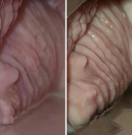
Case 2 (Fig. 4) shows the specular exam of a 44-year-old woman with premature menopause and a family history of breast cancer. The figure on the left shows the vaginal walls before the laser session: note the pallor, the dryness, and the absence of rugations. Three circumferential passes of the CO2 Femilift™ laser were performed, with the parameters: energy: 75 mJ per pixel, long-pulse, power: medium. The middle figure shows the vaginal wall immediately after the three laser passes: note the microthermal zones (MTZ) ablated in the mucosa, representing epithelium vaporization. After healing (right), see the improvement in pallor and rugations, 1 month after the first laser session.
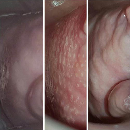
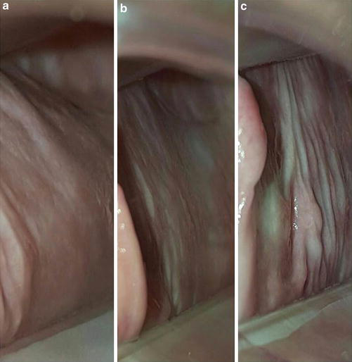
In case 3, we show the specular exam of a 58-year-old postmenopausal woman with vaginal dryness and dyspareunia (Fig. 5a–c). In Fig. 5a, the vaginal wall before any treatment: observe the pallor, the lack of rugae, and dryness. In Fig. 5b, the vaginal wall after the first session of CO2 laser, starting to present rugae and increased mucous secretion. In Fig. 5c, observe the vaginal wall 15 days after the second session of CO2, with healing areas (in white), a significant increase in vaginal rugae, and improvement in pallor and lubrication. The laser used was the Femilift™ with the parameters: energy: 100 mJ per pixel, long-pulse, power: high, three circumferential passes per section.
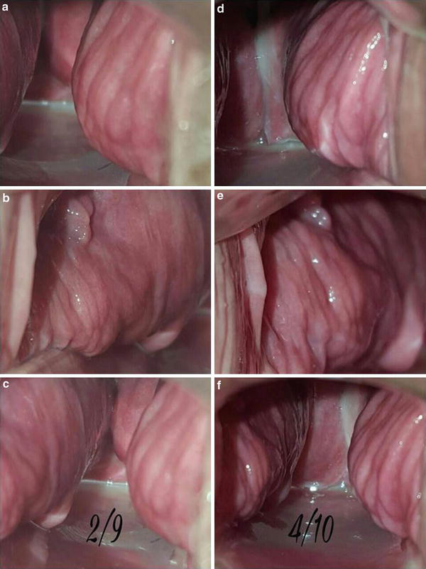
Case 4 (Fig. 6) illustrates a 57-year-old postmenopausal woman’s specular exam, showing, in Fig. 6a–c, the right, the left, and both vaginal walls, respectively, before the Er:YAG 2,940 nm Femina™ laser treatment. On the right, in Fig. 6d–f, we can see the same walls 30 days after one laser session, with the 360° scope in multiple micropulse mode, 1.7 J delivered per shot, 3 multishots, 3 passes per session.
Conclusion
According to the literature review and our experience, laser treatment is a new and effective tool for vaginal rejuvenation. It is able to improve symptoms such as vaginal dryness, itching, urinary incontinence, dysuria, as well as vaginal pain during sexual intercourse.
Take Home Messages
- Almost half of menopausal women experience some symptoms of the genitourinary syndrome of menopause (GSM), including vaginal dryness, dyspareunia, urinary incontinence, and loss of sexual pleasure.
- The GSM is due to ovarian failure and the consequent lack of estrogen, whose replacement is not always a comfortable and feasible option for the patient.
- The vaginal lasers were developed to stimulate, via deep tissue heating, the regrowth of epithelium and dermal structures, with reorganization of collagen and elastin fibers, regained mucosal thickness and irrigation, and recovery of vaginal wall elasticity and lubrication, with a consequent increase in sexual pleasure.
- The CO2 and the Er:YAG lasers are the most studied and are proven to be efficient technologies. Radiofrequency has also been used with promising results.
- The candidate for treatment must have a Pap smear at least 30 days before the first laser session, as well as provide assurance of non-pregnancy status on the days of the laser sessions.
- There cannot be any sign of active infection or neoplasia at the sessions, making the specular exam an essential tool before the laser procedure.
- Previous herpes simplex infections require prophylactic treatment, starting 24 h prior to the laser session and kept for 7 days (until re-epithelization).
- Patients are oriented to refrain from sexual intercourse for 3–5 days after the laser. No healing cream is needed. Tampons are not recommended.
- The treatment consists of three monthly laser sessions, with a maintenance protocol of single annual sessions.
References
Alexiades-Armenakas MR, Dover JS, Arndt KA. The spectrum of laser skin resurfacing: nonablative, fractional, and ablative laser resurfacing. J Am Acad Dermatol. 2008;58:719–37.
Anderson DJ, Marathe J, Pudney J. The structure of the human vaginal stratum corneum and its role in immune defense. Am J Reprod Immunol. 2014;71:618–23.
Athanasiou S, Pitsouni E, Antonopoulou S, et al. The effect of microablative fractional CO2 laser on vaginal flora of postmenopausal women. Climacteric. 2016;19(5):512–8.
Bachmann GA, Notelovitz M, Kelly SJ, et al. Long-term nonhormonal treatment of vaginal dryness. Clin Pract Sex. 1992;8:3–8.
Beeson WH, Rachel JD. Valacyclovir prophylaxis for herpes simplex virus infection or infection recurrence following laser skin resurfacing. Dermatol Surg. 2002;28(4):331–6.
Bygdeman M, Swahn M. Replens versus dienoestrol cream in the symptomatic treatment of vaginal atrophy in postmenopausal women. Maturitas. 1996;23:259–63.
Fisher JC. Photons, physiatrics, and physicians. A practical guide to understanding laser light interaction with living tissue, part 1. J Clin Laser Med Surg. 1992;10(6):419–26.
Gambacciani M, Levancini M. Short-term effect of vaginal erbium laser on the genitourinary syndrome of menopause. Minerva Ginecol. 2015;67(2):97–102.
Gambacciani M, Levancini M, Cervigni M. Vaginal erbium laser: the second-generation thermotherapy for the genitourinary syndrome of menopause. Climacteric. 2015;18:757–63.
Gaspar A, Addamo G, Brandi H. Vaginal fractional CO2 laser: a minimally invasive option for vaginal rejuvenation. Am J Cosmet Surg. 2011;28:156–62.
Gaviria JE, Lanz JA. Laser vaginal tightening (LVT) – evaluation of a novel noninvasive laser treatment for vaginal relaxation syndrome. J Laser Health Acad. 2012;2012(1):59–66.
Kaplan I, Goldman J, Ger R. The treatment of erosions of the uterine cervix by means of the CO2 laser. Obstet Gynecol. 1973;41(5):795–6.
Mehta A, Bachmann G. Vulvovaginal complaints. Clin Obstet Gynecol. 2008;51(3):549–55.
Murina F, Karram M, Salvatore S, Felice R. Fractional CO2 laser treatment of the vestibule for patients with vestibulodynia and genitourinary syndrome of menopause: a pilot study. J Sex Med. 2016;13:1915.
NAMS. Management of symptomatic vulvovaginal atrophy: 2013 position statement of the North American Menopause Society. Menopause. 2013;20(9):888–902.
Perino A, et al. Is vaginal fractional CO2 laser treatment effective in improving overactive bladder symptoms in postmenopausal women? Preliminary results. Eur Rev Med Pharmacol Sci. 2016;20(12):2491–7.
Portman DJ, Gass ML, Vulvovaginal Terminology Consensus Conference Panel. Genitourinary syndrome of menopause: new terminology for vulvovaginal atrophy from the International Society for the Study of Women’s Sexual Health and the North American Menopause Society. Climacteric. 2014;17:557–63.
Prignano F, Campolmi P, Bonan P, et al. Fractional CO2 laser: a novel therapeutic device upon photobiomodulation of tissue remodelling and cytokine pathway of tissue repair. Dermatol Ther. 2009;22(Suppl 1):S8–S15.
Salvatore S, Nappi RE, Zerbinati N, et al. A 12-week treatment with fractional CO2 laser for vulvovaginal atrophy: a pilot study. Climacteric. 2014;17:363–9.
Salvatore S, Maggiore ULR, Athanasiou S, Origoni M, Candiani M, et al. Histological study on the effects of microablative fractional CO2 laser on atrophic vaginal tissue: an ex vivo study. Menopause. 2015;22(8):845–9.
Santoro N, Komi J. Prevalence and impact of vaginal symptoms among postmenopausal women. J Sex Med. 2009;6:2133–42.
Sasaki GH, Travis HM, Tucker B. Fractional CO2 laser resurfacing of photoaged facial and non-facial skin: histologic and clinical results and side effects. J Cosmet Laser Ther. 2009;11:190–201.
Sokol ER, Karram MM. An assessment of the safety and efficacy of a fractional CO2 laser system for the treatment of vulvovaginal atrophy. Menopause. 2016;23:1102–7.
Sturdee DW, Panay N, International Menopause Society Writing Group. Recommendations for the management of postmenopausal vaginal atrophy. Climacteric. 2010;13(6):509–22.
Vanaman M, Bolton J, Placik O, Fabi SG. Emerging trends in nonsurgical female genital rejuvenation. Dermatol Surg. 2016;42(9):1019–29.
Vizintin Z, Rivera M, Fistonić I, Saraçoğlu F, Guimarães P, et al. Novel minimally invasive VSP Er:YAG laser treatments in gynecology. J Laser Health Acad. 2012;2012(1):46–58.
Vizintin Z, Lukac M, Kazic M, Tettamanti M. Erbium laser in gynecology. Climacteric. 2015;18(Suppl 1):4–8.
Zerbinati N, Serati M, Origoni M, et al. Microscopic and ultrastructural modifications of postmenopausal atrophic vaginal mucosa after fractional carbon dioxide laser treatment. Lasers Med Sci. 2015;30:429–36.







