
Laser for Hair Removal
Laser for Hair Removal
Patricia Ormiga and Felipe Aguinaga Patricia Ormiga, Rio de Janeiro, RJ, Brazil Felipe Aguinaga, Instituto de Dermatologia Professor Rubem David Azulay — Santa Casa de Misericórdia do Rio de Janeiro, Rio de Janeiro, RJ, Brazil
Abstract
Lasers for the removal of unwanted hair, using selective destruction of the hair follicle without damage to adjacent tissues, are a common procedure in the dermatologist’s practice. The diode laser, neodymium-doped yttrium-aluminum-garnet (Nd:YAG) laser, ruby laser, alexandrite laser, and intense pulsed light are some of the technologies available today for long-term hair removal. Proper selection of the wavelength, pulse duration, and fluence is crucial to the success and safety of the procedure. Treatment performed with laser and other light sources usually produces a complete temporary hair loss, followed by partial permanent hair removal. The patient’s phototype and hair color must be carefully evaluated as well as the presence of tanning, since the presence of epidermal melanin increases the risks and decreases the effectiveness of the procedure. The ideal patient for laser or IPL hair removal is the one with low skin phototype and dark hair. There is currently no consensus regarding the number of treatment sessions and the interval between them. Its determination must take into consideration the individual characteristics of hair growth, the body area, and the type of light source used. The most common complications are hyper- or hypopigmentation, which is temporary in most cases, but there are reports of permanent changes. In general, laser treatment for hair removal is considered to be both safe and effective.
Keywords EpilationIntense pulsed lightLaserPhotothermolysisHair removal
Introduction
The desire to permanently remove unwanted hair is becoming more prevalent every day. Devices using laser technologies and other light sources, such as intense pulsed light (IPL), are widely used for this purpose.
The term “hair removal” encompasses two different concepts: “temporary hair loss” and “permanent hair reduction.” “Temporary hair loss” defines a delay in hair growth, which can last for up to 3 months, according to the induction of telogen phase. “Permanent hair reduction” refers to a significant decrease in the number of terminal hair after treatment administration that remains stable for a longer period than the full cycle of hair follicles, which is now defined as 4–12 months (Dierickx and Grossman 2006; Klein et al. 2013).
Treatment performed with laser and other light sources usually produces a complete temporary hair loss (for up to 3 months), followed by partial permanent hair removal. Therefore, the expected result is an absolute reduction in hair count, while remaining hair becomes thinner, lighter, and grows more slowly.
The efficacy in achieving long-term hair removal, as well as an excellent safety profile, has been demonstrated in the medical literature for all treatment systems available for medical use (Gan and Graber 2013).
History
The earliest reports of skin treatments using lasers were made about 50 years ago (Goldman et al. 1963). But it was with the advent of the selective photothermolysis theory that the concept of achieving selected targets in the tegument was established (Anderson and Parrish 1983).
The neodymium-doped yttrium-aluminum-garnet (Nd:YAG) laser was the first device developed for laser hair removal. It was approved by the Food and Drug Administration (FDA) in 1996 (Grossman et al. 1996). The ruby (694 nm), alexandrite (755 nm), diode (800–810 nm), and Nd:YAG (1,064 nm) lasers and IPL (590–1,200 nm) are some of the technologies available today for long-term hair removal (Ibrahimi et al. 2011).
After the development of lasers with longer wavelength and pulse duration, all skin phototypes can now be handled safely (Vachiramon et al. 2012).
The Hair Follicle
The hair follicle is divided into three main areas: infundibulum, isthmus, and bulb. The germinal matrix of the hair follicle is located at the base of the bulb of the hair, about 2–7 mm below the skin surface, and it has a high concentration of melanin in comparison with the epidermis. The matrix is situated around the dermal papilla (or follicular papilla), which plays an important role in determining the thickness, length, and cycle of the hair (Krause and Foitzik 2006 Mar; Campos and Pitassi 2012).
Stem cells or totipotent cells are located in the “bulge,” a structure with a great capacity for regeneration located near the insertion of the arrector pili muscles. The bulge is supposed to be the real target of laser hair removal, according to the latest studies.
The hair follicle cycle can be divided into three distinct phases: anagen, when there is active growth; catagen, a regression phase; and telogen when the follicle interrupts its growth (Philpott et al. 1990).
During the anagen phase, the majority of matrix cells are replicating, and the amount of pigment present in the bulb is increased. Anagen duration depends on the activity of those cells.
The proportion of hair follicles in each phase varies in different body areas. Axillary and inguinal regions have more follicles in the anagen phase than the limbs (Kolinco 2000). The telogen phase can last a few weeks in the face and a few months in the limbs. The complete body hair cycle duration is approximately of 4–10 months. There are differences in hair growth between men and women, due to hormonal variations (Casey and Goldberg 2008).
There are three main types of hair: lanugo (very thin, identified in the neonatal period); vellus (slightly pigmented, approximately 30 μm diameter); and terminal hair (150–300 μm diameter). The hair type produced by each follicle can be changed according to different stimuli (Rosenfield 2005; Ibrahimi et al. 2011).
Mechanism of Hair Follicle Destruction
Light can destroy the hair follicles through three different mechanisms: thermal (local heating), mechanical (wave shocks or violent cavitations), and photochemical (generation of toxic mediators) (Dierickx 2000). Lasers and IPL promote photothermal tissue destruction, which is based on the selective photothermolysis principle, except for Nd:YAG devices equipped with the Q-switched mechanism, which promote photomechanical destruction.
Described by Anderson and Parrish in 1983, the selective photothermolysis principle postulates that there will be selective thermal damage of pigmented structures as long as light’s wavelength is selectively absorbed by a target with sufficient energy and for a duration shorter than, or equal to, its thermal relaxation time (TRT) (Anderson and Parrish 1983; Dierickx and Grossman 2006).
When light is applied to the skin, part of it is reflected, another part spreads, and only a portion is absorbed. Only the amount absorbed is converted into heat, causing thermal effects on the treated area.
Light’s wavelength is the factor that most influences its absorption. Each structure of the human body has affinity for certain wavelengths. This allows selective absorption of light energy and the treatment of specific targets without any damage to adjacent structures. Targets capable of absorbing specific wavelengths are called chromophores (Boechat 2002).
It is hypothesized that, in order to obtain permanent hair removal, the target of destruction is the stem cells of the follicle, located in the “bulge” area. However, the actual chromophore in hair follicles is the melanin in the matrix, its most pigmented area.
Because of the distance between the chromophore and the real target area, the extended photothermolysis theory has been proposed, which postulates that the heat generated on the chromophore is diffused to the target during the laser pulse (Altshuler et al. 2001).
The electromagnetic spectrum absorbed by melanin ranges from 350 to 1,200 nm. Thus, ruby, alexandrite, diode, and Nd:YAG lasers and IPL devices, which operate in this range, are able to produce epilation (Casey and Goldberg 2008; Ibrahimi et al. 2011; Campos and Pitassi 2012).
We know that, within the range of the electromagnetic spectrum with higher affinity to melanin, this affinity decreases as the wavelengths become larger. Thus, lasers with longer wavelengths have a greater penetration power in the tissue and a low affinity for melanin, with reduced risk of damage to the skin surface, being able to achieve dermal chromophores as the hair follicle bulb (Boechat 2002; Gan and Graber 2013).
Hair has melanocytes in greater number, size, and having more melanosomes compared to the epidermal melanocytes. Therefore, wavelengths near 700 nm are two to six times more absorbed by hair melanin than by the epidermal melanin, which allows the use of laser epilation even if there is little contrast between the color of the skin and the hair of the patient (Macedo 2012).
Thermal damage is more selective when pulse duration is close to the thermal relaxation time (TRT) of the target. TRT corresponds to the time required for the tissue to be cooled to half the temperature it has reached after a laser pulse.
If the pulse duration is longer than the TRT, the heat can dissipate beyond the target, before the thermal damage required occurs. If it is shorter, it can lead to excessive damage. Thus, the pulse duration should be immediately shorter than the TRT, so that the chromophore does not have its heat dissipated and thermal damage remains restricted to the target (Stratigos and Dover 2000; Gan and Graber 2013).
The TRT is directly related to the target dimensions. Long pulse lasers (measured in milliseconds) are closer to the TRT of the hair follicle (10–100 ms) (Anderson and Parrish 1983; van Gemert and Welch 1989; Ross et al. 1999).
There are two basic principles that should be observed when we evoke the selective photothermolysis concept. The first is to use an optimal wavelength to be selectively absorbed by the target tissue. The second is that the pulse should be long enough to produce an effect on the chromophore, but fast enough to cause minimal effect on adjacent tissues.
However, all technology that targets dermal structures hits the epidermis first, which has a melanin “barrier” in their basal layer. Therefore, there is always some attenuation of the energy used, with less tissue penetration and some risk of reaching the surrounding skin. The higher the phototype of the patient, the bigger is the risk, and, therefore, ideal patients for laser and LIP epilation are those of lower phototypes, with thick and pigmented hair (Boechat 2002).
It is important to note that LIP, which uses lower wavelength and shows higher affinity for melanin, needs greater contrast between the skin and hair colors, being less suitable for higher phototypes.
To solve the problem of presence of melanin in the epidermis, the most modern devices use high energy with cooling protection systems. Thus, the treatment effectiveness remains high, with low risk and less pain for the patients, also allowing treatment of higher phototype patients (Dierickx 2000; Zenzie et al. 2000; Mandt et al. 2005).
Parameters
The amount of energy delivered to the tissue is the factor that determines its temperature increases.
The energy is the amount of power dispensed by lasers in a given time interval, measured in Joules (J). The device power is measured in watts (W).
Another parameter required to determine the amount of energy delivered to the target is the spot size, which is measured in millimeters (Nouri et al. 2004).
Fluence is the energy density obtained in each shot, and this is measured in J/cm2.

The pulse duration is the time, in seconds, during which the tissue is exposed to light (Boechat 2002).
Thus, fluence and pulse duration are important parameters to determine the amount of heat absorbed. Fluence determines the peak temperature obtained on the target structures, and pulse duration determines the time during which the structure is exposed to a given temperature (Gan and Graber 2013).
Proper selection of the wavelength, pulse duration, and fluence is crucial to the success and safety of the procedure (Dawber 2005). To treat thin and light hair, higher fluence should be used. To treat high concentration of thick hair or higher phototypes, lower fluence is required (Macedo 2012). A good method to determine the appropriate fluence is to observe the immediate clinical response, which shows perifollicular erythema and edema on the treated area. Higher fluence is associated with increased effectiveness in hair removal; however, it may increase adverse effects (Ibrahimi et al. 2011).
Hair diameter can influence the structure’s TRT and, therefore, the pulse duration selection (Dierickx 2000). For thin hair, shorter pulses are used, around 7–10 ms, while for thick hair, pulses around 30–40 ms are preferable (Macedo 2012).
The matrix depth influences the wavelength, spot size, and energy choice, while the hair color is also decisive for appropriate parameters choice (Randall et al. 2006, Table 1).
| Parameter | Definition |
|---|---|
| Power (W or J/s) | Quantity of energy given per time unit |
| “Spot size” (mm) | Tip area |
| Pulse duration (s) | Time during which the tissue is exposed to light |
| Fluence (J/cm2) | Energy dispensed through the tip area |
Lasers and IPL Characteristics
Light emitted by laser has characteristics that differentiate it from other light sources. It is monochromatic, emitting photons with the same wavelength, and coherent, with all photons going in the same direction. It is also collimated, with photons moving in parallel between them and with minimal angular divergence (Boechat 2002). Diode and alexandrite lasers are considered, in general, the most effective for hair removal, followed by IPL and Nd:YAG devices (Campos and Pitassi 2012). Recent studies point that diode laser is more effective than IPL (Ormiga et al. 2014).
The diode laser (800 nm) can be safely used on I–V skin phototypes (Campos et al. 2000). The device’s pulse duration varies between 5 and 400 ms and its fluences, in general, between 10 and 60 J (Dierickx and Grossman 2006).
Latest devices have larger tips, about 22 × 35 mm, and a vacuum system that sucks the skin prior to light emission. The objective is to decrease the discomfort and to optimize the treatment of large areas. There are also devices with 10 × 50 mm tips, with which the treatment may be done continuously and scanned.
Another recent technology is the combination of radio frequency energy to the laser optics energy, in order to achieve higher target temperatures, even with the use of lower fluence, providing safety and effectiveness to the treatment.
Alexandrite laser (755 nm) has shorter pulse durations than diode laser, showing better results on thin hair. But most studies show similar results on hair removal produced by both technologies when used on skin phototypes I to IV. This technology should be used preferably in patients of lower phototypes, due to increased possibility of complications in higher phototypes.
The Nd:YAG (1,064 nm) has lower affinity for melanin. For this reason, the laser is considered the safer technology for patients of V and VI skin phototypes (Gan and Graber 2013) and also shows good results.
IPL has different characteristics because it is generated differently. The devices use a flash lamp controlled by a computer. The light is polychromatic, emitting various wavelengths. Filters placed in front of the lamps provide the wavelength selection. Another LIP characteristic is to be incoherent, emitted in several directions, being focused with reflective surfaces (Boechat 2002; Gan and Graber 2013).
IPL devices work on a range of 650–1,200 nm wavelength, emitting shorter wavelengths that do not penetrate so deeply into the skin, and are safer to use in lower phototypes patients, because of its increased risk of reaching the epidermal melanin (Ismail 2012). The IPL pulse duration is given in milliseconds. Because of its broad spectrum, emitting shorter wavelengths, IPL can be used on thin light hair, although with not so interesting results as those seen for dark hair (Campos and Pitassi 2012).
Patient Selection and Preparation
Patient selection should include a thorough clinical examination, hormonal evaluation, and medication use investigation.
The patient’s phototype must be carefully evaluated as well as the presence of tanning, since the presence of epidermal melanin increases the risks and decreases the effectiveness of the procedure (Gan and Graber 2013). Hair color evaluation is also important, because light hair hardly responds to therapy. Thus, the ideal patient for laser or IPL hair removal is the one with low skin phototype and dark hair (Dierickx and Grossman 2006).
Around 80% of women with polycystic ovary syndrome develop hirsutism, which may have good response to light treatment, if properly administered (McGill et al. 2007).
The presence of herpes virus infections should be investigated, and chemoprophylaxis is required for patients with recurrent episodes. The propensity to hypertrophic scars or keloids should also be investigated.
Although there is no evidence of risk, patients should not be treated during pregnancy. There is no consensus on the treatment during breastfeeding.
The presence of autoimmune disorders should be investigated, due to the predisposition to photosensitivity. Diseases that can trigger Koebner phenomenon, such as vitiligo or psoriasis, are also contraindications to the procedure.
Patients using medications with gold or photosensitizing drugs should not be treated, because of the risk of hyperpigmentation (Ibrahimi et al. 2011; Gan and Graber 2013).
There is no consensus on the use of oral isotretinoin and epilation, and although some studies suggest their safety, there is a risk of phototoxicity, skin fragility, scarring, and delayed re-epithelialization. It is proposed the interval of 6 months to 1 year after discontinuing the medication for treatment initiation (Khatri 2004; Cassano et al. 2005; Khatri and Garcia 2006). Topical retinoids may be suspended 1 or 2 days before the sessions.
Drugs such as cyclosporine, cortisone, phenytoin, and penicillamine, as well as hormonal imbalances such as polycystic ovary syndrome or menopause, can stimulate the growth of new hair, and patients may require maintenance treatment regime after completion of conventional treatment sessions (Fields and Pitassi 2012).
Patients should avoid sun exposure, in order to ensure the security of the procedure.
Any traction method for hair removal must be avoided in the 4 weeks prior to treatment, to ensure that the chromophore of follicles is present. The hair should be shaved or removed by depilatory creams, so that its shafts do not become targets in the surface of the skin, increasing the risk of burns.
Conducting Treatment
The parameters choice has already been explained above. However, it is important to discuss other issues to be considered during treatment.
More intense pigmented and thicker hair is more susceptible to treatments. Thus, the cycle hair phases should be considered in determining the frequency of treatment sessions, since the aim is to submit the follicle to thermal aggression in its anagen phase, when it is more pigmented. Furthermore, it is important that the hair is not completely removed from the follicle through traction methods, such as waxing, in order to preserve sufficient melanin for the light source action.
There is currently no consensus regarding the number of treatment sessions and the interval between them. What is known is that the cycle varies according to the anatomical location and gender of patients. Likewise, the time required for hair resurgence after treatment sessions also varies, with an average of 8 weeks. Thus, the ideal interval between sessions remains unclear, and the main authors estimate it to be between 4 and 8 weeks.
The ideal number of treatment sessions is also not well established. Its determination must take into consideration the individual characteristics of hair growth, the body area, and the type of light source to be used. Most authors agree that repeated treatments increase the effectiveness of the method, recommending three to eight sessions of treatment to achieve satisfactory results (Casey and Goldberg 2008). It is estimated that there is an average reduction of 20–30% in each treatment and that patients with lower phototypes and dark hair, the chance of long-term epilation is 80–89% depending on the apparatus used (Dierickx and Grossman 2006).
The use of proper eye protection is critical to the treatment safety. Each wavelength requires different types of lens, so different machines require the use of different glasses. Patients, applicators, and remaining people in the room must use protection. Treatment of the periocular area is not advised.
At the beginning of the procedure, a test with a few shots in a small area must be performed. Pigmented lesions and tattoos must be avoided during treatment.
After the session, ice packs may be used to decrease the occurrence of pain and edema. If there is evidence of excessive inflammation, with burning risk, high-potency topical corticosteroids may be used to minimize potential side effects (Gan and Graber 2013). Perifollicular erythema and edema are expected reactions, particularly with the laser use. This reaction will be less intense with the use of LIP, which works with longer pulse duration (Fields and Pitassi 2012).
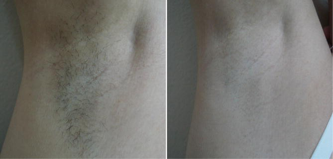
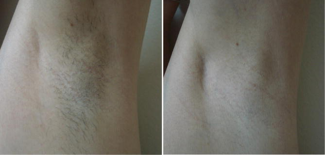
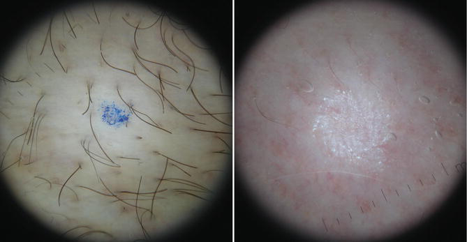
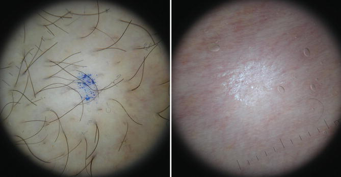
It is extremely important to avoid sun exposure for 6 weeks before and after each treatment session (Pictures 1, 2, 3, and 4).
Complications
The most common complications are hyper- or hypopigmentation, which is temporary in most cases, but there are reports of permanent changes (Ibrahimi et al. 2011). There may also be erythema, edema, pain, burns, blistering, and scarring (Lim and Lanigan 2006). The skin phototype and prior sun exposure of the treated area are crucial in the development of such complications. Protected areas, such as the armpits and groin, tend to have fewer side effects (Gan and Graber 2013).
Paradoxical hypertrichosis occurs in 0.6–10% of patients treated with LIP or lasers. The exact mechanism by which it occurs is unknown, and it is most common after treatment with IPL and alexandrite lasers. Factors associated with its development include the use of insufficient fluence, higher phototypes, hormonal changes, and the use of medications (such as finasteride or corticosteroids) or hormone supplements. Women seem to be most affected and areas such as the face and neck are more prone to this complication (Desai et al. 2010).
Severe and persistent urticaria can occur, even in patients without a history of physical urticaria, possibly caused by the use of cryogens or sensitivity to specific wavelength (Bernstein 2010).
Ocular complications such as cataract, atrophy and iris adhesions, iritis, uveitis, photophobia, pupil changes, and visual field alterations have been described, even in patients who used appropriate glasses. Wavelengths 400–1,400 nm, falling directly on the eyes, can cause burns on the retina and permanent visual disturbances, making mandatory the use of eye protection for patients and applicators (Shulman and Bichler 2009; Gan and Graber 2013).
Acne aggravation, development of lesions similar to those of rosacea, early follicles depigmentation, diffuse erythema, and inflammatory or pigmentary changes of pre-existing nevi were also reported as rare side effects (Rasheed 2009).
Devices for Home Use
Portable devices for domestic use (home devices) have been developed on a large scale and are gaining popularity because of their low cost and ease of use. They use laser and IPL technologies, with very low fluences when compared to the medical devices.
Although approved by the FDA, there are few controlled studies showing their effectiveness and safety (Thaysen-Petersen et al. 2012).
Cross-References
Biophotonics
Take Home Messages
- Laser hair removal has emerged as a leading treatment option for long-term hair reduction. Devices such as diode laser, Nd:YAG laser, and intense pulsed light (IPL) are in constant development, but there are few comparative studies between these technologies. Recent studies point that diode laser might be more effective than IPL.
- Proper selection of the wavelength, pulse duration, and fluence is crucial for the success and safety of the procedure. To treat thin and light hair, higher fluences are required. To treat high concentration of thick hair or higher phototypes, lower fluences might be used.
- The ideal patient for laser or IPL hair removal is the one with low skin phototype and dark hair.
- There is currently no consensus regarding the number of treatment sessions and the interval between them.
- Hyper- or hypopigmentation, burns, scarring, and paradoxical hypertrichosis are some of the adverse effects reported.
References
Altshuler GB, Anderson RR, Manstein D, Zenzie HH, Smirnov MZ. Extended theory of selective photothermolysis. Lasers Surg Med. 2001;29(5):416–32.
Anderson RR, Parrish JA. Selective photothermolysis: precise microsurgery by selective absorption of pulsed radiation. Science. 1983;220(4596):524–7.
Bernstein EF. Severe urticaria after laser treatment for hair reduction. Dermatol Surg. 2010;36:147–51.
Boechat A. Laser: princípios, efeitos e aplicações. In: Osório N, Torezan LAR, editors. Laser em dermatologia. São Paulo: Roca; 2002. p. 1–19.
Campos V, Pitassi L. Epilação no rosto e no corpo. In: Mateus A, Palermo E, editors. Cosmiatria e Laser: Prática no consultório médico. São Paulo: GEN; 2012. p. 489–503.
Campos VB, Dierickx CC, Farinelli WA, Lin TY, Manuskiatti W, Anderson RR. Hair removal with an 800-nm pulsed diode laser. J Am Acad Dermatol. 2000;43(3):442–7.
Casey AS, Goldberg D. Guidelines for hair removal. J Cosmet Laser Ther. 2008;10(1):24–33.
Cassano N, Arpaia N, Vena GA. Diode laser hair removal and isotretinoin therapy. Dermatol Surg. 2005;31(3):380–1.
Dawber RPR. Guidance for the management of hirsutism. Curr Med Res Opin. 2005;21:1227–34.
Desai S, Mahmoud BH, Bhatia AC, Hamzave IH. Paradoxical hypertrichosis after laser therapy: a review. Dermatol Surg. 2010;36:291–8.
Dierickx C. Laser-assisted hair removal: state of the art. Dermatol Ther. 2000;13:80–9.
Dierickx CC, Grossman MC. Epilação com laser. In: Goldberg DJ, editor. Laser e Luz, vol. 2. Rio de Janeiro: Elsevier; 2006. p. 63–79.
Gan SD, Graber EM. Laser hair removal: a review. Dermatol Surg. 2013;39(6):823–38.
Goldman L, Blaney DJ, Kindel DJ Jr, Franke EK. Effect of the laser beam on the skin. Preliminary report. J Invest Dermatol. 1963;40:121–2.
Grossman MC, Dierickx C, Farinelli W, Flotte T, Anderson RR. Damage to hair follicle by normal-mode ruby lasers pulses. J Am Acad Dermatol. 1996;35:889–94.
Ibrahimi OA, Avram MM, Hanke CW, Kilmer SL, Anderson RR. Laser hair removal. Dermatol Ther. 2011;24(1):94–107.
Ismail SA. Long-pulsed Nd:YAG laser vs. intense pulsed light for hair removal in dark skin: a randomized controlled trial. Br J Dermatol. 2012;166:317–21.
Khatri KA. Diode laser hair removal in patients undergoing isotretinoin therapy. Dermatol Surg. 2004;30(9):1205–7, discussion 1207.
Khatri KA, Garcia V. Light-assisted hair removal in patients undergoing isotretinoin therapy. Dermatol Surg. 2006;32(6):875–7.
Klein A, Steinert S, Baeumler W, Landthaler M, Babilas P. Photoepilation with a diode laser vs. intense pulsed light: a randomized, intrapatient left-to-right trial. Br J Dermatol. 2013;168(6):1287–93.
Krause K, Foitzik K. Biology of the hair follicle: the basics. Semin Cutan Med Surg. 2006;25(1):2–10.
Lim SPR, Lanigan SW. A review of the adverse effects of laser hair removal. Lasers Med Sci. 2006;21:121–5.
Macedo F. Luz intensa pulsada versus laser. In: Mateus A, Palermo E, editors. Cosmiatria e Laser: Prática no consultório médico. São Paulo: GEN; 2012. p. 508–18.
Mandt N, Troilius A, Drosner M. Epilation today: physiology of the hair follicle and clinical photo-epilation. J Investig Dermatol Symp Proc. 2005;10:271–4.
McGill DJ, Hutchinson C, McKenzie E, McSherry E, MacKay IR. A randomized, split-face comparison of facial hair removal with the alexandrite laser and intense pulsed light system. Laser Surg Med. 2007;39(10):767–72.
Nouri K, Chen H, Saghari S, Ricotti CA Jr. Comparing 18- vs. 12-mm spot size in hair removal using a GentleLase 755-nm alexandrite laser. Dermatol Surg. 2004;30(4 Pt 1):494–7.
Ormiga P, Ishida CE, Boechat A, Ramos-E-Silva M. Comparison of the effect of diode laser versus intense pulsed light in axillary hair removal. Dermatol Surg. 2014;40(10):1061–9.
Philpott MP, Green MR, Kealey T. Human hair growth in vitro. J Cell Sci. 1990;97:463–71.
Randall VA, Lanigan S, Hamzave I, James LC. New dimensions in hirsutism. Lasers Med Sci. 2006;21:126–33.
Rasheed AI. Uncommonly reported side effects of hair removal by long pulsed-alexandrite laser. J Cosmet Dermatol. 2009;8:267–74.
Rosenfield RL. Clinical practice. Hirsutism. N Engl J Med. 2005;353(24):2578–88.
Ross EV, Ladin Z, Kreindel M, Dierickx C. Theoretical considerations in laser hair removal. Dermatol Clin. 1999;17:333–55, viii.
Shulman S, Bichler I. Ocular complications of laser-assisted eyebrow epilation. Eye (Lond). 2009;23:982–3.
Stratigos AJ, Dover JS. Overview of lasers and their properties. Dermatol Ther. 2000;13:2–16.
Thaysen-Petersen D, Bjerring P, Dierickx C, Nash J, Town G, Haedersdal M. A systematic review of light-based home-use devices for hair removal and considerations on human safety. J Eur Acad Dermatol Venereol. 2012;26(5):545–53.
Vachiramon V, Brown T, McMichael AJ. Patient satisfaction and complications following laser hair removal in ethnic skin. J Drugs Dermatol. 2012;11(2):191–5.
van Gemert MJC, Welch AJ. Time constants in thermal laser medicine. Lasers Surg Med. 1989;9:405–21.
Zenzie HH, Altshuler GB, Smirnov MZ, Anderson RR. Evaluation of cooling methods for laser dermatology. Lasers Surg Med. 2000;26(2):130–44.







