
Intense Pulsed Light for Rosacea and Other Indications
Intense Pulsed Light for Rosacea and Other Indications
Juliana Merheb Jordão and Luiza Pitassi Skin and Laser Center of Boom, Boom, Belgium Department of Hospital Universitário Evangélico de Curitiba, Curitiba, PR, Brazil Division of Dermatology, Department of Medicine, State University of Campinas (UNICAMP), Campinas, SP, Brazil
Abstract
Intense pulsed light (IPL) treatment is one of the most effective procedures for patients with vascular and pigmented lesions as well as for photoaging. The nonablative nature of IPL makes it an increasingly attractive alternative for patients unwilling to accept the adverse effects associated with other procedures. IPL has been used widely to treat a number of lesions, including benign pigmented lesions, inflammatory acne, hypertrophic scars, hair removal, and a variety of vascular lesions such as port-wine stains, hemangiomas, telangiectasias, poikiloderma of Civatte, and rosacea. In the last years, many studies and case reports have been written with different indications for IPL expanding its use. Those studies showed that IPL represents a valid therapeutic support with excellent outcomes and low side effects. However, it should be underlined that the use and the effectiveness of IPL are strongly related to the operator’s experience.
Keywords Intense pulsed lightIPL technologyRosaceaVascular lesions
Introduction
Intense pulsed light (IPL) systems have evolved since they were introduced into medical practice 20 years ago. IPL devices use flashlamps and band-pass filters to emit noncoherent, noncollimated, polychromatic light energy at different wavelengths that target specific chromophores (González-Rodríguez and Lorente-Gual 2015; Babilas et al. 2010). IPL systems mainly target hemoglobin and melanin, with greatest efficacy in the treatment of color rather than texture, according to certain authors. However, despite discussion over whether they can remodel collagen, they have proved to be effective for skin rejuvenation (González-Rodríguez and Lorente-Gual 2015).
This selective targeting capability makes IPL a versatile therapy with many applications such as the treatment of unwanted hair growth, vascular lesions, pigmented lesions, and acne vulgaris and as a light source for photodynamic therapy (PDT) (González-Rodríguez and Lorente-Gual 2015; Babilas 2010).
Similar to lasers, the basic principle of IPL devices is the selective thermal damage of the target structure (Babilas 2010). The combination of prescribed wavelengths, fluences, pulse durations, and pulse intervals facilitates the treatment of this wide spectrum of skin conditions. The large spot size is also a great advantage in terms of treatment duration (Babilas et al. 2010).
IPL was first indicated for the treatment of vascular malformations (Babilas et al. 2010). Actually, it has been used widely to treat a number of vascular lesions, including port-wine stains, hemangiomas, telangiectasias, poikiloderma of Civatte, and rosacea (Ichikawa and Furue 2013). The Food and Drug Administration (FDA) has approved intense pulsed light devices for the treatment of this variety of benign pigmentary and vascular lesions, but the range of disease that can be treated with IPL continues to expand (Wat et al. 2014).
History
Muhlbauer et al. first described the use of polychromatic infrared light in 1976 for the treatment of vascular malformations (Babilas et al. 2010). The effects produced by applying a light source to tissue can be explained by the principle of selective photothermolysis, as described by Anderson and Parrish (1983). Thus, the energy supplied to a tissue has a selective action on a target molecule, denoted the chromophore, with no or minimal impact on the adjacent structures. This selective action is the underlying principle by which intense pulsed light works (González-Rodríguez and Lorente-Gual 2015).
In 1990, Goldman and Eckhouse described a new high-intensity flashlamp as a suitable tool for treating vascular lesions (Goldman and Eckhouse 1996; Babilas et al. 2010). In 1994, the Israeli engineer Shimon Eckhouse managed to produce a broad-spectrum stimulated light emission, thereby creating IPL. The US Food and Drug Administration approved the technique for therapeutic ends in 1997, after the first article on their use in dermatology, when Raulin et al. used them successfully to treat 14 patients with telangiectasias of the face and legs or with poikiloderma of Civatte. Shortly afterward, the same authors published two cases of permanent hair removal and subsequently have conducted several more standardized studies that have demonstrated the efficacy and safety of the technique (Raulin et al. 1997; González-Rodríguez and Lorente-Gual 2015).
First-generation IPL devices emit light of the infrared part of the spectrum, which prevalently led to epithelial damage and a high incidence of side effects. Second-generation IPL devices use water to filter off the infrared part of the light spectrum, keeping the wavelength spectrum between 515 and 950 nm. This reduces the risk of side effects (Babilas et al. 2010; González-Rodríguez and Lorente-Gual 2015). Cooling systems are used to protect the epidermis in contact with the crystal (González-Rodríguez and Lorente-Gual 2015). In IPL devices, similar to lasers, the basic principle is the absorption of photons by endogenous or exogenous chromophores within the skin, generating heat and, subsequently, destroying the target structure (Babilas et al. 2010).
In the following years, multiple technical modifications allowed an easier handling, increased safety, and amplifying the spectrum of potential indications. The emission spectrum of IPLs ranges from 400 to 1,300 nm, and pulse duration ranges from 2 to 200 ms. Convertible cutoff filters of IPL devices can be easily adapted to the desired wavelength, allowing certain versatility (Babilas et al. 2010; González-Rodríguez and Lorente-Gual 2015).
Most IPLs have a single pulse, and the energy is proportional to the pulse duration. Some IPLs have multiple sequential pulsing, and some have the ability to independently vary the pulse duration, the energy fluence, or both in each pulse. Other variables include the cutoff wavelengths, spectral output, and size of the delivered light (Wat et al. 2014).
Basic Concepts
Intense Pulsed Light Wavelengths
Traditional indications of IPL consist in its ability of targeting pigmented and vascular lesions.
In pigmented lesions, the target is melanosome, whose chromophore is melanin. This absorbs the appropriate wavelength, transforming the light into heat, causing target destruction. The transfer of melanosomes toward the upper layers accompanies these processes, where they are eliminated along with necrotic keratinocytes (González-Rodríguez and Lorente-Gual 2015).
In vascular lesions, IPL acts on three target chromophores: oxyhemoglobin (predominant lesions of red appearance), deoxyhemoglobin (predominant in blue lesions), and methemoglobin (González-Rodríguez and Lorente-Gual 2015).
Oxyhemoglobin contained in red blood cells within blood vessels has a maximum peak of absorption around 540 nm (alpha peak) and 580 nm (beta peak). This holds true of small superficial vessels mainly located on the face and the neck. Vessels on the legs are usually located deeper and contain more deoxyhemoglobin. This situation moves the absorption curve to the right, from 800 nm to 1,200 nm. Infrared wavelengths tend to be more effective in treating deeper blue vessels, while shorter wavelengths are more effective for superficial red telangiectasias (Adamic et al. 2007).
The longer the wavelength, the deeper it penetrates into the skin (Adamic et al. 2007). Although the peak of maximum absorption for oxyhemoglobin is around 418–540 nm, less penetration is achieved and a strong competition with melanin from epidermis occurs. At 577 nm, although absorption is low, the degree of penetration is greater, reducing melanin absorption and avoiding side effects, such as hypopigmentation. For this reason, current devices use longer wavelengths (515–600 nm) to ensure deeper penetration while still being absorbed by oxyhemoglobin (Fig. 4) (González-Rodríguez and Lorente-Gual 2015). The use of longer wavelengths ensures the safety of IPL in higher phototypes.
Among these wavelengths, the patient’s skin type, the skin condition, and the target present determine the choice of suitable cutoff filters and therefore the spectrum of wavelengths to be emitted (Babilas et al. 2010).
Pulse Duration
Pulse duration can be set in relatively wide ranges (depending on the particular device) in the millisecond range. Similar to laser devices, pulse duration should be equal to or lower than the thermal relaxation time (TRT) of the target structure to prevent unselective damage to the surrounding tissue (Babilas et al. 2010).
For IPL systems, usually pulse duration is determined in milliseconds (ms). For deep lesions, higher pulse durations are suggested. For superficial lesions, smaller pulse durations are recommended.
Selectivity
The key chromophores of the human skin (hemoglobin, melanin, water) show broad absorption spectrums. Thus, monochromaticity is not a requirement for photothermolysis. As IPL devices emit a spectrum of wavelengths, the three key chromophores can be activated with one single light exposure. This versatility implies a reduced selectivity (Babilas et al. 2010) and can explain the need of long-term treatment for vascular lesions (Ichikawa and Furue 2013).
Treatment Principles
Smaller vessels need shorter pulses; larger vessels need longer pulses.
The deeper the blood vessel is located in the dermis, the larger the spot size, the longer the wavelength, and the longer the pulse duration should be combined with cooling to protect the epidermis.
Darker skin types need longer pulses and longer wavelengths (Adamic et al. 2007).
Mechanisms of Action
Intense pulsed light systems use a flashlamp that emits high-intensity, noncoherent, and polychromatic broad-spectrum light. The emission spectrum of IPLs ranges from 400 to 1,200 nm. The light is emitted in pulses with various pulse durations and intervals in a large rectangular spot of up to 1 by 4 cm (Raulin and Greve 2003; Meesters et al. 2014).
The mechanism of action of IPL system utilizes the theory of selective photothermolysis (Anderson and Parrish 1983). The basic principle is the absorption of photons by skin chromophores, generating heat, which is responsible for target destruction and selective thermal damage of the target. Selective photothermolysis begins with local absorption of light energy in target chromophores such as hemoglobin in vascular lesions or melanin in pigmented lesions (Babilas et al. 2010).
The combination of prescribed wavelengths, fluences, pulse durations, and pulse intervals facilitates the treatment of a wide spectrum of skin conditions. Various different wavelengths of absorption are emitted to treat various targets, using different filters in order to optimize treatment of different conditions. By selecting a cutoff filter specific for the absorption wavelength of the chromophore, energy can be deposited in the blood vessels, preventing damage and subsequent scarring to the surrounding tissues (Kassir et al. 2011).
For vascular lesions, the mechanism of action of IPLs is related to their selective absorption by hemoglobin (oxyhemoglobin, deoxyhemoglobin, and methemoglobin) within the blood vessels with peaks of absorption at 418 nm (blue), 542 nm (green), and 577 nm (yellow). Successful treatment depends on the type and size of the vessels (Babilas et al. 2010; Barikbin et al. 2011; Goldberg 2012).
Shorter wavelengths may be preferred for superficial vascular lesions such as facial telangiectasias. Lower cutoff filters are effective in treating smaller caliber vessel. For standard-sized small vessels in the papillary dermis with a diameter of 0.1 mm, IPL has been shown to be an effective treatment. IPL is able to treat the surface area of telangiectasias and to diminish the intensity of erythema and flushing seen in patients with erythematotelangiectatic rosacea (Mark et al. 2003; Goldberg 2012).
Indications
The conditions treated with IPL include, mainly, vascular lesions and pigmented lesions, as well as skin rejuvenation, inflammatory dermatoses, acne vulgaris, hidradenitis suppurativa, hair removal, hypertrophic scars, and keloids (Babilas et al. 2010; Vrijman et al. 2011; Goldberg 2012; Wat et al. 2014).
The IPL device had been originally developed for the treatment of a wide range of benign vascular lesions, including telangiectasias and reticular varicose leg veins (Goldberg 2012; Meesters et al. 2014). There are multiple well-established IPL treatments for targeting blood vessels in the skin. IPL devices have been used effectively in the treatment of facial telangiectasias, erythematotelangiectatic rosacea, spider nevi, erythrosis, hemangiomas, venous and capillary malformations, and poikiloderma of Civatte (Acarturk and Stofman 2003; Clementoni et al. 2005; Adamic et al. 2007; McGill et al. 2008; Li et al. 2010a; Nymann et al. 2010; Campolmi et al. 2011; Dan 2011; Barikbin et al. 2011; Meesters et al. 2014).
Successful treatment of vascular lesions depends on the type and size of the vessels. Fodor et al. conducted a comparative study of IPL and Nd:YAG laser and obtained better results with IPL for more superficial and smaller lesions. A study conducted by Murray et al. in 2012 showed an improvement in systemic sclerosis-related telangiectasias; however, these improvements were not sustained during follow-up, suggesting that other treatments should be added.
IPL can also be useful in infants with superficial hemangiomas or rapidly growing lesions >1 cm (González-Rodríguez and Lorente-Gual 2015).
Rosacea
Intense pulsed light therapy is a safe and effective treatment for the signs and symptoms of rosacea. Several studies have demonstrated the successful use of intense pulsed light for the vascular components of erythematotelangiectatic rosacea, including facial telangiectasia and papular lesions (Schroeter et al. 2005; Papageorgiou et al. 2008; Bae et al. 2009; Babilas et al. 2010; Dahan 2011; Kassir et al. 2011; Liu et al. 2014).
Rosacea is a chronic cutaneous disease that manifests as facial flushing, persistent erythema, telangiectasia, papules, and pustules.
Erythematotelangiectatic rosacea is the most common and may have the strongest vascular component among the four subtypes (erythematotelangiectatic, papulopustular, phymatous, and ocular) (Crawford et al. 2004; Lim et al. 2014). Advantages of IPL include short downtime, mild adverse reactions, and short duration of procedure due to the large application area (Kawana et al. 2007; Babilas et al. 2010; Lim et al. 2014).
The ability to choose the duration of pulses makes IPL a versatile tool in the treatment of rosacea. The possibility of different filter settings allows a wider selection of the range color of the vascular system (Schroeter et al. 2005; Piccolo et al. 2014). IPL can improve rosacea treating abnormal vessels and inducing collagen remodeling (Piccolo et al. 2014).
Various studies have demonstrated the effectiveness of IPL in reducing blood flow, telangiectasia, and severity of erythema in individuals with rosacea (Wat et al. 2014) (Fig. 1). Taub noticed a reduction of 83% of erythema, a reduction of 75% of flushing, and an improved skin texture (Taub 2003).
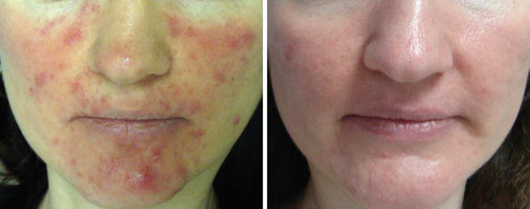
A prospective trial involving 60 patients analyzed the effect of IPL on facial telangiectasias and found that 77.8% of patients achieved greater than 50% reduction of vessels after treatment. These results were maintained during a 3-year posttreatment follow-up period (Schroeter et al. 2005).
Papageorgiou and colleagues confirmed the efficacy of IPL in the treatment of rosacea disease of phase I. It showed a significant improvement of erythema, telangiectasias, and flushing. The severity of rosacea was reduced more than 50%, and the results were sustained at 6 months (Papageorgiou et al. 2011).
IPL appears to be an effective, well-tolerated treatment option for rosacea (level 2a evidence), with efficacy equal to that of pulsed dye laser. Typical reported improvement was in the 50% range (Neuhaus et al. 2009; Wat et al. 2014).
Other Vascular Lesions
Hemangioma
IPL has been shown to be applicable in infants with superficial hemangiomas and appears as one of the effective and safe treatments. Treating the vascular tumor at the earliest stage probably controls the excess in endothelial growth. IPL is limited by the deep and mixed types of the vascular tumors and a size over 5 cm at initiation of treatment, as well as some locations including the eyelids, lips, nostril, and genitalia (Paquet et al. 2014). Angermeier studied patients with different vascular lesions with IPL. It demonstrated 75–100% of clearance after one or two treatments with IPL in 174 of 188 patients with vascular lesions (45 cases of them had facial hemangiomas) (Angermeier 1999).
IPL has notably been used in a study of 62 patients with infantile hemangiomas who received four to five IPL treatments at 4-week intervals with a clearance rate of more than 80% (Li et al. 2010a).
Treatment with IPL suggested that lesions larger than 1 cm in diameter at the first visit or those rapidly growing more than 0.5 cm over the first 6–8 months of life must be treated in order to avoid further physical and psychological sequelae. Treatment in an early stage of development with IPL appeared to stop the rapid growth and initiated involution in three treatment sessions, thus preventing any unpredictable disfiguring impairment (Rafiq et al. 2014).
Poikiloderma of Civatte
Because of their ability to target vascular and pigment components simultaneously, IPL sources (IPLs) have been utilized in the treatment of poikiloderma of Civatte (Fig. 2).
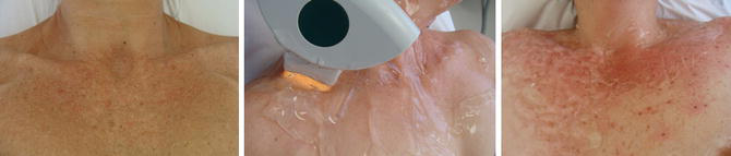
Weiss and coworkers have reported their 5-year experience in treating poikiloderma of Civatte of the neck and chest in more than 100 patients. A clearance of more than 75% of telangiectasias and hyperpigmentation was observed, with a 5% incidence of side effects including pigment changes (Weiss et al. 2000; Goldman and Weiss 2001).
In 2008 Rusciani et al. studied 175 patients with poikiloderma of Civatte of the neck and chest treated with various IPL settings. Eighty percent of the vascular and the pigmented components were cleared. Less than 5% of the individuals had side effects, which were minimal and transient (Rusciani et al. 2008). Weiss et al. succeeded in reaching a clearance rate of 75–100% in 82% of 135 patients after three treatments with PhotoDerm VL (Weiss et al. 2000).
Goldman et al. achieved a 50–75% improvement in the extent of telangiectasias and hyperpigmentation after an average of 2.8 treatments (Goldman and Weiss 2001). Schroeter et al. even had clearance rates of 90% in the vascular part in 15 patients (Schroeter and Neumann 1998).
IPL can be considered a safe and effective therapy for poikiloderma of Civatte due to its wide range of wavelengths, which allows treating both the pigmentation and the telangiectasia at the same time (Barikbin et al. 2011).
Take care in darker skin phototypes treatment, and during cervical area, treatment is necessary to avoid dyschromia and scars. When correctly performed, IPL procedure can be considered a safe and effective therapeutic option for poikiloderma of Civatte, allowing a marked improvement of vascular and pigmented lesions with minimal side effects (Rusciani et al. 2008).
Nasal Telangiectasia
Nasal telangiectasia can be treated with IPL. High fluences and stacking pulses should be necessary in resistant telangiectasias. For smaller vessels, short wavelengths and low pulse duration are suggested. For thicker telangiectasias, bigger wavelengths and high pulse durations are more effective.
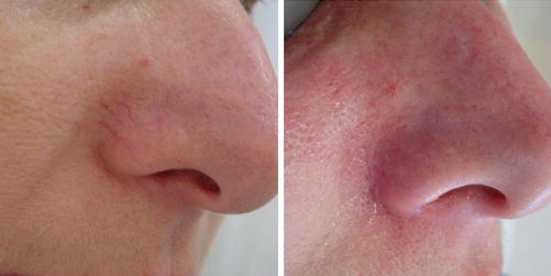
To be sure that the telangiectasia was effectively removed, it is necessary to see the telangiectasia immediately disappearing (Fig. 3) or an edema in the path of the vessel or a purpura in its place. Repeated sessions with 3 week intervals are indicated as some telangiectasias reopen after few days.
Leg Telangiectasia
Studies demonstrated that IPL could be used for multiple forms of telangiectasia treatment. However, clinical evidence for IPL in the treatment of leg veins is scarce. In a multicenter trial of 159 patients, Goldman reported 90% clearance rate with vessels smaller than 0.2 mm and 80% clearance rate with vessels 0.2–1 mm in diameter. Schroeter et al. reported immediate clearing in 73.6% of patients and in 84.3% of patients after 4 weeks (Goldman et al. 1996; Schroeter et al. 2013; Meesters et al. 2014). Doctors have to take into account the skin phototype to avoid side effects.
Striae Distensae (SD)
The exact mechanism of action of IPL in the treatment of SD is unknown, but it is probably related to dermis remodeling, in which the activity of fibroblasts is increased and more collagen fibers are synthesized or rearranged within the stroma (Goldberg 2000; Al-Dhalimi and Abo Nasyria 2013).
Previous studies have demonstrated that IPL induces neo-collagen formation and potentially improves epidermal atrophy and dermal elastosis. Pérez et al. showed that SD improved clinically and microscopically after IPL with minimal side effects (Pérez et al. 2002).
A comparative study of the effectiveness of intense pulsed light in the treatment of striae distensae showed that both of the wavelengths (650 nm and 590 nm) were effective in the treatment of striae distensae and both of them resulted in statistically significant reduction in the number, the lengths, and the maximum widths of striae. For all of these assessed parameters, the wavelength 590 nm was more effective (Al-Dhalimi and Abo Nasyria 2013).
Port-Wine Stain (PWS)
Raulin and Goldman reported the first successful treatment of an adult port-wine stain (PWS) with IPL (Raulin et al. 1997).
The pulsed dye laser represents the most commonly used laser for treatment of PWS and has the most published literature supporting its use. IPL has also been reported to be an effective alternative to PDL for treatment of PWS. Although IPL has been shown to be effective in the clearance of pink and red PWS, a head-to-head trial comparing the efficacy of IPL against PDL determined that the median clinical improvement was significantly better for PDL (65%) than IPL (30%) (Faurschou et al. 2009). Nevertheless, IPL can be considered for treating PDL-resistant PWS (Ho et al. 2004; Ozdemir et al. 2008; Li et al. 2010b; Adatto et al. 2010; Chen et al. 2012).
Bjerring and colleagues treated 15 patients with PWS that had previously been resistant to PDL and found that IPL was able to achieve 75–100% clearance in 46.7% of cases (Bjerring et al. 2003).
A randomized, controlled, single-blind head-to-head trial comparing PDL with IPL for the treatment of PWS showed that both modalities were effective but that PDL was superior in terms of median clinical improvement and patient preference (Wat et al. 2014).
Recently, Wang and colleagues confirmed the efficacy of IPL in the treatment of facial and extrafacial PWS in Chinese patients (Wang et al. 2013).
There is reasonable evidence to suggest that IPL is an effective, safe modality for the treatment of capillary malformations (level 2a evidence). It may be especially useful for darker lesions that have greater vascularity but minimal nodularity (Wat et al. 2014).
Hypertrophic Scars and Keloids
A recent report of IPL in 109 patients with hypertrophic scars and keloids (due to traumatic wounds, burns, and surgical incisions) demonstrated improvement in 92.5% of subjects, in terms of scar height, erythema, and hardness, with a high level of patient satisfaction (Erol et al. 2008).
In Fig. 4, it is possible to see a good result of a traumatic scar treated with eight sessions of IPL 540 nm.
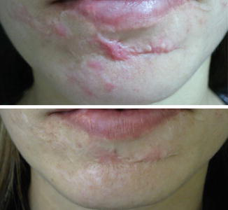
Hyperpigmented, erythematous, and proliferative scars all demonstrated greater than 50% improvement after a mean of 2.97 sessions, whereas atrophic scars did not respond (Kontoes et al. 2003). A very recent pilot study has demonstrated the effectiveness of IPL in wound healing after suture removal. The basic mechanism is not yet fully understood, but most probably an action on vascular proliferation, essential for the growth of collagen, and on pigmentation resulting from scar formation is involved (Erol et al. 2008; Piccolo et al. 2014).
Kontoes et al. reported an improvement of more than 75% in the pigmentation of hypertrophic scars, 50% higher than that in the scars from asphalt, and 50% reduction in the size and thickness of hypertrophic scars. This is probably due to the inhibition of the action of the vessel caused by IPL on scar tissue and on the subsequent proliferation of collagen (Kontoes et al. 2003).
Contraindications
Pregnancy, breastfeeding, the intake of retinoids or photosensitizing medications, diseases or genetic conditions causing photosensitivity or tending to aggravate after light exposure (Roelandts 2000), as well as suntan are exclusion criteria for IPL treatment. Patients suffering from long-term diabetes, hemophilia, or other coagulopathies and patients with implants in the treatment area or with a heart pacemaker should be treated with special care. Patients with a history of herpes simplex require an antiviral prophylaxis for holohedral facial treatments (Adamic et al. 2007).
The skin type of the patient has to be documented according to the Fitzpatrick scale because photophysical parameters need to be adjusted depending on the individual patient’s skin type. It is recommended to avoid sunlight, in addition to applying a broad-spectrum sunscreen. When treating areas close to tattoos, definitive makeup, ephelides, and nevus, caution should be taken to avoid a possible color change or scar injury after treatment with intense pulsed light at the site.
Recently, a series of home-use intense pulsed light devices has been developed. These devices, in spite of being FDA approved, have scarce controlled studies related to the method’s safety and efficacy. All systems tested are attractively packaged with clear educational material for the customer regarding contraindications to treatment such as too dark skin types, active suntan, and medications (Town and Ash 2010).
Side Effects and Their Managements
After the administration of high fluences and short pulse times, there have been reports of transient or permanent pigment changes, edema, erythema, purpura, mild discomfort, blistering, and crusting. These findings typically resolved within 1–48 h but sometimes lasted up to 1 week. These side effects are much more common with older, first-generation flashlamps which emit a higher proportion of infrared light (Wat et al. 2014).
The most common complication after IPL treatment is changed pigmentation, with possible hyper- or hypopigmentation. These are most commonly seen in patients with a darker or recently tanned skin. The skin must be sufficiently cooled during treatment to protect the epidermis from being burned; the cooling can be performed using cooling gels, ice gels, contact spray cooling, or special cooling handpieces (Raulin and Greve 2003).
Blistering and crusting are signs of overfluenced treatment; in case of blisters and crusts, patients must strictly avoid scratching, which may result in infections and scar formation. Antimicrobial ointments help loosening the crusts and prevent bacterial superinfections. Potential side effects that might last longer or may even be irreversible are pigmentary changes, such as hypopigmentation or hyperpigmentation. Adjusting wavelengths and fluences to the patient’s skin type and treatment area can mostly prevent these side effects. Unsuitable patients (due to suntan or skin type) are excluded from therapy as well as patients who are unable or unwilling to strictly avoid postoperative UV exposition. Scarring occurs rarely and is almost always evoked by overfluenced treatments or by crusting with subsequent manipulation and infection. In general, the most important measure to prevent side effects is the application of test shots for every chosen set of parameters and even for the same set of parameters applied at different parts of the body (Babilas et al. 2010).
Safety and efficacy with IPL devices rely on the appropriate use of device settings and sufficient operator experience (Wat et al. 2014).
Take Home Messages
- The main skin chromophores present in the skin are melanin and hemoglobin.
- Delay the treatment in tanned skins.
- Photograph areas before treatment.
- The informed consent form should be provided.
- Use sunscreens with a high protection factor.
- In brunette skins, perform a previous test to increase procedure safety.
- Eye protection is mandatory, both for the physician and the patient.
- Use the highest amount of safe energy for best results.
- Scars are extremely rare, but these may occur when excessive energies are used.
References
Acarturk TO, Stofman GM. Treatment of the vascular lesions of the face and neck using selective vascular photothermolysis with intense pulse light. Eur J Plast Surg. 2003;26:319–23.
Adamic M, Troilius A, Adatto M, Drosner M, Dahmane R. Vascular lasers and IPLS: guidelines for care from the European Society for Laser Dermatology (ESLD). J Cosmet Laser Ther. 2007;9(2):113–24.
Adatto MA, Luc-Levy J, Mordon S. Efficacy of a novel intense pulsed light system for the treatment of port wine stains. J Cosmet Laser Ther. 2010;12:54–60.
Al-Dhalimi MA, Abo Nasyria AA. A comparative study of the effectiveness of intense pulsed light wavelengths (650 nm vs 590 nm) in the treatment of striae distensae. J Cosmet Laser Ther. 2013;15:120–5.
Anderson RR, Parrish JA. Selective photothermolysis: precise microsurgery by selective absorption of pulsed radiation. Science. 1983;220(4596):524–7.
Angermeier MC. Treatment of facial vascular lesions with intense pulsed light. J Cutan Laser Ther. 1999;1:95–100.
Babilas P. Light-assisted therapy in dermatology: the use of intense pulsed light (IPL). Med Laser Appl. 2010;25:61–9.
Babilas P, Schreml S, Szeimies RM, Landthaler M. Intense pulsed light (IPL): a review. Lasers Surg Med. 2010;42:93–104.
Bae YI, Yun SJ, Lee JB, Kim SJ, Won YH, Lee SC. Clinical evaluation of 168 Korean patients with rosacea: the sun exposure correlates with the erythematotelangiectatic subtype. Ann Dermatol. 2009;21:243–9.
Barikbin B, Ayatollahi A, Hejazi S, Saffarian Z, Zamani S. The use of intense pulsed light (IPL) for the treatment of vascular lesions. J Lasers Med Sci. 2011;2(2):73–81.
Bjerring P, Christiansen K, Troilius A. Intense pulsed light source for the treatment of dye laser resistant port-wine stains. J Cosmet Laser Ther. 2003;5:7–13.
Campolmi P, Bonan P, Cannarozzo G, Bruscino N, et al. Intense pulsed light in the treatment of non-aesthetic facial and neck vascular lesions: report of 85 cases. J Eur Acad Dermatol Venereol. 2011;25:68–73.
Chen JK, Ghasri P, Aguilar G, van Drooge AM, Wolkerstorfer A, Kelly KM, Heger M. An overview of clinical and experimental treatment modalities for port wine stains. J Am Acad Dermatol. 2012;67(2):289–304.
Clementoni MT, Gilardino P, Muti GF, Signorini M, et al. Facial teleangectasias: our experience in treatment with IPL. Lasers Surg Med. 2005;37:9–13.
Crawford GH, Pelle MT, James WD. Rosacea: I. etiology, pathogenesis, and subtype classification. J Am Acad Dermatol. 2004;51:327–41.
Dahan S. Laser and intense pulsed light management of couperose and rosacea. Ann Dermatol Venereol. 2011;138:S219–22.
Erol OO, Gurlek A, Agaoglu G, Topcuoglu E, Oz H. Treatment of hypertrophic scars and keloids using intense pulsed light (IPL). Aesthetic Plast Surg. 2008;32(6):902–9.
Faurschou A, Togsverd-Bo K, Zachariae C, Haedersdal M. Pulsed dye laser vs intense pulsed light for port-wine stains: a randomized side-by-side trial with blinded response evaluation. Br J Dermatol. 2009;160:359–64.
Goldberg DJ. New collagen formation after dermal remodeling with an intense pulsed light source. J Cutan Laser Ther. 2000;2:59–61.
Goldberg D. Current trends in intense pulsed light. Clin Aesth. 2012;6:45–53.
Goldman MP, Eckhouse S. Photothermal sclerosis of leg veins. Dermatol Surg. 1996;22(4):323–30.
Goldman MP, Weiss MP. Treatment of poikiloderma of Civatte on the neck with an intense pulse light source. Plast Reconstr Surg. 2001;107:1376–81.
González-Rodríguez AJ, Lorente-Gual R. Current indications and new applications of intense pulsed light. Actas Dermosifiliogr. 2015;106(5):350–64.
Ho WS, Ying SY, Chan PC, Chan HH. Treatment of port wine stains with intense pulsed light: a prospective study. Dermatol Surg. 2004;30:887–91.
Ichikawa R, Furue M. Successful treatment of scrotal angiokeratomas (Fordyce type) with small-spot narrow-band intense pulsed light. Dermatol Surg. 2013;39(10):1547–8.
Kassir R, Kolluru A, Kassir M. Intense pulsed light for the treatment of Rosacea and Telangiectasias. J Cosmet Laser Ther. 2011;13:216–22.
Kawana S, Ochiai H, Tachihara R. Objective evaluation of the effect of intense pulsed light on rosacea and solar lentigines by spectrophotometric analysis of skin color. Dermatol Surg. 2007;33:449–54.
Kontoes PP, Marayiannis KV, Vlachos SP. The use of intense pulsed light in the treatment of scars. Eur J Plast Surg. 2003;25:374–7.
Li DN, Gold MH, Sun ZS, Tang AR, et al. Treatment of infantile hemangioma with optimal pulse technology. J Cosmet Laser Ther. 2010a;12:145–50.
Li G, Lin T, Wu Q, Zhou Z, Gold MH. Clinical analysis of port wine stains treated by intense pulsed light. J Cosmet Laser Ther. 2010b;12:2–6.
Lim HS, Lee SC, Won YH, Lee JB. The efficacy of intense pulsed light for treating erythematotelangiectatic rosacea is related to severity and age. Ann Dermatol. 2014;26(4):491–5.
Liu J, Ren Y, Li B, Lu S. Comparative efficacy of intense pulsed light for different erythema associated with rosacea. J Cosmet Laser Ther. 2014;16(6):324–7.
Mark KA, Sparacio RM, Voigt A, Marenus K, Sarnoff DS. Objective and quantitative improvement of rosacea-associated erythema after intense pulsed light treatment. Dermatol Surg. 2003;29(6):600–4.
McGill DJ, MacLaren W, Mackay IR. A direct comparison of pulsed dye, alexandrite, KTP and Nd:YAG lasers and IPL in patients with previously treated capillary malformations. Lasers Surg Med. 2008;40:390–8.
Meesters AA, Pitassi LHU, Campos V, Dierickx C, Wolkerstorfer. Transcutaneous laser treatment of leg veins. Lasers Med Sci. 2014;29(2):481–92.
Murray AK, Moore TL, Richards H, Ennis H, et al. Pilot study of intense pulsed light for the treatment of systemic sclerosis-related telangiectases. Br J Dermatol. 2012;167:563–9.
Neuhaus IM, Zane LT, Tope WD. Comparative efficacy of nonpurpuragenic pulsed dye laser and intense pulsed light for erythematotelangiectatic rosacea. Dermatol Surg. 2009;35(6):920–8.
Nymann P, Hedelund L, Haedersdal M. Long-pulsed dye laser vs. intense pulsed light for the treatment of facial telangiectasias: a randomized controlled trial. J Eur Acad Dermatol Venereol. 2010;24:143–6.
Ozdemir M, Engin B, Mevlitoglu I. Treatment of facial port-wine stains with intense pulsed light: a prospective study. J Cosmet Dermatol. 2008;7:127–31.
Papageorgiou P, Clayton W, Norwood S, Chopra S, Rustin M. Treatment of rosacea with intense pulsed light: significant improvement and long-lasting results. Br J Dermatol. 2008;159(3):628–32.
Paquet P, Caucanas M, Piérard-Franchimont C, et al. Intense pulsed-light in infantile hemangiomas. Open Access J Sci Technol. 2014;2:1–6.
Pérez EH, Charrier EC, Ibiett EV. Intense pulsed light in the treatment of striae distensae. Dermatol Surg. 2002;28:1124–30.
Piccolo D, Di Marcantonio D, Crisman G, et al. Unconventional use of intense pulsed light. Biomed Res Int. 2014;2014:618206.
Rafiq U, Shah AA, Rizwan M. Treatment of infantile hemangioma with intense pulsed-light: a case report. J Pak Assoc Dermatol. 2014;24(3):267–9.
Raulin C, Greve B. IPL technology: a review. Lasers Surg Med. 2003;32(2):78–87.
Raulin C, Goldman MT, Weiss MA, Weiss RA. Treatment of adult port-wine stains using intense pulsed light therapy (PhotoDerm VL): brief initial clinical report. Dermatol Surg. 1997;23(7):594–7.
Roelandts R. The diagnosis of photosensitivity. Arch Dermatol. 2000;136(9):1152–7.
Rusciani A, Motta A, Fino P, Menichini G. Treatment of poikiloderma of civatte using intense pulsed light source: 7 years of experience. Dermatol Surg. 2008;34:314–9.
Schroeter CA, Neumann M. An intense light source. The PhotoDerm VL-flashlamp as a new treatment possibility for vascular skin lesions. Dermatol Surg. 1998;24:743–8.
Schroeter CA, Haaf-von Below S, Neumann HA. Effective treatment of rosacea using intense pulsed light systems. Dermatol Surg. 2005;31:1285–9.
Schroeter C, Wilder D, Reineke T, Thürlimann W, Raulin C, Neumann HAM. Clinical significance of an intense, pulsed light source on leg telangiectasias of up to 1 mm diameter. Eur J Dermatol. 2013;7(1):38–42.
Taub AF. Treatment of rosacea with intense pulsed light. J Drugs Dermatol. 2003;2(3):254–9.
Town G, Ash C. Are home-use intense pulsed light (IPL) devices safe? Lasers Med Sci. 2010;25(6):773–80.
Vrijman C, van Drooge AM, Limpens J, Bos JD, van der Veen JPW, Spuls PI, Wolkerstorfer A. Laser and intense pulsed light therapy for the treatment of hypertrophic scars: a systematic review. Br J Dermatol. 2011;165(5):934–42.
Wang B, Wu Y, Zhu X, Xu XG, et al. Treatment of neck port-wine stain with intense pulsed light in Chinese population. J Cosmet Laser Ther. 2013;15:85–90.
Wat H, Wu DC, Rao J, Goldman MP. Application of intense pulsed light in the treatment of dermatologic disease: a systematic review. Dermatol Surg. 2014;40:359–77.
Weiss RA, Goldman MP, Weiss MA. Treatment of poikiloderma of civatte with an intense pulsed light source. Dermatol Surg. 2000;26:823–8.







