
Biophotonics
Biophotonics
Álvaro Boechat BLB Fotomedicina LTDA, São Paulo, Brazil
Abstract
Light is one of the most beautiful forms of pure energy; we know some of its therapeutic properties, but there is still much to be explored. The aim of this chapter is to provide a better understanding of the best-known light tools used in modern medicine, such as the laser, intense pulsed light, the advent of fractional systems, radio frequency, and hybrid systems which combine light and radio frequency — how they work, how to select which device will be better for your application, and how light and RF interact with the skin. This will enable the improvement of current treatment techniques as well as broaden the horizons of applications of these devices.
Keywords Dermatological laserLaser physicsTypes of lasersPulsed lightIPLTreatment platformsLight–tissue interactionSelective photothermolysisRelaxation timeRadio frequencyFractional lasersPenetration depthAblative laserNon-ablative laserSublativeFractional radio frequencyELOS
Introduction
The laser and pulsed light are simply sources of light. The visible light that we experience in our day-to-day is only one facet of a much broader physical phenomenon known as “electromagnetic radiation.”
As shown in Fig. 1, the electromagnetic spectrum (Siegman 1986) includes several well-known phenomena, such as TV and radio waves, microwaves, infrared, and, on the other side of the spectrum, ultraviolet and X-rays. However, our eyes are sensitive to only a very narrow range of the spectrum, which forms the visible light from violet to red. It is important to realize that each visible color or each emission spectrum is associated with a frequency or wavelength.
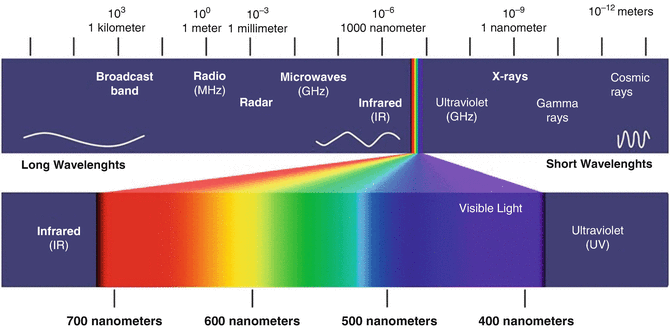
The differentiation between blue and green, for example, is related to their frequencies. It is similar to musical notes: the difference of the note “do” (C) from the note “sol” (G) or “fa” (F) is their frequencies; one is low-pitched and the other high-pitched. Drawing a parallel with them, in the light spectrum the higher frequencies correspond to blue and violet and, on the other side of the spectrum, the lower frequencies correspond to red. As light frequencies are very high, of the order of millions of hertz, they are characterized by their wavelength — the distance between two adjacent peaks in the wave, illustrated in Fig. 2 (Siegman 1986; Arndt et al. 1997).
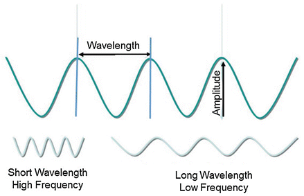
Light radiation may be defined as point-to-point power transmission in space, regardless of the medium in which it is propagated. Light or electromagnetic radiation propagates at high speed in open space independent of the transmission medium, in the form of waves that can travel in a vacuum or in spaces containing matter, such as gases, liquids, or solids. As it enters, or moves from, a different medium, it undergoes changes in the direction and speed of propagation.
Lasers are sources of electromagnetic radiation, or light, with special characteristics that differ from other light sources, such as a car headlight or a lamp.
The word laser is an acronym for light amplification by stimulated emission of radiation. We can divide this acronym into two well-defined parts: the stimulated-emission phenomenon and light amplification.
Stimulated Emission
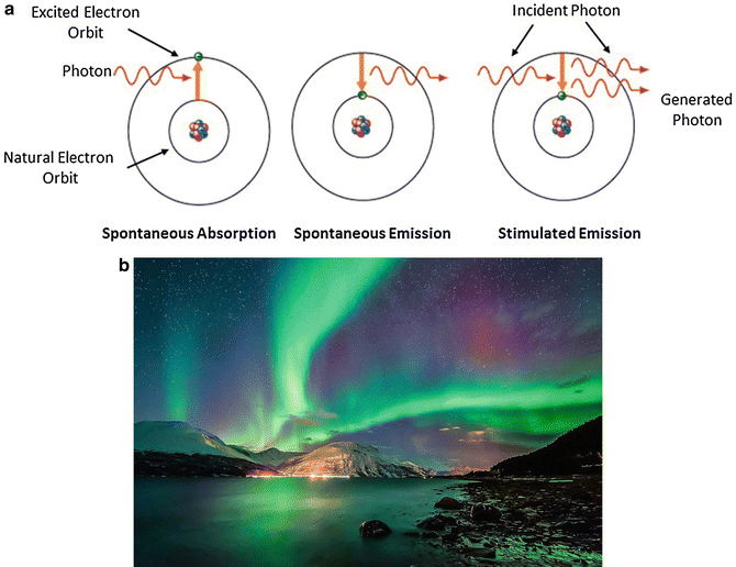
Light is a form of energy generated, emitted, or absorbed by atoms or molecules. To emit energy, the atom or molecule is raised to an excitation energy level above its natural resting state, in which there is excess energy to be discharged. Atoms cannot maintain this excitement for long periods. Consequently, they have a natural tendency to eliminate the excess energy in the form of emission of particles or packets of light waves called photons (Fig. 3a). This phenomenon is called spontaneous emission of light. The wavelength (λ), or frequency of the emitted photons, is related to the photon energy through the relationship:
- h — Planck’s universal constant = 6.6260693 × 10⁻³⁴ J·s
- c — speed of light = 300,000 km/s
- λ — wavelength of the light (nanometers, nm)
We can draw an important conclusion from this equation: long wavelengths of light, such as red, carry less energy than shorter wavelengths, such as blue, at the other end of the spectrum.
Each atom or molecule in nature has different energy levels of excitement. Consequently, each element emits photons with different energies and wavelengths (frequencies). All these primary radiations are monochromatic. The fact that sunlight is polychromatic indicates that it is composed of a mixture of several distinct elements.
Another important relationship is that of frequency with wavelength (Siegman 1986):
- f — frequency of the light wave (Hz)
- c — speed of light = 300,000 km/s
- λ — wavelength of the light (nanometers, nm)
These two quantities are inversely proportional; that is, the higher the frequency, the smaller the wavelength. For example, the frequency of visible light is very high, of the order of terahertz, and its wavelength is the size of a molecule. As an analogy, an FM radio wave, of the order of megahertz, has a wavelength the size of a two-story house.
Atoms can be excited by different mechanisms: heat, mechanical shock with other particles such as an electrical discharge (collision with electrons), or when they selectively absorb electromagnetic radiation energy from other photons. This is a natural process that occurs all the time around us, but as its magnitude is very small and very narrow in the visible spectrum, we cannot see it. A location on Earth where we can more easily observe this phenomenon is near the North Pole, with the famous Northern Lights or auroras. It is produced by the impact between air molecules and cosmic particles from the Sun that constantly bombard Earth, producing a phenomenon of luminescence in the upper atmosphere (Fig. 3b).
However, atoms can also decay producing light radiation in a stimulated form. In 1917, Albert Einstein postulated and proved the existence of this mechanism (Siegman 1986; Wright and Fisher 1993; Arndt et al. 1997). When an excited atom collides with a photon, it instantly emits a photon identical to the first (Fig. 3a). This stimulated emission follows two basic laws:
- The stimulated photon travels in the same direction as the incident photon.
- The stimulated photon synchronizes its wave with the incident; in other words, the waves of the two photons align their peaks, adding their magnitudes and thereby increasing the intensity of the light. Photons with aligned peaks produce coherent (organized) light. In a coherent beam, light travels in the same direction, at the same time, and with the same energy.
The end result of stimulated emission is a pair of photons that are coherent and travel in the same direction. Stimulated emission of light is the working principle of the laser, invented more than 50 years after Einstein’s discovery.
Light Amplification
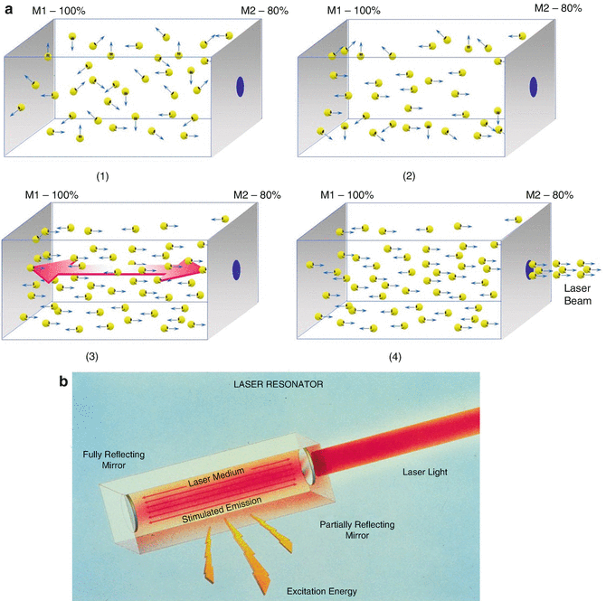
To illustrate the generation of light inside a laser, let us first imagine a rectangular box or a tube like a straight cylinder, containing a large number of identical atoms or molecules — for example, a fluorescent lamp tube with its gas. At each end of the tube we place mirrors, which by construction are parallel to one another. At one end the mirror is totally reflective (100% mirror), and at the other end — the exit window of the light, or output coupler — the mirror is partially reflective (80% mirror), so that part of the light is reflected back into the tube and part is transmitted through the mirror to the outside (Wright and Fisher 1993; Kulick 1998; Boechat 2009; Raulin and Karsai 2011; Kaminsky Jedwab 2010).
Let us also imagine that the atoms are excited to a higher-energy level by an external source (a light source or an electrical discharge), as if we had flipped the switch turning on the lamp. Through the mechanism of spontaneous emission, which takes place completely randomly, the atoms emit photons that begin traveling in various directions within the tube. Those hitting the tube wall are absorbed and lost as heat, disappearing from the scene; in the case of a lamp, they leave the tube into the environment, illuminating the room. On the other hand, photons traveling parallel to the tube axis are likely to find other excited atoms and thus stimulate the emission of additional photons, consistent with the stimulating photon and traveling in the same direction — i.e., along the longitudinal axis of the tube. These two photons continue their journey, again likely to stimulate two additional photons through a similar process — all consistent with each other and traveling on the same axis. The progression continues indefinitely, and 8, 16, 32, 64, etc. photons are produced, all traveling in the same direction, as illustrated in Fig. 4.
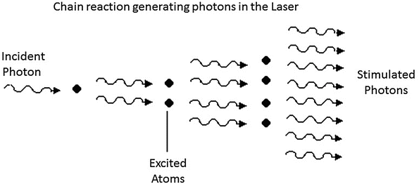
A light amplification process is clearly established, generating a large luminous flux in the longitudinal direction of the tube.
The mirrors perpendicular to the tube axis reflect the photons back, intensifying this amplification effect. Each reflected photon traveling along the axis in the opposite direction contributes to the chain reaction, generating a stream of coherent photons. When they reach the partially reflecting mirror, 80% of the photons return to the tube, continuing the amplification effect. The remaining 20% emerge, forming the laser beam (Fig. 5a, b). In absolute terms they represent a very intense beam of photons produced by the amplification effect. The tube and its excited medium, together with the mirrors, are called the resonator (or oscillator) — the basic component of a laser in addition to the excitation source.
Characteristics of Laser Light
As described above, laser light has unique properties that make it different from other light sources (Goldman and Fitzpatrick 1994; Arndt et al. 1997; Kaminsky Jedwab 2010; Sardana and Garg 2014):
- Monochromatic: it is generated by a collection of identical atoms or molecules; thus all photons emitted have the same wavelength, a single frequency. This feature is important because of the selective absorption of human tissue, presented in the next section.
- Coherent: because of stimulated emission and the way the light is amplified, only in the longitudinal direction inside the resonator, the photons are organized like soldiers marching in a military parade. This is called spatial and temporal coherence. At any point of a laser beam, the photons:
- have the same power;
- travel in the same direction;
- travel at the same time.
Being coherent, light from a laser is called collimated. Traveling parallel to the tube axis, the laser beam has a very small divergence angle — the light does not spread; the photon beam is collimated (parallel). The small divergence allows a lens system to concentrate all the laser energy precisely on a small focal spot (spot size), achieving a greater concentration of light energy, or brightness. Optical laws tell us that the smaller the divergence, the smaller the focal point. When we focus a common, incoherent light source such as a lamp, the focal point is large and imprecise, whereas with a laser we obtain a very fine, extremely precise focal point and therefore a much more intense effect on the tissue.
Energy, Power, and Fluence
The increase in temperature, or the treatment effect on the tissue, depends on the amount of energy it receives. Energy, power, and fluence (energy density) are the physical parameters that control the treatment effect and determine the eventual temperature increase.
- Energy is measured in joules (J).
- Power is measured in watts (W).
These are different parameters, related through the following equation:
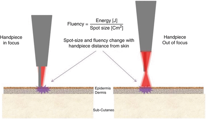
Energy is thus the amount of power delivered to the tissue in a given time, or the laser pulse duration. The thermal effect of the laser is highly localized. The physical quantity that governs the thermal response of the tissue is the amount of energy delivered to a certain area — the overall size of the application area, or “spot size,” produced by the laser handpiece. The energy density, or fluence, is measured in J/cm2:
The higher the fluence, the faster the temperature rises in the tissue and, consequently, the greater the intensity of the desired effect. The treatment effect is achieved both by varying the laser output energy and by varying the laser pulse duration over the application area. All commercial lasers allow the energy to be changed easily and continuously.
For a fixed operating power, fluence in the tissue can be varied by changing the application area (spot size — changing the lens that focuses the laser beam in the handpiece) or by varying the distance of the handpiece from the tissue in a “focused” handpiece.
When we work with the light in focus (Fig. 6), power density is at its maximum because all the laser energy is concentrated in a small focal point (usually of the order of 0.1–1 mm), called the “spot size.” At the focal point the tissue can be cut precisely, and the application has its maximum effect. When the handpiece is moved away from the tissue into a defocused, or out-of-focus, position, the application area becomes larger, reducing power density (fluence) and the temperature increase in the tissue. In this position the effect becomes milder, producing a superficial effect of vaporization and coagulation (used in skin rejuvenation — skin resurfacing).
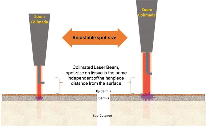
Another widely used laser handpiece is called “collimated.” Here the laser beam remains parallel (collimated) and constant regardless of distance from the tissue. It is used in hair-removal systems and in various skin treatments, such as tattoo and melasma removal (Fig. 7).
It is important to note how the cutting effect is controlled with a laser. The surgeon is accustomed to controlling the depth of a cut by the pressure of the blade against the tissue. With a laser, as there is no mechanical contact with the tissue, the cut is determined by two factors:
- hand movement speed;
- laser energy.
Speed is linked to tissue exposure time: if the laser keeps acting on a point indefinitely, it begins to vaporize layer upon layer of tissue, increasing the depth of the cut. Thus, for constant power, moving the hand slowly produces a deep cut. Likewise, at constant speed, cutting is deeper at greater energy.
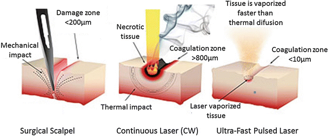
Laser exposure time also governs the amount of adjacent tissue that may be affected. Modern laser systems have mechanisms that quickly deliver energy to the tissue while minimizing the thermal effect in adjacent areas. These mechanisms can be ultrafast pulses (“ultrapulse” lasers) or computerized rapid laser-beam scanning systems (fractional scanners), used in skin rejuvenation treatments and, more recently, in fractional treatment systems. The scanner divides and moves the laser beam at high speed to position it over the skin, minimizing damage to adjacent tissue. Scanners are computer-controlled and can execute different types of scanning, with great precision and control over the amount of tissue vaporized (Goldman and Fitzpatrick 1994; Arndt et al. 1997; Kulick 1998; Alster and Apfelberg 1999; Alster 1997).
Operating Modes of a Laser
Depending on the treatment effect we want to obtain on the tissue, laser systems can operate in the following modes (Boechat 2009; Raulin and Karsai 2011; Kaminsky Jedwab 2010; Sardana and Garg 2014):
- Continuous mode (CW): in this mode (also known as continuous wave), the laser stays on, like an ordinary lamp, and emits a beam of constant energy as long as the system remains powered through the foot switch or the power button on the handpiece (available on some devices). It is widely used in surgery for coagulation or vaporization of tissue.
- Pulsed mode: this mode works as if a lamp were switched on and off. The laser is pulsed electronically, with the pulse times and intervals controlled by the equipment computer and selected from the panel. The repetition rate, or frequency (given in Hz), can also be programmed. Most lasers used in dermatology work with ultrafast pulses, to vaporize tissue faster than the skin’s thermal-diffusion time and so minimize damage to adjacent tissue, resulting in safe and effective treatments (Fig. 8).
According to laser pulse duration, pulsed systems can be classified into:
- Long pulses — millisecond (ms), 10−3 s: hair removal, varicose veins;
- Quasi-CW — microsecond (μs), 10−6 s: skin rejuvenation, onychomycosis, inflammatory acne;
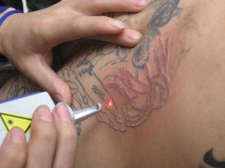
- Q-switched — nanosecond (ns), 10−9 s: treatment of melasma, tattoo removal;
- Mode-locked — picosecond (ps), 10−12 s: tattoo removal and pigmented lesions;
- Femto — femtosecond (fs), 10−15 s: refractive surgery in ophthalmology.
Q-Switched: Nanosecond Laser
This mode is achieved by placing an optical accessory inside the resonator, next to the laser crystal, whose purpose is to pulse the light optically (Siegman 1986; Goldman 1967; Raulin and Karsai 2011). It is generally used in crystal lasers such as ruby, alexandrite, and Nd:YAG, described below. The goal is to accumulate laser energy at very high levels and release it in extremely rapid pulses. The result is a very-high-peak-power laser pulse (often higher than an ordinary pulse), which can penetrate deep into the tissue with minimal side effects. A shockwave-induced mechanical action caused by the impact of the laser pulse on the target tissue then causes its fragmentation. In the long-pulse and quasi-CW modes, the effect is purely thermal.
The Q-switch can be passive, using a crystal called a “saturable absorber” that produces rapid pulses, or active, using an electronic modulator crystal called a “Pockels cell.”
Passive systems using a saturable absorber are generally simpler and more compact, resulting in small portable devices or systems built into handpieces attached to a platform. They are more limited: the stability of the fast pulse cannot be efficiently controlled; the crystal is sensitive to higher energies, limiting the maximum working energy; the application spot size is limited to a few millimeters (1–3 mm); and they cannot achieve high pulse repetition rates, working at a maximum of 2–3 Hz.
The active Q-switch uses a Pockels cell, a crystal subjected to a high electric field and electronically controlled to produce very fast, stable light switching. The result is faster pulses with very high peak powers not attainable with passive systems. They can therefore handle high energy, larger spot sizes (10 mm), and faster repetition frequencies of 2–20 Hz. Equipment with an active Q-switch can also be switched off, allowing the laser to work in quasi-CW mode with micropulses, giving the system greater flexibility (Fig. 9).
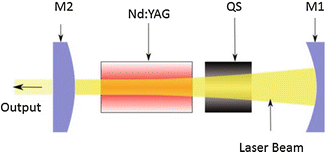
The classic application is tattoo removal and treatment of pigmented skin lesions such as dark circles, postinflammatory hyperpigmentation, and melasma (Goldman 1967; Reid and Muller 1978; Raulin et al. 1998; Chang et al. 1996; Shimbashi et al. 1997; Reid et al. 1983, 1990; Stafford et al. 1995; Ogata 1997; Chan et al. 1999; Jeong et al. 2008; Mun et al. 2010) (Fig. 10).
Mode-Locked: Picosecond Laser
To achieve picosecond pulses, a technique called “mode-locking” is used (Siegman 1986; Raulin and Karsai 2011; Sardana and Garg 2014). The base is a Q-switch system as described above, in which nonlinear effects of the Q-switch crystal are stimulated and modulated inside the resonator to create faster pulses, with a technique in which only those pulses are amplified. It is more commonly used in crystal lasers such as alexandrite and Nd:YAG.
There is passive mode-locking with a saturable absorber and active mode-locking with an electronically controlled Pockels cell. The limitations and benefits of each are the same as in Q-switched systems.
Picosecond lasers for dermatology provide pulses ranging from 375 to 760 ps.
To understand the advantages of the picosecond laser over a nanosecond device, we return to the relationship between energy, power, and pulse duration described above. Peak power is inversely proportional to pulse duration. In other words, faster (shorter) pulses generate higher power for the same energy:
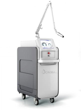
A picosecond laser generates very high peak power, making photomechanical fragmentation of the target tissue — and consequently the treatment — more efficient. It also does not need high energy levels; working at very low energy results in milder treatments and faster recovery. In tattoo removal, for example, a picosecond laser needs fewer sessions than a nanosecond system, and applications can be performed every 15 days, whereas nanosecond sessions are 45–60 days apart. The faster the system, the milder and more effective the treatment — which is why industry has been investing in the development of these ultrafast devices (Fig. 11).
As seen in the following chapter, pulse duration governs the way light interacts with tissue (selective photothermolysis), and by varying pulse duration the laser application in dermatology can be completely changed.
Laser Types
All laser devices consist of the following parts (Siegman 1986; Goldman and Fitzpatrick 1994; Boechat 2009; Kaminsky Jedwab 2010):
- the resonator/oscillator — with mirrors (total and partial reflectors) and an active medium which, when excited, produces the light and thus determines the wavelength;
- the excitation source (also called pumping) — which delivers power to the active medium, producing photons;
- the laser-beam delivery system, from the source to the hand of the operator;
- the handpiece, with a focusing lens or a scanning system.
Industry uses various elements to manufacture laser sources in order to cover a growing range of electromagnetic wavelengths. Today there are ultraviolet, visible-light, and infrared lasers. To this end, gases, liquids, crystals, fiber optics, and semiconductors (electronic components) are used. Pumping of each element also varies: electrical discharges, radio frequency, and light sources such as flash-lamps or even other lasers are used.
To carry laser light from the resonator to the user, various mechanisms are used depending on the wavelength and energy of the equipment. The most common are:
- Articulated arm — a set of multiple mirrors positioned at the corners of articulated pipes, allowing freedom of movement in all directions (Fig. 12).
- Optical fiber — a thin waveguide with a quartz core covered with a thin layer of slightly different material (cladding), encapsulated with plastic and metal coatings to give flexibility. It delivers the laser beam by multiple internal reflections: light enters the fiber, reflects at the core/cladding interface, and keeps moving until it exits. Note that at the fiber output the laser beam has wide divergence and is no longer collimated; the beam spreads, losing part of its coherence (Boechat et al. 1991, 1993) (Figs. 13 and 14).
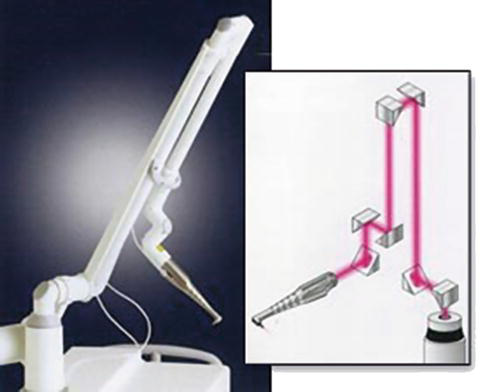
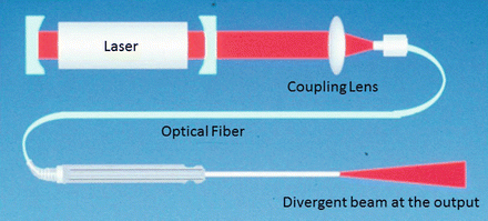
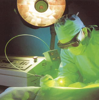
A handpiece is placed at the end of the beam delivery system, whether articulated arm or optical fiber. It contains the lens system that focuses the laser light on the working area, facilitating handling of the laser during treatment, as already described. In fractional laser devices, described below, the handpiece holds scanning systems (scanners) in addition to lenses.
Below we describe typical commercial laser systems used in medicine, grouped according to the laser medium (Alster and Apfelberg 1999; Alster 1997; Boechat 2009; Raulin and Karsai 2011; Kaminsky Jedwab 2010; Sardana and Garg 2014).
Gas Lasers
Excimer
Gas molecules that exist only in the excited state, called “dimers,” form the active medium; examples are molecules of halogens combined with noble gases (ArF, KrF, XeCl, XeF). The word “excimer” is an abbreviation of “excited dimer.” Emission covers several wavelengths in the ultraviolet range, such as 193 nm ArF, 222 nm KrCl, 248 nm KrF, and 308 nm XeCl. Pumping is usually by electrical discharge or by the shock of electrons with gas molecules. Quartz optical fibers are used as the beam-delivery system. Because the wavelength is very short and carries high energy, these lasers are widely used for high-precision incisions or tissue ablation, such as in ophthalmic refractive surgery (myopia). In dermatology, this system has shown excellent results in the treatment of psoriasis and vitiligo (Zelickson et al. 1996; Guttman 2000).
Argon Ion
Excited ionized argon gas, Ar+, forms the laser medium. Pumping is by electrical discharge. The wavelength can vary between 488 nm (blue) and 514 nm (green). It uses a quartz optical fiber as the delivery system (Siegman 1986; Boechat 2009).
Helium–Neon (He–Ne)
The active medium is a mixture of helium and neon gases, also pumped by electrical discharge. The wavelength is in the visible range, 632.8 nm — red. These systems are generally used for low-power applications such as cell stimulation, laser pointers, or aiming systems for invisible infrared lasers. They use quartz optical fibers (Siegman 1986; Boechat 2009).
Carbon Dioxide (CO2)
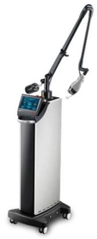
CO2 is still one of the most used lasers in surgery, dermatology, and industrial applications. Its power may vary from a few kW up to MW, delivered continuously or in pulses. The laser medium is a gas mixture including N2 (nitrogen, 13–45%), He (helium, 60–85%), and CO2 (1–9%). Pumping is achieved by high-voltage electrical discharge or radio frequency (RF). The CO2 molecule is excited by mechanical collision with electrons from the N2 and He molecules. The wavelength is in the infrared range at 10,640 nm. This is a relatively efficient laser (30% electro-optical conversion) and consequently has low power consumption and maintenance. It uses an articulated arm and special dielectric-coated flexible hollow waveguides (Siegman 1986; Kulick 1998; Alster and Apfelberg 1999; Alster 1997) (Fig. 15).
Liquid Lasers
Dye Laser
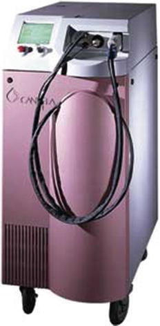
It uses a liquid Rhodamine solution (R6G), a fluorescent dye, as the laser medium. It is pumped by a flash-lamp or another laser. The wavelength can vary continuously from 300 to 1,000 nm, and the resonator can be tuned. It is most commonly used in yellow (585–600 nm). Its main application is treatment of vascular lesions and inflammatory processes of the skin. It uses a quartz optical fiber (Siegman 1986; Reichert 1998; McMillan et al. 1998; Reyes and Geronemus 1990) (Fig. 16).
Solid-State Lasers (Crystal)
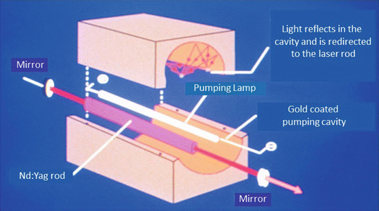
Figure 17 shows a schematic of the most common solid-state laser systems on the market. The mirrors, the laser rod (the crystal), and the flash-lamp used for pumping, inside a cavity of coated elliptical reflecting material — usually ceramic or a high-resistance metal such as gold — compose the resonator (Siegman 1986; Boechat 2009).
Ruby: Cr3+:Al2O3
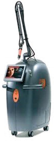
This was the first laser, developed by Maiman in 1961 (Siegman 1986; Goldman and Fitzpatrick 1994; Arndt et al. 1997), although some time passed before the system was used in medicine. The medium is an ionized ruby crystal, pumped by a flash-lamp. The wavelength is in the red range, 694 nm. The nature of the crystal requires high pumping energy, or high-power flash-lamps. It uses fiber optics and an articulated arm for delivery. It is generally used for treatment of pigmented lesions and for hair and tattoo removal (Goldman 1967; Reid and Muller 1978; Raulin et al. 1998; Chang et al. 1996; Shimbashi et al. 1997; Yang et al. 1996; Ono and Tateshita 1998; Reid et al. 1983, 1990) (Fig. 18).
Alexandrite: Cr:BeAl2O4
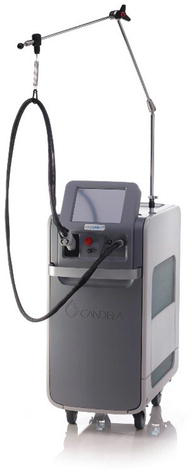
The gain medium is chromium-doped chrysoberyl — the semiprecious stone alexandrite, ionized. It is pumped by a flash-lamp. The wavelength is at the end of the red range, 755 nm. It uses flexible optical fibers or an articulated arm. This crystal has better optical properties, enabling faster, more efficient operation in a smaller device than ruby. It is widely used for hair removal and treatment of pigmented lesions (Siegman 1986; Finkel et al. 1997; Stafford et al. 1995; Chan et al. 1999; Alster 1997) (Fig. 19).
YAG Family
YAG is short for yttrium aluminum garnet, a synthetic crystalline structure serving as host to the ion that produces radiation at the desired wavelength. It is pumped by laser diodes or a flash-lamp and operates in the near-infrared spectrum. It uses optical fiber and, in some cases (high-energy pulsed lasers — Q-switched), an articulated arm as the delivery system. The most common members are (Siegman 1986; Goldman and Fitzpatrick 1994; Kulick 1998; Wong and Goh 1998; Ogata 1997; Chan et al. 1999; Boechat 2009; Raulin and Karsai 2011; Kaminsky Jedwab 2010):
Nd:YAG
Uses the neodymium ion, with wavelengths of 1,064 nm and 1,320 nm; the latter is used for non-ablative skin rejuvenation (Muccini et al. 1998; Goldberg 1999, 2000b).
Nd:YAG/KTP
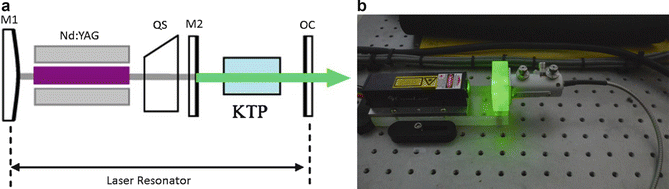
By placing a second crystal in the resonator, generally the well-known potassium-titanyl-phosphate (KTP), a frequency-doubled Nd:YAG laser is generated, with a green wavelength at 532 nm. It is used for removal of superficial pigmented and vascular lesions (Figs. 20 and 21).
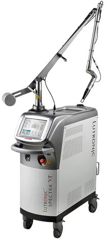
Nd:YAG/KTP with Crystal-Dye Handpiece
A solid-state fluorescent dye handpiece can be added to these lasers to obtain different wavelengths, such as 595 nm (yellow) and 650 nm (red), making the machine extremely versatile for treatment of pigmented lesions and removal of light-colored tattoos at different depths (Fig. 22).
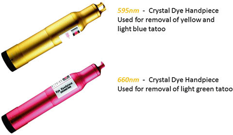
Ho:YAG
Uses holmium ions, with a wavelength of 2,100 nm. Excellent for treatments in bone and cartilage and for fragmentation of kidney stones.
Er:YAG
Uses erbium ions, with a wavelength of 2,940 nm. Well known for its use in skin resurfacing (skin rejuvenation) (Fleming 1999; Weinstein 1998).
Tm:YAG
Uses thulium ions, with a wavelength of 1,927 nm. Used for non-ablative skin rejuvenation with a more superficial action (Fig. 23).
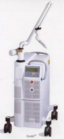
Nd:YAP
Uses neodymium ions in a yttrium aluminum perovskite crystal, with a wavelength of 1,340 nm. Used for non-ablative skin rejuvenation and chronic inflammatory diseases such as hidradenitis (Milanic and Majaron 2013; Antonio et al. 2015).
Er:Glass
The gain medium changes to crystal glass, which hosts the erbium ion. The wavelength shifts to 1,540 nm, in the near infrared. Used for deeper skin rejuvenation and employed in fractional laser systems (Mordon et al. 2000) (Fig. 24).
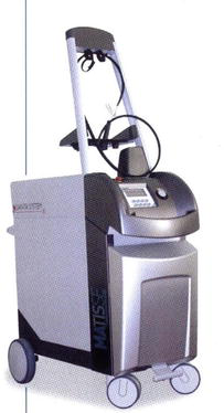
Er:YSGG
The gain medium is similar to the YAG crystal, using the erbium ion in a yttrium scandium gallium garnet (YSGG) host. The wavelength is also in the near infrared, 2,790 nm, used in the Pearl™ handpiece of Cutera. The main application is fractional skin rejuvenation; it is an alternative to Er:YAG laser skin resurfacing.
Semiconductor Lasers
Diode
The laser medium is a semiconductor — an electronic component — pumped by electric current. Changing the semiconductor provides a wide range of wavelengths, from visible 450 nm to near infrared 1,400 nm. The most common are aluminum gallium arsenide (AlGaAs), with wavelengths from red to near infrared, 620–900 nm, and gallium arsenide (GaAs), in the near infrared, 830–920 nm. It has very efficient electro-optical conversion (greater than 50%) and is therefore generally a small, greatly simplified system. It uses optical fibers or simply free handheld devices. Some manufacturers provide systems with one or more laser diodes of different wavelengths, increasing flexibility. Diodes are widely used for hair removal, non-ablative skin rejuvenation, and treatment of vascular lesions. They are also used to pump other lasers such as Nd:YAG, Nd:YAG/KTP, and fiber-optic lasers, as seen below (Siegman 1986; Goldberg 2000a; Ross and Hardway 2000; Lou et al. 2000) (Fig. 25).
Optical Fiber Lasers
Extremely robust, long-lasting, and highly reliable, this technology — employed in undersea optical telecommunications cables — has found medical application in the development of fractional lasers (Manstein et al. 2004; Geronemus 2006; Raulin and Karsai 2011).
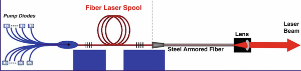
Y, Er:FIBER — the gain medium is a quartz optical fiber only 150 μm in diameter, containing erbium and yttrium ions. It is pumped by laser diodes. The wavelength is 1,550 nm. The system needs no optical components such as mirrors, output couplers, flash-lamps, or a cooling system, significantly reducing maintenance needs and cost. This technology produces microscopic focal points on the skin, of the order of 100 μm (approximately the thickness of a human hair), since the light source itself is a microscopic fiber — leading to the advent of fractional skin treatment (Figs. 26 and 27).
LED: Light-Emitting Diode
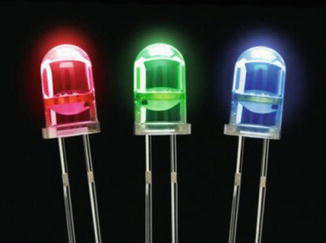
LEDs are electronic components — semiconductor diodes — that emit light when stimulated by electric current. They may be considered related to laser diodes, since they are manufactured from the same materials, such as GaAs, GaAlAs, and GaInPAs, and thus provide the same wavelengths. However, they have no light-amplification effect from a laser resonator. They produce incoherent monochromatic light that diverges in various directions, like a low-intensity lamp (Boechat 2009; Raulin and Karsai 2011; Kaminsky Jedwab 2010).
To concentrate and direct the emitted light, they are manufactured with a parabolic plastic housing that functions as a small lens (Fig. 28).
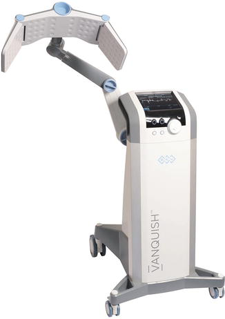
LED treatment systems use panels of 1,000–2,000 components to extend and optimize the application area. Depending on the application or treatment, the panel can be changed for one of a different wavelength. Some manufacturers integrate LEDs of different wavelengths on the same panel, avoiding the need to change them (Fig. 29).
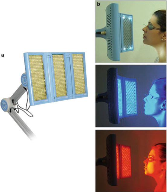
Some of the most common applications are biomodulation of cells, described below and in following chapters, such as anti-inflammatory effects and improved wound healing. LEDs are also used in photodynamic therapy and teeth whitening.
Intense Pulsed Light
This system employs a flash-lamp for many applications, but it is not a laser: pulsed light, or intense pulsed light (IPL). Dr. Shimon Eckhouse at ESC Medical in Israel developed the concept in the 1990s (Boechat 2009; Raulin and Karsai 2011; Kaminsky Jedwab 2010).
It uses an electronically controlled intense flash-lamp, and therefore has distinct characteristics from a laser source:
- Polychromatic: it emits a broad spectrum of wavelengths, generally in the range 400–1,200 nm. Band-pass filters placed in front of the lamp are used for wavelength selection. These filters remove a band of wavelengths, generally those below the filter specification, letting through all wavelengths above it. Some machines use a more complex filter that narrows emission to a range of wavelengths, as illustrated in Fig. 30. Even with a narrow emission spectrum limited by filters, the emitted energy is dispersed among several wavelengths — some absorbed by the tissue to be treated and others having no effect. The selectivity and effectiveness of treatment are thus reduced compared with a laser, with 100% of its energy concentrated at a single wavelength.
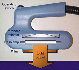
- Incoherent: unlike a laser, IPL energy is emitted in all directions; it spreads. Mirrored surfaces placed behind the lamp, similar to the reflectors used in car headlights, concentrate and direct the light. It has a more superficial, milder effect on tissue because it is less intense than laser light. The application is also less painful.
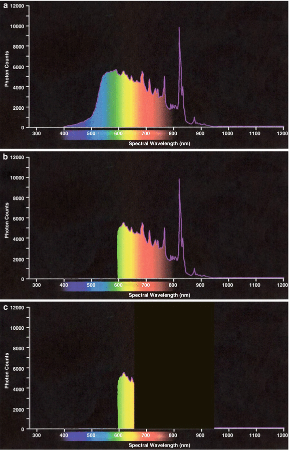
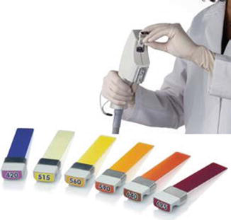
The multiplicity of emitted wavelengths makes these systems very versatile, able to perform several applications — hair removal, pigmented lesions, non-ablative rejuvenation, and vascular lesions — by simply changing the filter and pulse duration (Fig. 31).
These systems generally have a fixed pulse duration set by the manufacturer according to the application. To change pulse duration, it is generally necessary to change the entire handpiece. Pulse duration is restricted to the millisecond range because of the lamp characteristics, but this suits most skin applications.
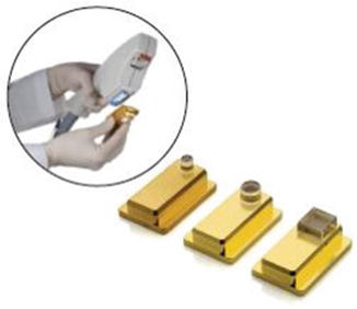
Figure 32 illustrates an IPL with various application filters. To change spot size, it is necessary to change the handpiece for one with a smaller application area, or to use physical filters — such as a plate with a hole of different sizes placed in front of the lamp, as shown in Fig. 33. The plate limits the application area but significantly reduces treatment energy. In this they differ from lasers, in which a lens system in the handpiece changes spot size more versatilely while preserving the total energy of the beam.
Treatment Platforms
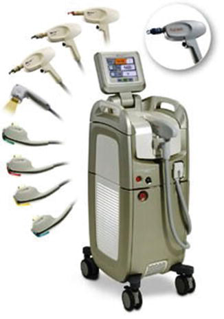
Following the market trend toward increasingly compact systems providing various applications, the laser industry developed the concept of the multi-application platform. These systems consist of a base (platform) carrying the energy source and cooling system; several handpieces can then be connected to the base, each providing a different application. Each handpiece may contain an IPL or a laser system. The most frequent applications are hair removal, skin rejuvenation, treatment of pigmented and vascular lesions, and tattoo removal (Raulin and Karsai 2011; Kaminsky Jedwab 2010; Sardana and Garg 2014).
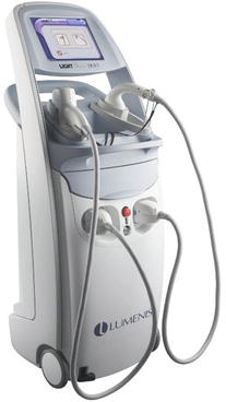
Treatment platforms became very popular because of their excellent cost/benefit and the versatile combination of intense pulsed light and laser in the same equipment. There are platforms with only lasers or only IPL, and others that add an RF handpiece for skin tightening.
One limitation of this design is that it does not allow simultaneous treatments; for example, hair removal must be finished before skin rejuvenation can be performed. For busy clinics performing several applications at the same time, separate pieces of equipment are a better alternative for increasing revenues.
Another important point: when a laser is used as a platform handpiece, it suffers from limitations in energy and versatility. An Er:YAG device with its articulated arm, for example, can provide more power, versatility, spot-size range, and pulse-duration options than when installed in a platform handpiece. The same happens with a Q-switched laser, as it is very difficult to adapt an active Q-switch into a simple handpiece; on a platform, Q-switched devices are therefore usually limited to the passive design (Fig. 34).
After becoming familiar with these technologies and their operating principles, a question comes to mind: when and how should each of these systems be used?
The application of each laser, IPL, or LED in dermatology depends on the response of the tissue to the wavelength used.
Light–Tissue Interaction
Light can interact with living tissue in the following forms (Anderson and Parrish 1981; Goldman and Fitzpatrick 1994; Arndt et al. 1997; Kulick 1998):
- Photothermal: light energy is absorbed by the target tissue (chromophore) and transformed into heat, causing coagulation or vaporization.
- Photomechanical: fragmentation by a mechanical effect, as in the Q-switch described above.
- Photochemical:
- direct breaking of chemical bonds between the atoms of a molecule — produced, for example, by an ultraviolet excimer laser when sculpting a cornea, with great accuracy;
- light activates a chemical reaction producing reactive free radicals, as in photodynamic therapy (PDT), described in a following chapter.
- Photobiomodulation: light is used to modulate intra- and intercellular activities. It employs low-power lasers and LED panels; it has anti-inflammatory action and effects on wound healing and tissue regeneration (Lopes 1999).
- Selective photothermolysis: the art of combining wavelength, pulse duration, and energy to obtain the desired effect on target tissue while preserving adjacent areas, as described below.
When a beam of light hits tissue (Fig. 35), it is partially transmitted, reflected, spread (scattered), or absorbed.
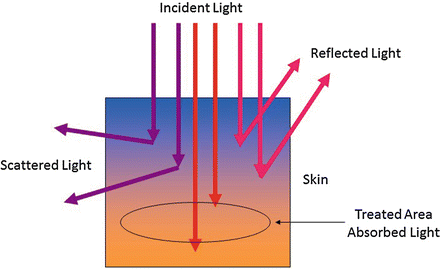
Laser light produces a therapeutic effect only if the target tissue is “in tune” with the energy used, as with a mobile phone. At any moment thousands of mobile-phone waves pass around us, but the phone does not ring; it is triggered only when the emitted wave is in tune with the device. Similarly, several wavelengths of light can be placed in the skin, but the target tissue absorbs only a specific one. In particular, the energy deposited by the most commonly used medical lasers is transformed into heat, producing a temperature increase in the chromophore.
The effect of temperature increase in tissue can be summarized in the following ranges:
- 37–43 °C: accelerated cell metabolism, stimulation, contraction of elastic fibers, and skin tightening. The effect is limited and reversible.
- 44–45 °C: exponential increase in the acceleration of cell metabolism, protein changes, collagen stimulation, and cell apoptosis with long application times, due to hyperthermia.
- 50–70 °C: protein denaturation; coagulation of collagen (which must be replaced through regeneration), cell membranes, and hemoglobin; permanent contraction of collagen fibers.
- 90–100 °C: formation of extracellular vacuoles, evaporation of liquids.
- Above 100 °C: vaporization of tissue, charring.
The laser parameter that most influences the absorption factor — the “tuning” effect — is the wavelength of the light (its color, its frequency). Each part of the organism, each component of the skin, responds differently or has an affinity for a particular wavelength. Certain tissues are transparent to a given laser; others absorb it completely. The necessary thermal effect can therefore be induced selectively at a specific point without affecting the surrounding tissue, giving rise to the theory of “selective photothermolysis,” developed by Dr. Rox Anderson and colleagues in Boston, USA (Anderson and Parrish 1983, 1981; Goldman and Fitzpatrick 1994).
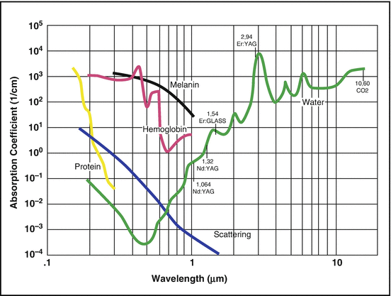
The graph in Fig. 36 represents the fundamental result of Anderson et al.’s publication. It shows how the absorption coefficients of certain skin components — melanin, hemoglobin, and water — vary with wavelength. Melanin has high absorption for visible-range lasers, such as green (KTP), which can be used, for example, in treatment of pigmented lesions. Hemoglobin has an absorption peak in the yellow range (dye laser), making it a good option for treating vascular lesions. The ruby laser, in the red range, is well absorbed by melanin and dark pigment in the skin; on the other hand, it sits at a minimum for hemoglobin absorption, partly explaining the difficulty these systems have removing red pigments in tattoo treatment and vascular lesions (low coagulation effect).
When the light of these lasers enters the skin in fast — or rather, ideal — pulses, it can cross the skin without causing damage and is absorbed only by the target tissue with which it has an affinity. These components are referred to in the literature as “chromophores.”
The graph also shows that melanin absorption in the visible and near infrared (invisible) is very wide, allowing a number of different lasers to be used effectively for pigmented lesions and hair removal, such as the 810 nm diode laser and the 1,064 nm Nd:YAG. Because of its longer wavelength, Nd:YAG penetrates deeper into the skin, described below, and its absorption coefficient for melanin is lower than that of visible lasers such as green. These properties make such lasers suitable for a variety of treatments: they present a reduced risk of damage to the skin surface from melanin absorption, and are effective for dermis treatments such as deep vascular lesions and melasma.
The Er:YAG (2,940 nm) and CO2 (10,600 nm) lasers, in the infrared, have high absorption coefficients for water. As water is the major component of cellular structures, its interaction with these wavelengths predominates. The first cell layers therefore rapidly absorb the energy of these lasers, raising their temperature to vaporization level and making them excellent tools for cutting or for precise, superficial tissue removal, such as laser skin resurfacing or fractional laser skin resurfacing. The Er:YAG wavelength sits at the peak of water absorption, with a coefficient at least ten times higher than the CO2 laser. Since its light is absorbed more rapidly, energy penetrates less, giving it a more superficial action than CO2. Treatment also has less thermal effect, being gentler on the skin (Chernoff et al. 1995; Alster et al. 1999; Weinstein 1998).
Another important aspect of light–tissue interaction is laser pulse duration (pulse length, or exposure time). It must be such that the energy produces a temperature increase confined (concentrated) to the target tissue, with minimal dispersion to surrounding areas. In other words, pulse duration must be long enough to raise the temperature of the target tissue to destruction level, while being short enough not to radiate heat into surrounding tissue. A similar situation occurs when testing whether an iron is hot enough: a finger is placed on the iron long enough to check the heat, but removed very quickly to avoid a burn.
To achieve correct pulse duration, the thermal relaxation time (TRT) of the target tissue must be observed. The TRT is the time the tissue takes to cool after being intensely heated. Following physical principles, chromophores with large volumes or cross-sections take longer to cool and therefore have a higher TRT. A thick hair, for example, has an average TRT of 40 ms, whereas a thin hair has a TRT of 1–3 ms.
For a flat object, such as a melanosis, TRT can be estimated by the ratio (Anderson and Parrish 1981; Goldman and Fitzpatrick 1994; Arndt et al. 1997; Lapidoth et al. 2015):
where d is the thickness of the material and a its thermal conductivity.
For a cylindrical object, such as a hair or a vein:
where d is the diameter of the object.
Thus, to confine energy or heat in the target tissue, pulses must be less than or equal to the chromophore’s TRT.
An important conclusion follows: with the same wavelength (the same laser), different treatments can be performed simply by changing pulse duration. With a long-pulse Nd:YAG laser, in milliseconds, varicose veins can be treated or hair removed; installing a Q-switch, with nanosecond pulses, allows tattoo or melanosis removal, since the melanosome TRT is of the order of 100 ns.
The basic principles of selective photothermolysis are therefore (Anderson and Parrish 1983; Goldman and Fitzpatrick 1994):
- an ideal wavelength, absorbed only by the target tissue or chromophore;
- an ideal pulse duration, sufficient to produce the desired effect on any target tissue but fast enough to cause minimal effect on surrounding tissue — i.e., confining the energy in the chromophore;
- energy sufficient to achieve the treatment effect.
In summary, the great majority of photomedicine treatments occur as follows:
- light is absorbed by the target tissue or chromophore;
- absorption causes selective heating of the target while preserving surrounding tissue;
- selective heating of the chromophore causes its coagulation or vaporization, achieving the treatment goal.
The Melanin “Curtain”
The absorption curve in Fig. 36 (Anderson and Parrish 1983, 1981) shows that skin melanin presents absorption for most lasers in the visible and near-infrared range. In applications where the target tissue lies below the papillary dermis (hair, vascular lesions, pigmented lesions, etc.), or below the melanin layer where greater light penetration is needed, energy is always attenuated, reducing treatment efficiency. The melanin in the upper skin layers acts like a window curtain. Absorption by this melanin “curtain” increases with darker-skinned patients or with patients at higher positions on the Fitzpatrick scale. Absorbed energy generates local heat, which when excessive can produce unpleasant adverse effects such as burns, hypochromic spots, or stimulation of melanocytes producing hyperchromic spots.
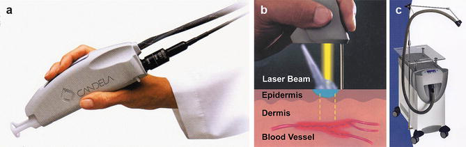
Lasers and IPLs that operate at higher energies therefore employ systems to protect the epidermis, ranging from simple solutions, such as cooling the area with cold gel or ice packs, to sophisticated cooling systems coupled to the handpiece. All are necessary to dissipate some of the heat generated by light absorption in the first skin layer.
Epidermal cooling systems can be (Waldorf et al. 1997; Alster and Apfelberg 1999; Goldberg 2000a; Ross and Hardway 2000; Klavuhn 2000):
- Static: the handpiece has a sapphire window cooled with water or cryogenic gas, which remains in contact with the skin, removing excess heat during the laser pulse.
- Dynamic: also coupled to the handpiece, the system fires a cryogenic gas jet that freezes the skin immediately before the laser pulse, after which skin temperature rapidly returns to normal. The duration of the gas jet (according to phototype and patient discomfort) and the interval between cryogenic spray and laser pulse can be varied.
- Continuous/independent: a separate device providing a cold-air blast that cools tissue during the procedure, operating independently from the laser or pulsed light.
The advantages of these devices are:
- the possibility of using higher energies, increasing treatment efficacy;
- reduction of patient discomfort and of the risk of adverse effects;
- the possibility of treating darker skin types (Fig. 37).
Light Penetration Depth
Given the importance of treatment effectiveness, light penetration depth is governed primarily by wavelength, with the following factors observed (Anderson and Parrish 1981; Raulin and Karsai 2011; Sardana and Garg 2014; Lapidoth et al. 2015):
- scattering of light in the visible part of the spectrum;
- absorption by water in skin cells, particularly epidermal ones, in the near-infrared range;
- for a given wavelength, the higher the energy, the deeper it reaches.
Returning to the absorption-coefficient graph in Fig. 36, light scattering (the blue curve) becomes stronger at shorter wavelengths in the visible range. For this part of the spectrum, then, regardless of the energy used, penetration is usually very shallow, as shown in Fig. 38. Scattering begins to reduce in red light (700 nm) and practically disappears in the near infrared, around 900–1,100 nm, allowing these wavelengths to penetrate deeply into tissue. Beyond 1,200 nm, absorption by water — the abundant component in skin cells — starts to become significant, again reducing light penetration.
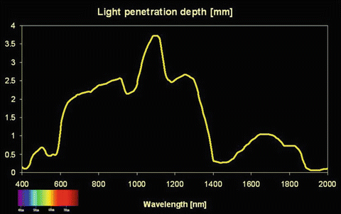
As light penetrates the skin, its energy is absorbed and scattered along the way, its intensity decreasing until it disappears. Power distribution along the light path reduces with penetration; the energy at the surface is always higher than at any point within the tissue. For a given wavelength, then, light with higher surface energy yields a slight increase in tissue penetration.
In summary, visible wavelengths are ideal for treatment of superficial lesions, such as spots or port-wine stains. It is common in treatment of superficial pigmented lesions to observe that some spots do not clear completely; this can indicate that part of the spot lies in a deeper layer light cannot reach.
Wavelengths in the 900–1,100 nm range, near infrared, should be used for treatment of deep lesions such as varicose veins or hemangiomas, and for dermal melasma.
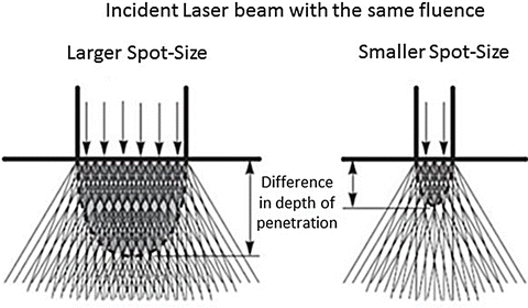
Another mechanism controls light penetration depth: for a given wavelength and fluence (energy/application area), greater energy penetration can be achieved by increasing spot size. Figure 39 illustrates the effect of spot size. With a small spot size, light cannot be concentrated deep in the skin because of scattering; with a larger spot size, dispersion is the same, but it is compensated by a greater concentration of energy deeper in the skin. The larger the spot size, therefore, the greater the energy concentration — the deeper the penetration. This effect is important, for example, in laser hair removal, treatment of dermal melasma, and tattoo removal (Raulin and Karsai 2011; Kaminsky Jedwab 2010; Sardana and Garg 2014).
“Ablative” and “Non-ablative” Skin Rejuvenation
The wide application of lasers working in the near and mid-infrared, from 900 to 10,000 nm, and of intense pulsed light, has revolutionized skin-rejuvenation technique. By definition, an ablative laser removes the skin surface and produces controlled coagulation of the tissue underneath. Non-ablative systems produce only tissue coagulation, keeping the skin surface intact (Muccini et al. 1998; Goldberg 2000b; Khan et al. 2005; Munavalli et al. 2005).
In the near-infrared range, melanin absorption reduces drastically while water absorption increases exponentially. Through appropriate selection of wavelength and pulse duration, the intensity of the heat generated in the skin can therefore be varied, changing from a non-ablative to an ablative interaction.
The difference between a non-ablative and an ablative laser is therefore simply its wavelength and the consequent intensity of interaction with the water in the chromophore. The graph in Fig. 40 shows the water-absorption curves for two lasers used in skin rejuvenation, Er:Glass (1,540 nm) and CO2 (10,600 nm) (Anderson and Parrish 1983, 1981).
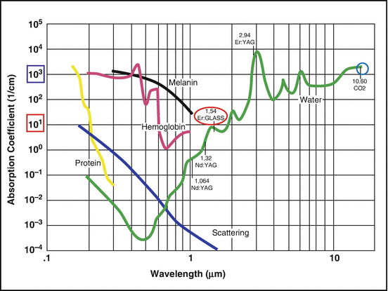
The water-absorption coefficient at the Er:Glass wavelength is approximately 10, while at the CO2 wavelength it is approximately 3,000 — a 300-fold difference. For two lasers with the same fluence, Er:Glass heats the skin to an average temperature of 60 °C, where only coagulation (cell death) occurs, while the CO2 laser raises tissue temperature to 180 °C, leading to vaporization, since this value is far above the boiling point of water. The histology in Fig. 41 shows the difference in laser–tissue interaction for two fractional lasers, described below.
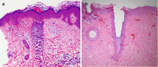
Non-ablative treatment promotes regeneration of deep tissue, with collagen remodeling, promoting a natural filling effect of the skin. It has a fast recovery time with little interference in the patient’s routine. Treatment takes four to five monthly sessions. It improves fine lines, open pores, skin texture, and overall skin quality.
Ablative treatment promotes removal of the skin surface (resurfacing), regeneration of the tissue underneath (the same as the non-ablative effect), and skin tightening, because of the high temperature reached and the removal of a percentage of skin by laser vaporization. It provides a more complete treatment than the non-ablative approach, but with a longer recovery time. Treatment takes two to three sessions 45–60 days apart. It shows good results for deep wrinkles, scars, tightening, texture, and overall skin quality (Pitanguy et al. 1996; Chernoff et al. 1995; Alster et al. 1999; Lask 1995).
As IPL systems employ a range of wavelengths from visible to near infrared, they can treat many types of skin lesions simultaneously: near-infrared wavelengths improve skin quality, while the visible part of the spectrum removes superficial pigmented lesions and small surface telangiectasias. They show milder, more superficial effects than lasers, however.
Several manufacturers produce laser systems and pulsed light for this purpose; some examples are:
- CO2 (10,600 nm) — produces a balanced mix of ablation and coagulation leading to a complete rejuvenation result, still the gold standard of the market.
- Er:YAG (2,940 nm) — has a ten-times-greater water-absorption coefficient than the CO2 laser and thus produces more ablation than coagulation. Ablation is shallower and milder, with less collagen remodeling in the dermis.
- Nd:YAG (1,064 nm and 1,320 nm) — a line of Nd:YAG lasers, typically producing 1,064 nm, with the resonator altered to produce the new wavelength. Non-ablative effect only, coagulating the dermis (Muccini et al. 1998; Goldberg 1999, 2000b; Goldberg and Whitworth 1997).
- Tm:YAG — thulium laser working at 1,927 nm; non-ablative effect.
- Diode — a compact system at 1,450 nm; non-ablative effect (Goldberg 2000a; Ross and Hardway 2000).
- Er:Fiber or Er:Glass — using the erbium ion on a different material, an optical fiber or glass crystal, with wavelengths of 1,550 nm or 1,540 nm. Both non-ablative (Mordon et al. 2000).
- Intense pulsed light — the versatility, milder treatment, and good cost–benefit of these devices have led to various devices in recent years dominating the landscape of non-ablative skin rejuvenation, although the effect is generally superficial.
Fractional Laser Systems
To appreciate the revolution introduced by fractional laser technology, imagine a patient seeking aesthetic improvement of the skin as a family photograph that needs some finishing touches. Today a photograph is digitally altered pixel by pixel to improve the appearance of objects in the image; likewise, damaged paintings are restored gently, a small area at a time.
This same concept is employed in systems using fractional photothermolysis. The laser produces microscopic thermal injuries called microthermal zones (MTZs), approximately 100–150 μm in diameter — the thickness of a human hair — and 0.2–2.4 mm deep (IPL devices generally achieve a maximum of 0.3 mm below the surface). These MTZs are surrounded by unaffected healthy tissue that helps recovery of the micro-damaged area; the surrounding tissue is also mobilized in the overall skin-regeneration process. The resulting rejuvenation effect is comparable to deep chemical peels or dermal mechanical abrasion, but with minimal side effects and little downtime (Fig. 42).
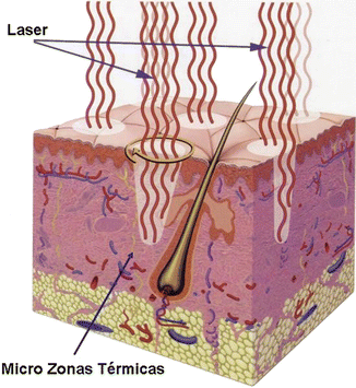
An intelligent scanning system (optical scanner) in the handpiece ensures even distribution of MTZs. The operator can choose directly on the laser panel the number of MTZs applied to the skin, or the percentage of total skin area stimulated, controlling treatment aggressiveness. More MTZs mean greater stimulus — a more aggressive application and consequently more results. This yields an application control that previously did not exist in dermatological treatments.
The method was developed by the originators of selective photothermolysis, Drs. Rox Anderson and Dieter Manstein, at the Wellman Laboratories in Boston, USA. The first fractional laser system, Fraxel SR, was presented by Reliant Technologies Inc. at the congress of the American Society for Lasers in Medicine and Surgery (ASLMS) in April 2004 (Laubach et al. 2005; Geronemus 2006; Raulin and Karsai 2011).
The system follows the principles of selective photothermolysis, with a wavelength around 1,550 nm, where the water in the chromophore is present in skin cells. In its original design, fractional systems perform non-ablative treatment, preserving the skin surface, with tissue temperature rising only to the coagulation point, producing microthermal zones. Treatment takes three to five monthly sessions.
Beyond minimal recovery time, advantages include the possibility of treating other body regions besides the face safely and efficaciously, and removal of deep pigmented lesions; there are also surprising improvements in unaesthetic and acne scars. Following chapters discuss fractional-treatment applications in detail.
Some commercial non-ablative fractional systems are:
- Fraxel re:Store — 1,550 nm, Er:Fiber laser. Its handpiece has an intelligent continuous scanning system that measures the operator’s application speed to distribute MTZs homogeneously.
- Palomar Lux1540 — fractional handpiece of the Palomar StarLux platform, employing an Er:Glass laser. It uses a fixed filter at the output to split the beam and generate the fractional effect; the number of MTZs and application area are fixed.
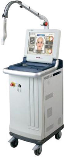
- Lutronic Mosaic — fractional system from the Korean company Lutronic Inc., also employing an Er:Fiber laser at 1,550 nm. It uses an intelligent scanner in the handpiece; the number of MTZs (treatment density) can be chosen, and application can be in static or continuous scanning mode, like Fraxel (Fig. 27).
The great success of fractional technology led to diversification and improvement of the method, originating ablative fractional treatment. The laser drills micro-holes of controlled depth in the skin, with a thin tissue-coagulation zone around them. The surface is damaged, producing small crusts and more persistent erythema. Recovery time is longer, and there are restrictions on skin type and the body areas treated. The histology in Fig. 41 clearly demonstrates the difference between non-ablative and ablative technology.
Ablative fractional treatment brought the CO2 laser back to the rejuvenation scene, because it gave control, safety, and fewer restrictions to the already efficient CO2 laser skin resurfacing that remains the gold standard of skin rejuvenation. Another advantage is that it can also be used for precise cuts and vaporization in minor surgery.
Examples of commercial fractional ablative lasers are shown below:
- Lutronic eCO2 — a fractional CO2 laser with static and dynamic scanning systems (Fig. 15). The diameter and density of MTZs can be programmed.
- Fraxel re:pair — a CO2 laser using the same fractional technology as the non-ablative Fraxel re:Store, with an intelligent continuous scanning system.
- Lumenis Total Active FX — also uses a CO2 laser with intelligent scanners allowing static and dynamic scanning modes. The diameter and density of MTZs can be programmed.
- DEKA SmartXide DOT/RF — a CO2 laser with scanners and radio-frequency technology integrated in the handpiece (Boechat et al. 1991).
- Alma Pixel CO2 — a CO2 laser with an array of micro-lenses at the tip to split the beam and produce the fractional effect. The number and diameter of MTZs are fixed, although handpieces of different sizes are offered.
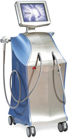
- Alma Pixel Handpiece — a fractional handpiece for the multi-platform Harmony, employing an Er:YAG wavelength of 2,940 nm and a filter effect to split the beam. The MTZs are thicker than in other devices, of the order of millimeters in diameter, and more superficial, reaching only the epidermal layer, because of the wavelength and energy of the system.
Radio Frequency
Radio frequency (RF) consists of a high-frequency electric current, of the order of 1 MHz, and has been used in medicine for several years. For comparison, household appliances such as TVs and refrigerators work at 50 or 60 Hz, low frequencies.
Returning to the electromagnetic-spectrum chart in Fig. 1, RF occupies the kHz-to-GHz range, used for radio communication, which gave it its name. Medical equipment uses a portion of this range — 200 kHz to 40 MHz — in different applications. In this frequency range the stimulation effects on nerves and muscles decrease, so energy can be applied gently to achieve different levels of tissue heating (Lapidoth et al. 2015).
The basic idea behind RF on skin is the ability to deliver volumetric heat in depth. Movement (current) of electrons (ions) causes tissue heating, unlike light, in which temperature rises through absorption of photon energy. There is no selectivity: the high-frequency current heats tissue as a whole regardless of skin type. There are also no losses from reflection or scattering as with laser light. It is safe for dark skin types and effective for light-colored chromophores. Energy diffusion depends only on tissue conductivity.
As seen earlier, the concentration of light energy (fluence), or power density, controls the tissue effect. The same is true with RF: high power applied over a large area through large electrodes causes mild heating, while concentration in a small area, such as a needle-shaped electrode, causes tissue ablation.
Penetration of RF energy into tissue — or rather, attenuation of energy as it penetrates — depends on the fluence used, the electrode configuration (monopolar, bipolar, or unipolar), the anatomy of the treated area, and the tissue’s conductivity characteristics.
RF systems can be monopolar, bipolar, multipolar, and unipolar (Lapidoth et al. 2015).
Monopolar RF
These devices use an active electrode, in the form of a handpiece, to apply RF to the treatment area, and a return electrode — usually a grounding pad with a large contact area — placed far from the treatment zone (Fig. 43).
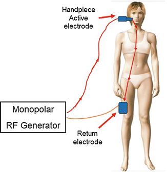
A high RF-current density is created at the active electrode, and the current diverges as it penetrates tissue toward the large return electrode. Heat is therefore generated near the active electrode and does not depend on the size, shape, or position of the return electrode.
The RF current diverges rapidly away from the electrode, so the heating effect decreases. At a distance equal to the electrode size, heating becomes insignificant; the heat zone can be estimated as half the size of the electrode. By controlling RF power and the geometry and size of the electrode, penetration depth and tissue effect can therefore be controlled.
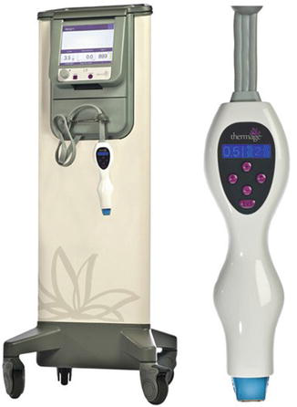
Popular monopolar uses in surgery are cutting and coagulation of blood vessels. In dermatology there is application for skin tightening and collagen remodeling, as the geometry of the large electrode targets the deeper tissues of the dermis (Fig. 44).
Bipolar RF
This configuration uses two electrodes placed close together, in contact with the treatment zone. RF current flows between the electrodes and does not spread to other parts of the body as in the monopolar configuration. This geometry creates more uniform heating of the treatment zone compared with monopolar devices (Fig. 45).
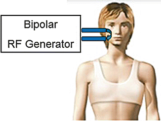
Both electrodes create an equal thermal effect near them, and divergence of the RF current is reduced because of the small distance between them. Most heat is therefore concentrated near the electrodes, allowing greater control over the size of the treated volume.
Penetration depth is a function of electrode size and the distance between them. Increasing electrode separation drives the RF current deeper, but divergence also increases, reducing the desired heating effect. If separation is too large relative to electrode size, the heating profile becomes similar to two monopolar electrodes. When electrode separation is comparable to electrode size, penetration depth is approximately half the distance between them (Lapidoth et al. 2015).
Penetration depth can also be controlled by varying the operating frequency: treatment can occur at different depths within the limits imposed by electrode separation, as shown in Fig. 46. The higher the frequency, the shallower the heating effect.
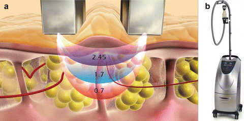
Folding the skin between the electrodes, for example by applying negative pressure (vacuum), allows uniform heating of a large tissue volume that can reach a few centimeters. This technique is used in body-contouring and cellulite devices such as Viora’s Reaction™ (Fig. 46b) and VelaShape™, which uses the Electro-Optical Synergy (ELOS) technology developed by Syneron Candela, described below.
Bipolar RF also presents reduced energy loss because of electrode proximity, and reduced energy density at the treatment area, subsequently reducing the risk of overheating and burning of the skin below the electrode. Application is better tolerated and causes less pain.
It also allowed development of fractional bipolar RF technology, described below.
Multipolar RF
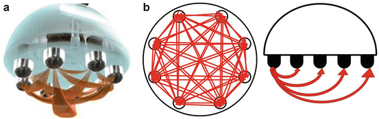
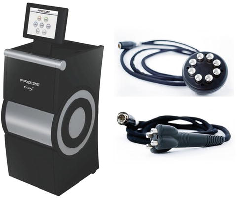
An interesting approach to bipolar RF geometry. In this case a series of bipolar electrodes is used in a circular or linear configuration. RF current flows between them, producing a more homogeneous heating effect over a larger tissue volume and at variable penetration depths, as shown in Fig. 47. The desired treatment-endpoint temperature is also reached quickly, since more electrodes are used simultaneously (Fig. 48).
Unipolar RF
This RF configuration uses a single electrode that works, in some ways, as an antenna for electromagnetic-energy coupling in the skin. It differs from monopolar RF, which uses one active and one return electrode: in unipolar RF the current flows into the skin (Fig. 49).
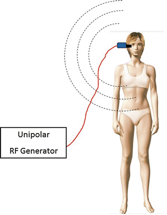
Electromagnetic-field coupling in human tissue produces heat. The RF heating effect depends on the device’s operating frequency. There are two mechanisms of heating biological tissue containing water: ionic current, produced by moving charged particles (electrons), and rotation of the dipoles of water molecules. These two forms of interaction lead to heating and a consequent temperature increase in tissue.
The RF configurations described so far — monopolar, bipolar/multipolar — use lower frequencies, 1–3 MHz, and the dominant mechanism is ionic current striking skin molecules, which in turn vibrate, producing heat.
At frequencies around 10 MHz and above, rotation of water molecules starts to be noticeable, and above 30–40 MHz this mechanism is the predominant cause of tissue heating (Lapidoth et al. 2015).
Unipolar devices use high RF frequencies, 20–40 MHz, so that the electromagnetic field produces molecular rotation to induce tissue heating.
Penetration depth in this case depends on operating frequency, electrode geometry and configuration, input power, and the time and mode of treatment (stationary — using a panel with electrodes over the treatment area — or in motion, moving the electrode over the skin).
As with ionic-current heating, penetration depth in unipolar RF also decreases at higher frequency. By operating at different frequencies, a key volume of energy can therefore be concentrated in a specific skin layer. Working at high frequency, such as 40 MHz, the main effect is skin tightening, since energy is concentrated mainly in the dermal area. Lower frequencies, such as 27 MHz, deposit energy in a deeper layer, which works for body contouring and fat destruction (Fig. 50).
It is important to note that the treatment effect (skin tightening, collagen remodeling, and fat reduction) is a function not only of temperature but also of the length of time the temperature is applied, or RF pulse duration. Exposure to 70–90 °C for a few milliseconds causes tissue coagulation, while application of a lower temperature such as 42 °C for tens of minutes also causes irreversible damage to sensitive cells. Fat cells, for example, are especially sensitive to temperature change. Using the correct electrode geometry, low input power (so as not to overheat the skin surface), and long application times (several minutes), apoptosis of fat cells can be caused in body-contouring procedures. The skin-damage function can be described by the Arrhenius equation (Lapidoth et al. 2015):
The degree of damage (D) is a linear function of exposure time, or pulse duration (t), and an exponential function of tissue temperature (T) (Fig. 51).
Fractional RF
Fractional bipolar RF (FRF) was developed following the same concepts and success as fractional lasers, described previously, and is gaining great popularity in dermatology. The procedure is based on heating or ablating multiple small points in the skin (MTZs), with a spot size of 100–400 μm, leading to improved skin quality, wrinkle reduction, treatment of acne scars, and stretch marks (Lapidoth et al. 2015; Brightman et al. 2009; Rongsaard and Rummaneethorn 2014).
RF can provide patterns of energy and heat distribution different from the MTZ shapes produced by fractional lasers. In contrast to lasers, where the thermal effect is limited to the periphery of the ablation crater (ablative procedure) or the coagulated column (non-ablative procedure), RF energy flows through the entire dermis, adding volumetric heating to fractional treatment. This produces a more effective skin-tightening effect.
There are mainly two types of fractional RF technology:
- a matrix of bipolar microelectrodes applying RF energy from the surface;
- a grid of microneedles delivering RF energy internally, within the dermis.
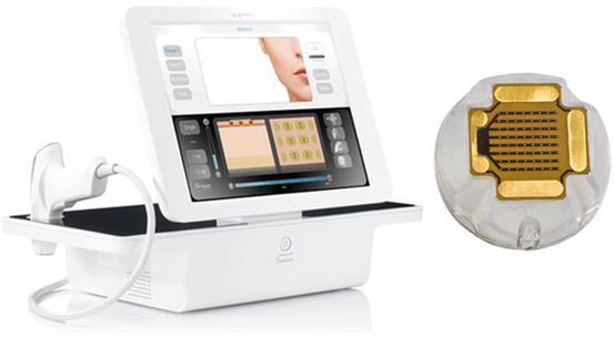
Surface electrodes provide a more superficial effect, improving texture and lines, treating stretch marks, and smoothing acne scars. Bipolar RF is applied with a matrix of active microelectrodes, as shown in Fig. 52.
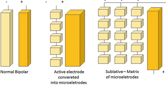
A normal bipolar device, described above, uses large-area electrodes and has low power density, with current and subsequent heating limited to the tissue between the electrodes. In FRF the active electrode is converted into a series of microelectrodes, increasing energy density and producing an ablation effect near the electrode; as the energy flows toward the large return electrode, it spreads, reducing the effect, which is limited to skin coagulation and skin tightening (Fig. 53). This action resembles a water nozzle: close to the nozzle the water jet is concentrated and can make us excessively wet; moving away, the water spreads and only a few drops reach us.
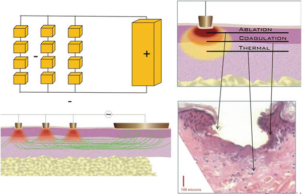
An interesting variation of fractional bipolar RF was developed by Syneron Candela: the “Sublative RF,” used in the Matrix RF and eMatrix devices. The proposal is to deliver heat energy to the dermal layer with minimal epidermal damage. By controlling RF-current energy and the delivery pulse, epidermal defects can be corrected and aggressive remodeling of the deeper dermis promoted. Since the effect on the epidermis is minimal, recovery time is shorter, and the risk of infection and pigmentary changes is also reduced (Fig. 54).
The RF-microneedle approach is based on introducing a set of fine, dielectric-coated needle electrodes deep into the skin, then activating them to deliver energy and produce strong dermal remodeling. Since energy is deposited directly into the deep dermis, there is no effect on the epidermis, which is preserved. Side effects and recovery time are minimal. Compared with surface fractional RF, microneedles can produce higher temperatures in the deep dermis and therefore stronger collagen contraction, leading to improvement of deep wrinkles and skin tightening (Lapidoth et al. 2015).
RF-energy penetration depth is controlled by adjusting needle size, and the effect is confined to the skin between the electrodes (Fig. 55).
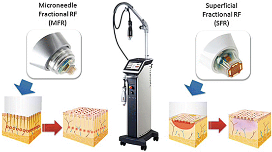
The combination of a superficial fractional treatment (Sublative) — improving the epidermis and collagen remodeling in the upper dermis — with the deep dermal remodeling produced by the microneedle device offers high potential for complete skin improvement with minimal adverse effects and recovery time.
Fractional RF is often said to be safe for all skin types because of its “color-blind” characteristic. It should be noted, however, that while RF interaction with skin does not depend on the presence of melanin or any other chromophore, darker skin types and tanned skin remain susceptible to postinflammatory hyperpigmentation (PIH). FRF heats the skin and induces a wound-healing response; high-risk skin types are therefore wisely treated with greater caution.
Another skin characteristic influencing RF-current treatment is conductivity, or resistance (impedance). RF current behaves somewhat like water: it flows through the path of least resistance. A piece of metal is an excellent electrical conductor (low impedance/resistance); current flows easily even at low energy, with little heat dissipation. At the other extreme, a piece of plastic does not conduct electricity (high impedance). Skin lies somewhere between metal and plastic, and several characteristics can change its impedance: young skin, well vascularized and well moisturized, is a good conductor (like metal), while aged, dry, poorly vascularized skin behaves more like plastic; in that case higher energy is needed for current to flow, and there is heat dissipation.
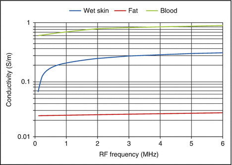
The graph in Fig. 56 shows how conductivity changes for some skin components as a function of RF operating frequency. Blood is a better conductor than wet skin, and fat cells are poor conductors (Lapidoth et al. 2015).
Temperature also changes skin conductivity, or impedance. RF current prefers heated, warm tissue, as shown in Fig. 57 (Lapidoth et al. 2015).
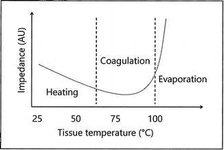
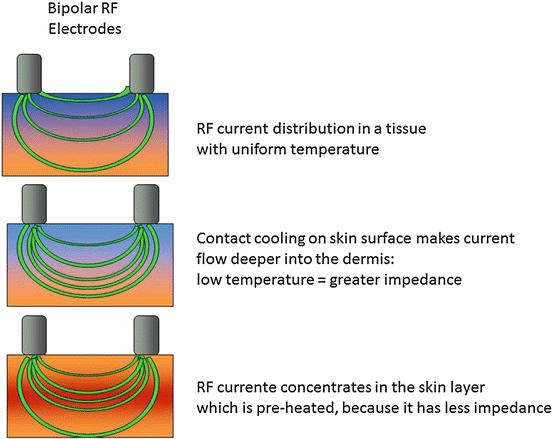
By changing tissue temperature, the RF current can therefore be “directed”; it can be forced to flow or concentrate in given parts or layers. Using a cooling contact tip on the skin surface, for example, RF current flows deeper into the dermis. A certain selectivity can also be generated, as the current concentrates or flows preferentially in whichever skin layer or tissue is hotter. This technique is the basis of the Electro-Optical Synergy (ELOS) system developed by Syneron Candela, presented below (Fig. 58).
Hybrid Systems
Seeking to overcome limitations and expand the safety and efficacy of treatments with laser or intense pulsed light, industry has diversified technology by associating light with other forms of energy, creating so-called hybrid systems.
A highly successful example of this diversification is Electro-Optical Synergy (ELOS™) technology — the synergy of light with radio frequency, developed by the inventor of intense pulsed light, Dr. Shimon Eckhouse, at Syneron Candela, Israel (Doshi and Alster 2005; Sadick et al. 2005; Lapidoth et al. 2005; Sadick and Trelles 2005).
ELOS™ technology employs bipolar RF with a water-cooled tip simultaneously with the laser or IPL pulse, as illustrated in Fig. 59.
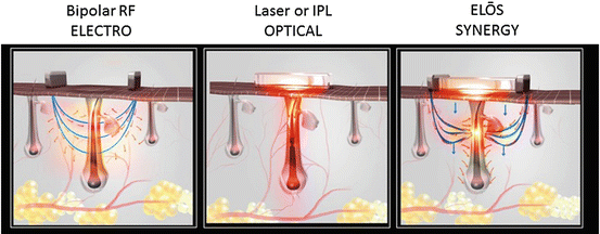
Following the principle of selective photothermolysis, light heats the chromophore while preserving surrounding tissue. A cool tip protects the skin surface and “pushes” RF into deeper layers. The RF concentrates in heated tissue because of its better conductivity, causing the chromophore to overheat and producing the desired therapeutic effect.
Figure 59 illustrates this synergic effect in hair-removal treatment, but the same occurs in treatment of pigmented and vascular lesions, skin rejuvenation, and tightening — in which an infrared light source in the 700–2,000 nm range is used with bipolar RF (Fig. 60).
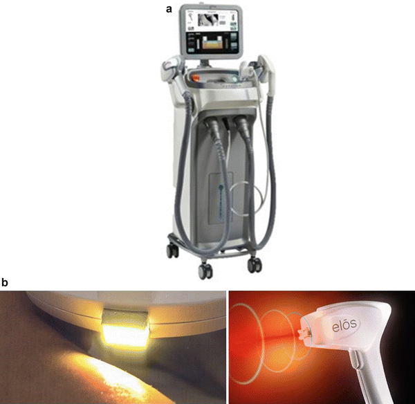
The main advantage of ELOS™ is the reduced optical fluence needed for treatment, minimizing patient discomfort during sessions and increasing safety for darker skin types. Because of RF action, simultaneous effects such as skin tightening during treatment of pigmented lesions can also occur.
Other applications of this technology are circumferential reduction, cellulite treatment, and skin tightening. In this case bipolar RF is associated with an infrared source (a lamp emitting 700–2,000 nm, or a high-power 870 nm LED), rotating cylinders that produce massage, drainage, and suction. The cylindrical rollers are the RF electrodes; suction creates a skin fold, increasing penetration of RF and light as explained earlier (Alster and Tanzi 2005; Wanitphakdeedecha and Manuskiatti 2006; Boechat 2009) (Fig. 61).
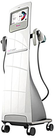
The goal is to raise the temperature of deep tissue to 43 °C, which accelerates fat-cell metabolism and thereby reduces their size, leading to circumferential reduction. With longer exposure and higher temperature (45 °C), apoptosis of fat cells can be produced, since they are more sensitive than skin cells, reducing localized fat. The skin-tightening effect occurs through stretching of elastic fibers and collagen remodeling, improving overall skin quality.
Conclusion
Laser and intense pulsed light systems are pure light sources with important properties that allow accurate, selective treatment of different types of tissue damage while preserving surrounding healthy tissue. Synergy with radio frequency shows how this equipment can still evolve, becoming safer and more efficient. With the advent of fractional skin treatment, a new horizon of applications at once gentle and effective has emerged in dermatology.
In many applications light appears as the only effective solution, as with flat vascular lesions of the face or port-wine stains. It has brought rapid, long-lasting results for unwanted-hair removal, treatment of pigmented lesions, and tattoo removal. It is used in skin tightening, cellulite treatment, circumferential reduction, and localized-fat reduction. In several dermatological applications light is an important complement to existing techniques, as with rhytidectomy in plastic surgery; it also improves body areas not normally treated by surgery, such as the neck, chest, hands, and arms, using ablative or non-ablative fractional lasers.
The future will certainly bring more efficient, compact devices, with a wider range of applications — among them, development of lasers able to act at the cellular level, stimulating production of enzymes whose purpose is preventing skin aging and skin cancer. Systems with subcutaneous fat as a chromophore can open a new horizon of applications for circumferential reduction, cellulite treatment, and improved skin quality; diagnostic medicine will also benefit from this development.
The more we study the effects of light interacting with living tissue, the more we learn to appreciate the variety and complexity of these critical interactions. The result will certainly open the door to a large number of remarkable applications in the years ahead.
We only have to “tune in” with the energy of light!
About the Author
Álvaro Boechat, M.Sc., Ph.D., is an electronics engineer graduated from the Technological Institute of Aeronautics (ITA), São José dos Campos, São Paulo, Brazil; he holds a master’s degree (M.Sc.) in optoelectronics and laser devices and a Ph.D. in laser engineering from Heriot-Watt University in Edinburgh, Scotland. With specialization courses in medical lasers from Laser Industries in Tel Aviv, Israel, he currently works as a photomedicine consultant.
Take Home Messages
- The visible light we experience day-to-day is only one facet of a much broader physical phenomenon known as “electromagnetic radiation.” The difference between each laser, every color of light we see, is its wavelength or frequency.
- All laser devices consist of the resonator/oscillator — with the active medium that produces the light and thus determines the wavelength; the excitation source (pumping), which delivers power to the active medium to produce photons; the laser-beam delivery system, from source to the operator’s hand; and the handpiece, with focusing lens or scanning system.
- The best way to determine which laser or IPL is best for a given application is to use the principles of selective photothermolysis: the wavelength absorbed only by the target tissue, a pulse duration confining heat to the chromophore, and sufficient energy to achieve the desired effect.
- The radio-frequency effect on skin does not depend on chromophore absorption, which makes it safe for treating all skin types.
- The RF effect in skin is a linear function of time and an exponential function of temperature. A small change in treatment temperature of a few degrees increases tissue effect severalfold; on the other hand, application time must be increased by several minutes or hours to obtain the same effect.
References
Alster TS. Manual of cutaneous laser techniques. Philadelphia: Lippincott-Raven; 1997.
Alster TS, Apfelberg DB. Cosmetic laser surgery — a practitioner’s guide. 2nd ed. New York: Wiley-Liss; 1999.
Alster TS, Tanzi EL. Cellulite treatment using a novel combination radiofrequency, infrared light, and mechanical tissue manipulation device. J Cosmet Laser Ther. 2005;7:81–5.
Alster TS, Nanni CA, Williams CM. Comparison of four carbon dioxide resurfacing lasers: a clinical and histopathologic evaluation. Dermatol Surg. 1999;25(3):153–9.
Anderson R, Parrish J. The optics of human skin. J Invest Dermatol. 1981;77:13.
Anderson R, Parrish J. Selective photothermolysis: precise microsurgery by selective absorption of pulsed radiation. Science. 1983;220:524.
Antonio CR, Oliveira GB, Coura MGG, Trídico LA, Pereira LR, D’Ávila SCGP. The use of a 1,340 nm Nd:YAP laser to treat hidradenitis. Surg Cosmet Dermatol. 2015;7(1):46–9.
Arndt KA, Dover JS, Olbricht SM. Lasers in cutaneous and aesthetic surgery. Philadelphia: Lippincott-Raven; 1997.
Boechat AAP. Fotomedicina: Princípios, Efeitos e Aplicações. In: Osório N, Torezan L, editors. Laser em Dermatologia: conceitos básicos e aplicações. Chapter 1. 2nd ed. São Paulo: Rocca; 2009.
Boechat AAP, Su D, Hall DR, Jones JDC. Bend loss in large-core multimode optical fiber beam delivery systems. Appl Opt. 1991;30:321–7.
Boechat AAP, Su D, Jones JDC. Dependence of the output near-field profile on launching conditions in graded-index optical fibers used in delivery systems for Nd:YAG lasers. Appl Opt. 1993;4:72–5.
Brightman L, Goldman MP, Taub AF. Sublative rejuvenation: experience with a new fractional radiofrequency system for skin rejuvenation and repair. J Drugs Dermatol. 2009;8(11):S9–13.
Chan HH, King WW, Chan ES, Mok CO, Ho WS, Van Krevel C, Lau WY. In vivo trial comparing patients’ tolerance of Q-switched alexandrite and Q-switched neodymium:yttrium-aluminum-garnet lasers in treatment of nevus of Ota. Lasers Surg Med. 1999;24(1):24–8.
Chang CJ, Nelson JS, Achauer BN. Q-switched ruby laser treatment of oculodermal melanosis (nevus of Ota). Plast Reconstr Surg. 1996;98(5):784–90.
Chernoff GW, Schoenrock RD, Cramer H, Wand J. Cutaneous laser resurfacing. Int J Facial Restor Surg. 1995;3(1):57–68.
Doshi SN, Alster TS. Combination radiofrequency and diode laser for treatment of facial rhytides and skin laxity. J Cosmet Laser Ther. 2005;7:11–5.
Finkel B, Eliezri YD, Waldman A, Slatkine M. Pulsed alexandrite laser technology for noninvasive hair removal. J Clin Laser Med Surg. 1997;15(5):225–9.
Fleming D. Controversies in skin resurfacing: the role of erbium. J Cutan Laser Ther. 1999;1:15–21.
Geronemus RG. Fractional photothermolysis: current and future applications. Lasers Surg Med. 2006;38:169–76.
Goldberg DJ. Non-ablative subsurface remodeling: clinical and histologic evaluation of a 1320 nm Nd:YAG laser. J Cutan Laser Ther. 1999;1(3):153–7.
Goldberg DJ. Smoothbeam™, non-ablative dermal remodeling with a 1450 nm diode laser in combination with DCD. Candela Corporation Clin Appl Notes. 2000a;1(1):123–7.
Goldberg DJ. Full-face nonablative dermal remodeling with a 1320 nm Nd:YAG laser. Dermatol Surg. 2000b;26(10):915–8.
Goldberg DJ, Whitworth J. Laser skin resurfacing with the Q-switched Nd:YAG laser. Dermatol Surg. 1997;23(10):903–6.
Goldman L. Laser treatment of tattoos. J Am Med Assoc. 1967;201:163.
Goldman MP, Fitzpatrick RE. Cutaneous laser surgery — the art and science of selective photothermolysis. Boston: Mosby; 1994.
Guttman C. Excimer laser system can safely target psoriatic plaques. Dermatol Times. 2000.
Jeong SY, Chang SE, Park HN, et al. New melasma treatment by collimated low-fluence Q-switched Nd:YAG laser. Korean J Dermatol. 2008;46:1163–70.
Kaminsky Jedwab SK. Laser e outras tecnologias na dermatologia. São Paulo: Editora Santos; 2010.
Khan MH, Sink RK, Manstein D, Eimerl D, Anderson RR. Intradermally focused infrared laser pulses: thermal effects at defined tissue depths. Lasers Surg Med. 2005;36:270–8.
Klavuhn KG. Epidermal protection: a comparative analysis of sapphire contact and cryogen spray cooling. Laser Hair Removal Tech Note. 2000;1:1–7.
Kulick MI. Lasers in aesthetic surgery. New York: Springer; 1998.
Lapidoth M, Halachmi S, editors. Radiofrequency in cosmetic dermatology. Aesthetic Dermatology, vol. 2. Goldberg DJ, editor. Basel: Karger; 2015.
Lapidoth M, Yaniv E, Ben-Amital D, Raveh E, Kalish E, Waner M, David M. Treatment of facial venous malformations with combined radiofrequency current and a 900 nm diode laser. Dermatol Surg. 2005;31:1308–12.
Lask G. Laser resurfacing in pigmented skin. J Dermatol. December 1995.
Laubach HJ, Tannous Z, Anderson RR, Manstein D. Skin responses to fractional photothermolysis. Lasers Surg Med. 2005;36:1–8.
Lopes LA. Análise in vitro da proliferação celular de fibroblastos de gengiva humana tratados com laser de baixa potência. M.Sc. thesis, Postgraduate Program in Biomedical Engineering, Universidade do Vale do Paraíba; 1999.
Lou WW, Quintana AT, Geronemus RG, Grossman MC. Prospective study of hair reduction by diode laser (800 nm) with long-term follow-up. Dermatol Surg. 2000;26:428–32.
Manstein D, Herron GS, Sink RK, Tanner H, Anderson RR. Fractional photothermolysis: a new concept for cutaneous remodeling using microscopic patterns of thermal injury. Lasers Surg Med. 2004;34:426–38.
McMillan K, et al. A 585 nm pulsed dye laser treatment of laryngeal papillomas: preliminary report. Laryngoscope. 1998;108:968.
Milanic M, Majaron B. Energy deposition profile in human skin upon irradiation with a 1,342 nm Nd:YAP laser. Lasers Surg Med. 2013;45(1):8–14.
Mordon S, Capon A, Creusy C, Fleurisse L, Buys B, Faucheux M, Servell P. In vivo experimental evaluation of non-ablative skin remodeling using an Er:Glass laser with contact cooling. Lasers Surg Med. 2000;27(1):1–9.
Muccini JA, O’Donnell FE, Fuller T, Reinisch L. Laser treatment of solar elastosis with epithelial preservation. Lasers Surg Med. 1998;23(3):121–7.
Mun JY, Jeong SY, Kim JH, Han SS, Kim IH. A low-fluence Q-switched Nd:YAG laser modifies the 3D structure of melanocytes and melanosome ultrastructure by subcellular-selective photothermolysis. J Electron Microsc. 2010:1–8.
Munavalli GS, Weiss RA, Halder RM. Photoaging and nonablative photorejuvenation in ethnic skin. Dermatol Surg. 2005;31:1250–61.
Ogata H. Evaluation of the effect of Q-switched ruby and Q-switched Nd:YAG laser irradiation on melanosomes in dermal melanocytosis. Keio J Med. 1997;46(4):188–95.
Ono I, Tateshita T. Efficacy of the ruby laser in treatment of Ota’s nevus previously treated using other therapeutic modalities. Plast Reconstr Surg. 1998;102(7):2352–7.
Pitanguy I, Machado BH, Carneiro LVF Jr. Peeling a laser de dióxido de carbono. Rev Bras Cir. 1996;86(6):313–25.
Raulin C, Karsai S. Tecnologias laser e IPL em dermatologia e medicina estética. Rio de Janeiro: Di Livros; 2011.
Raulin C, Schonermark MP, Greve B, Werner S. Q-switched ruby laser treatment of tattoos and benign pigmented skin lesions: a critical review. Ann Plast Surg. 1998;41(5):555–65.
Reichert D. Evaluation of the long-pulse dye laser for treatment of leg telangiectasias. Am Soc Dermatol Surg. 1998;24:737.
Reid R, Muller S. Tattoo removal by laser. Med J Aust. 1978;1:389.
Reid WH, McLeod PJ, Ritchie A, Ferguson-Pell M. Q-switched ruby laser treatment of black tattoos. Br J Plast Surg. 1983;36:455.
Reid WH, Miller ID, Murphy MJ, Paul JP, Evans JH. Q-switched ruby laser treatment of tattoos: a 9-year experience. Br J Plast Surg. 1990;43:663–9.
Reyes BA, Geronemus RG. Treatment of port-wine stains during childhood with the flash-lamp-pumped dye laser. J Am Acad Dermatol. 1990;23:1142–8.
Rongsaard N, Rummaneethorn P. Comparison of a fractional bipolar radiofrequency device and a fractional erbium-doped glass 1,550 nm device for treatment of atrophic acne scars: a randomized split-face clinical study. Dermatol Surg. 2014;40:14–21.
Ross EV, Hardway CA. Sub-surface renewal by treatment with a 1450 nm diode laser in combination with dynamic cooling. Candela Corp Clin Appl Notes. 2000;1(2):1–4.
Sadick NS, Trelles M. A clinical, histological, and computer-based assessment of the Polaris LV, a combination diode and radiofrequency system, for leg-vein treatment. Lasers Surg Med. 2005;36:98–104.
Sadick NS, Alexiades-Armenakas M, Bitter P, Mulholland RS. Enhanced full-face skin rejuvenation using synchronous intense pulsed optical and conducted bipolar radiofrequency energy (ELOS): introducing selective radiophotothermolysis. J Drugs Dermatol. 2005;4:181–6.
Sardana K, Garg VK, editors. Lasers in dermatological practice. New Delhi: Jaypee Brothers Medical Publishers; 2014.
Shimbashi T, Hyakusoku H, Okinaga M. Treatment of nevus of Ota by Q-switched ruby laser. Aesthetic Plast Surg. 1997;21(2):118–21.
Siegman AE. Lasers. London: Oxford University Press; 1986.
Stafford TJ, Lizek R, Boll J, Tan OT. Removal of colored tattoos with the Q-switched alexandrite laser. Plast Reconstr Surg. 1995;95(2):313–20.
Waldorf HA, et al. Effect of dynamic cooling on 585 nm pulsed dye laser treatment of port-wine stain birthmarks. Dermatol Surg. 1997;23:657–62.
Wanitphakdeedecha R, Manuskiatti W. Treatment of cellulite with a bipolar radiofrequency, infrared heat, and pulsatile suction device: a pilot study. J Cosmet Dermatol. 2006;5:284–8.
Weinstein C. Computerized scanning with an erbium:YAG laser for skin resurfacing. Dermatol Surg. 1998;24:83–9.
Wong SS, Goh KS. Successful treatment of traumatic tattoos with the Q-switched neodymium:YAG laser: a report of two cases. J Dermatol Treat. 1998;9:193–5.
Wright VC, Fisher JC. Laser surgery in gynecology. Toronto: W. B. Saunders; 1993.
Yang HY, Lee CW, Ro YS, Yu HJ, Kim YT, Kim JH. Q-switched ruby laser in treatment of nevus of Ota. J Korean Med Sci. 1996;11(2):165–70.
Zelickson BD, et al. Clinical and histologic evaluation of psoriatic plaques treated with a flashlamp pulsed dye laser. J Am Acad Dermatol. 1996;35:64–8.







