
Fractional Ablative and Non-Ablative Lasers for Ethnic Skin
Fractional Ablative and Non-Ablative Lasers for Ethnic Skin
Paulo Roberto Barbosa, Tais Valverde, Roberta Almada e Silva and Fabiolla Sih Moriya Brazilian Society of Dermatology, Clínica de Dermatologia Paulo Barbosa – Centro Odontomédico Louis Pasteur, 5° andar, sala 505 a 507, CEP 41.850-000 Itaigara, Salvador, BA, Brazil
Abstract
Ethnic skin is a term used to define the darker skin corresponding to Fitzpatrick’s IV, V, and VI skin types. Patients with ethnic skin have an increased risk of problems related to pigmentation, such as post-inflammatory hyperpigmentation or hypopigmentation. For a long time, ethnic skin treatment with lasers was a big challenge, especially when referring to ablative technologies, a technique so widely used and widespread in dermatological procedures. Over time, laser devices have evolved, and recent techniques make its use safer, with less downtime and, consequently, with less damage and risk of post-inflammatory pigmentation. When performed by skilled and well-trained dermatologists, the use of fractional ablative and non-ablative lasers can be considered safe and viable for many different treatments. This chapter is going to discuss ethnic skin and the peculiarities of laser treatment.
Keywords Ethnic skinLaserAblative technologiesCO2 laserErbium lasersFractional and non-fractional ablative lasers
Introduction
The color or race of the Brazilian population is diverse. Patients with dark color skin are more frequent in the North and Northeast of our country, due to our colonization, and most of them are concentrated at the north and northeast of the country. They have an ethnic skin, a term used to define darker skin and non-Caucasian, corresponding to skin types IV, V, and VI of the Fitzpatrick classification (IBGE 2000).
For a long time, treating ethnic skin with laser was a challenge, especially when referring to ablative technologies. Although the main chromophore of this technique is the water and not melanin, the heat generated by them can have serious postoperative complications if not used by trained professionals, making it necessary to have thorough knowledge of the physical basics of lasers (Battle and Hobbs 2003).
The number of melanocytes and the thickness of the skin are approximately the same in all races, but ethnic skin has its own characteristics. We can find in ethnic skin more and higher fibroblasts and bi- or multinucleated and hyperactive fibroblasts. This hyperactivity may explain the predisposition to keloid formation. It is also richer in sebaceous glands and has more collagen (Mateus and Palermo 2012).
The range is in melanin production speed, number, morphology, size, density, and distribution of melanosomes. Ethnic skin patients have a higher risk to pigmentation problems because the skin melanin competes with the main chromophore, increasing the risk of postoperative hyperpigmentation and hypopigmentation. Moreover, the risk of pigmentary changes can also be correlated with the depth of the laser injury as well as their effects on dermal heating. Post-inflammatory hyperpigmentation is the most common complication of facial resurfacing in patients with skin-type IV (Sriprachya-anunt et al. 2002; Wat et al. 2017).
Fractional Ablative Lasers
Carbon dioxide (CO2) lasers are the main representative of ablative lasers and, although devised in 1968, are still regarded as the gold standard treatment for photoaging. Novel technologies are now able to produce a column of ablation and coagulation with rigorously controlled depth, providing increased safety and reduced recovery time. The method is based on the principle of selective photothermolysis, developed by Parish and Anderson in 1983. The principle is the selective and specific destruction of a target in the skin with minimal thermal damage to the components in neighboring tissues. To achieve selective photothermolysis, the appropriate wavelength primarily absorbed by the target tissue or chromophore should be carefully chosen. For ablative lasers, the main chromophore is water (Riggs et al. 2007; Tierney et al. 2011).
However, CO2 lasers show intense residual thermic effect, leading to a final result much broader than the tissue ablation observed at the end of the procedure. Conversely, water is absorbed 13 times less efficiently with CO2 than with Erbium lasers, which are more superficial and cause less thermal damage (Kalil and Campos 2012).
A novel treatment concept was described by Manstein et al. in 2004: fractional photothermolysis. In this procedure, the laser produces microscopic lesions, ranging from 100 to 150 μm in thickness and 0.2–2.4 mm in depth, termed thermal microzones (TMZ), which are merely highly controlled microperforations in the epidermis and dermis that are, then, replaced by new organized tissue rich in collagen. Fractional photothermolysis revolutionized laser treatments, allowing for a more efficacious dermal coagulation without major damage to the epidermal layer, thus decreasing the risk of nonaesthetic scarification and reduced recovery time when compared to traditional ablative procedures. Several ablative fractional photothermolysis devices are available, in three different wavelengths: 2790, 2940, and 10,600 nm (Kalil and Campos 2012; Xu et al. 2011; Macrene et al. 2012; Manstein et al. 2004; Wat et al. 2017).
In 2007, Hantash et al. described the use of a new CO2 ablative fractional laser that generates, by their TMZ, ablation and coagulation columns extending through the stratum corneum, epidermis, and dermis. A CO2 laser beam is able to remove from 25 to 50 μm of tissue in each application, raising the temperature up to 100 °C in 640 ms. The heat leads to an immediate contraction in the skin, although partly caused by water evaporation-driven dehydration and by collagen contraction, which retracts in temperatures higher than 60 °C (Ciocon et al. 2011).
Despite long-lasting clinical improvement, the best results can be seen after 3 months posttreatment, mostly because of persistent inflammatory responses (demonstrated by the presence of heat-shock protein 47) and by the continuous collagen remodeling observed in histological and immunohistochemical studies. Ortiz et al. reported that patients maintain 74% of improvement in the first 1–2 years of following-up (Ortiz et al. 2010).
Cellular markers for neocollagenesis, such as procollagen III and collagen III, are found in treated areas, and the increase of both collagen density and elastic fibers can be observed up to 1 year posttreatment (Xu et al. 2011; Kim et al. 2013; Orringer et al. 2004).
Currently there are various CO2 fractional devices in the market, which are characterized by adjustable fluence and pulse length, allowing for precise control of the level and depth of heat in the dermis. These parameters – fluence, pulse length, potency, energy, and density – will determine the effectiveness and safety of the treatment (Kadunc et al. 2012).
The erbium:yttrium–aluminum–garnet (Er:YAG) laser was approved by the FDA in 1996 for skin resurfacing. Because its wavelength (2940 nm) is the closest to water absorption peak (3000 nm), all its energy is practically absorbed by the epidermis and the papillary dermis, causing superficial ablation and less thermal damage than CO2 lasers. Each Er:YAG short-pulse application (250–350 ms) ablates approximately 20–25 μm, utilizing 5 J/cm2 of energy. Thermal damage reaches 30–50 μm in depth with a fluence of 5–8 J/cm2, which is smaller when compared to those cause by CO2 lasers (50–200 μm in depth with a fluence of 3.5–6.5 J/cm2). Even with multiple applications, thermal damages caused by Er:YAG lasers are limited to 50 μm (Riggs et al. 2007).
This technology, which acts within the infrared spectrum, proved its higher ablation effect causing negligible thermal damage (5 μm per application). Because of its wavelength with maximum water absorption, short pulse length, and adequate energy, Er:YAG lasers are the preferred technology for fine wrinkles, photoaging, and treatments that demand higher phototypes. Injuries to neighboring tissues are minimum (maximum temperature reaches 30 °C), and bleeding spots started manifesting only after several applications (4–5, depending of the size treated), indicating that the dermis–epidermis junction was reached and therefore the laser application should be discontinued. Time for re-epithelization is around 2–4 days (Riggs et al. 2007).
Another new type of laser, the Er:YSGG (erbium:yttrium, scandium, gallium, garnet) was launched in 2007. It emits energy in the 2790 nm wavelength and presents a coefficient of water absorption of 5,000/cm2, an intermediate value between those for CO2 and Er:YAG (1,000 and 12,500/cm2, respectively). Thus, the ablation capacity of Er:YSGG lasers is higher than those of CO2 and lower than those of Er:YAG, with intermediate potential for thermal damage among the ablative lasers. It treats the whole epidermis and causes homeostatic thermal stimuli, which cannot be obtained by Er:YAG lasers. In addition, it does not cause the common side effects observed during CO2 laser applications, such as thermal damage and long recovery time (Macrene et al. 2012).
The 2790 nm laser allows for two types of ablation (continuous and fractional) in the same session. This combination causes both vaporization of the epidermal surface and coagulation zones in the dermis–epidermis (Munavalli et al. 2011).
The fractional technique is able to produce ablation columns of 300 μm diameter, with a depth of 300–1500 μm, and 40–60 μm of residual thermal damage (Munavalli et al. 2011).
Fractional Non-Ablative Lasers
After the establishment of the principle of fractional photothermolysis in 2004, there were the first lasers not ablative fractionated approved by the FDA (Food and Drug Administration). The primary chromophore is water and includes wavelengths ranging from 1064 to 1550 nm. These devices differ from the ablative technology, which promotes epidermal vaporization. The laser works with irreversible thermal damage, inducing a healing response in the papillary dermis and upper reticular dermis (coagulation zones), reaching temperatures between 50 and 70 degrees. The coagulation generated by denaturing collagen induces a located necrosis, with consequently formation of a new collagen. Once this kind of lasers generate heat into the deeper layers of tissue without affecting the integrity of the epidermal barrier, this technique can be considered more secure when used in higher phototype patients, reducing risks of unwanted complications when compared to ablative techniques. Coagulation columns can reach great depths in the skin, depending on the amount of energy used (fluence), the density of thermal microzones per application, and the number of passes (Macrene et al. 2012).
Indications for Laser Treatment in Ethnic Skin
Pigmentary Disorders
Pigmentary disorders in ethnic skin are one of the most common complaints in dermatological offices (Ortiz et al. 2010). Among them, melasma and post-inflammatory hyperchromia are very frequent.
The treatment of melasma (chapter “Q-Switched Lasers for Melasma, Dark Circles Eyes, and Photorejuvenation” in volume “Daily Routine in Cosmetic Dermatology”) is still a challenge for dermatology despite of the skin phototypes, but it is still worse in ethnic skin. It is not uncommon not to have any improvement after treatment, as well as darkening of the area treated. Post-inflammatory hyperchromia is a relative common side effect after laser treatment (Jackson 2003). Even though, the use of non-ablative fractionated lasers, Q-switched Nd-YAG laser, with low thermal damage, can be an option for melasma.
Microneedling technique with or without drug delivery (TED) (chapter “Microneedling for Transepidermal Drug Delivery on Stretch Marks,” this volume) is an excellent option for pigmentary disorders, melasma, or post-inflammatory hyperchromia. When associated with drug delivery, hydroquinone, kojic acid, azelaic acid, and tranexamic acid are applied just before the microneedling. These substances can be used isolated or combined. Light erythema is observed after treatment. It lasts less than 24 h, with very rapid recovery. Microneedling treatment or TED with microneedling can be associated with QS laser treatment alternating the sessions every 2–4 weeks.
Some superficial chemical peels, combining retinoic acid or alpha hydroxy acid with lightening medications (hydroquinone, kojic acid, and tranexamic acid) can also be used as a complementary treatment alternating with lasers. It is also possible to apply this peel above the skin, just after QS laser treatment.
Acne
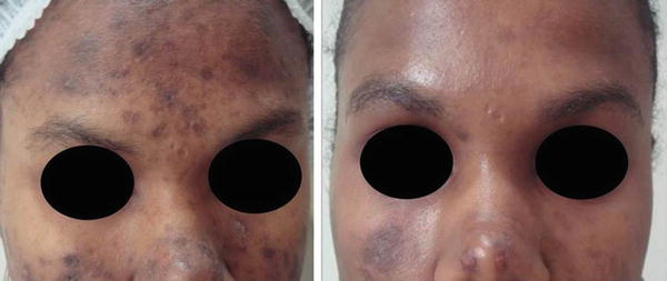
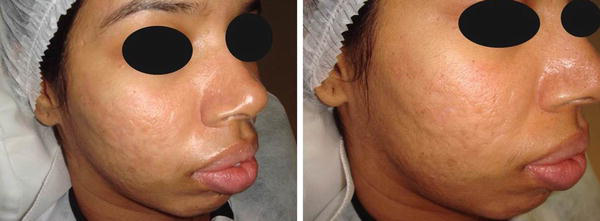
Acne commonly causes post-inflammatory pigmentation and atrophic scars (Taylor et al. 2002). Fractionated non-ablative laser is very well indicated to induce skin remodeling, sparing the epidermis (Jackson 2003). This procedure has a shorter downtime and a fewer risk of dyschromias; however it requires a greater number of sessions when compared to ablative lasers. We also use ablative lasers, although conservative parameters (low energy and low density) should be applied. Controlled radio-frequency microneedles and fractional ablative radio frequency are also excellent options to treat darker skin.
Benign Cutaneous Tumors
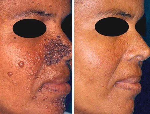
Ablative lasers can be used to remove facial angiofibromas, syringomas keratosis seborrheic, as well as dermatosis papulosa nigra, keloidal folliculitis of the neck, and dermatosis with higher prevalence in ethnic skin (Jackson 2003).
In these cases, we use CO2 or erbium YAG laser in surgical mode of operation, choosing the proper spot diameter according to the lesion dimension, avoiding perilesional thermal damage (Cole et al. 2009).
Stretch Marks
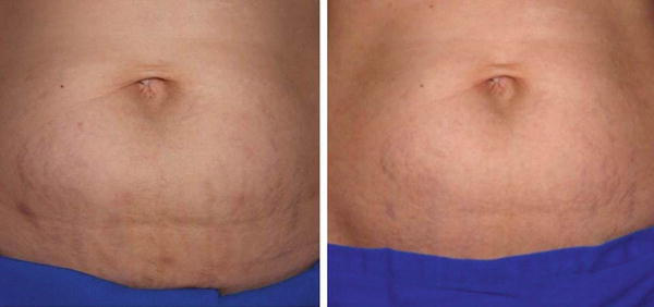
The literature reports numerous treatment options for stretch marks, such as intense pulsed light, pulsed dye laser, ablative radio frequency, Er:Glass, Er:YAG, and CO2. However, very few discuss those treatments on ethnic skin (Aldahan et al. 2016).
Some studies in Asians (phototype VI) suggest that ablative and non-ablative fractionated lasers are promising technologies in the treatment of this condition (Kim et al. 2008; Lee et al. 2010). In 2011, Yang and Lee (2011) compared the 1550 nm (Er:YAG) laser to fractionated CO2 in a study involving 24 Asian patients with white abdominal stretch marks. Treatments were randomized to each side of the body with three sessions every 4 weeks. Clinical improvement was somewhat higher on the side whose treatment was performed with fractionated CO2, despite of more adverse events in this group. Clinically, there was a reduction in the width of the striae, and an increase in the number of collagen and elastic fibers was observed in the histology study (Yang and Lee 2011).
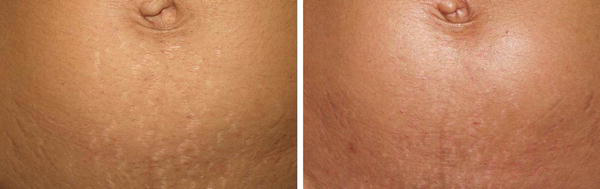
We have experience with fractionated ablative lasers (CO2 and Erbium 2940) in treatment of stretch marks in phototypes V. Our protocol includes testing two different parameters in a small area 30 days before the first session; using cold air cooling before, during, and after procedure; and applying low energy and low density (Kono et al. 2007). Post-inflammatory hypochromia is a common and mostly transient adverse event.
Rejuvenation
Although relatively more risks of pigmentary disorders and scars are described, effective treatment with lasers can be achieved in patients with higher phototypes (Bhatt and Alster 2008; Shah and Alster 2010). As an alternative to reduce these risks, non-ablative fractionated lasers are an excellent option. The correct parameters are essential to reduce side effects. Both technologies can be used in different sessions, respecting the interval of 4 weeks between the applications.
My Experience with Lasers for Ethnic Skin
Treating ethnic skin with lasers is always challenging. The first step is to evaluate the patient and the dermatosis to be treated and choose the best technology to be applied. We often perform a test treatment in a small area of the skin using two different parameters to check the effectiveness and to avoid side effects. This test is usually done 30 days before treatment.
The use of lightening cream, as hydroquinone isolated or associated with retinoic acid, to prepare the skin before laser application is controversial, but it is part of our routine. We also use these medications 3–4 weeks after treatment.
All patients are photographed before the procedure. Topical anesthetics are indicated to reduce the pain. During the procedure we use skin cooling equipment, as we believe that it can minimize inflammation, burning, and pain.
We routinely use conservative parameters at the first session, making adjustments in the subsequent sessions. Low fluence and low density are indicated to reduce thermal effects, avoiding dyschromia.
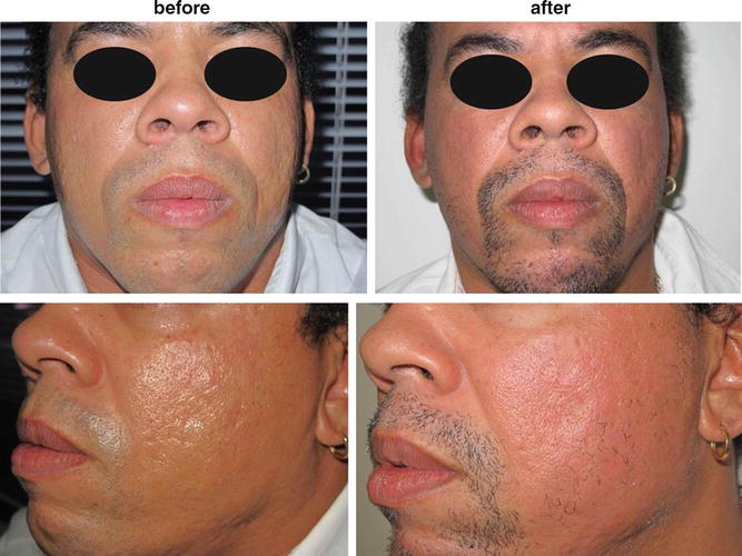
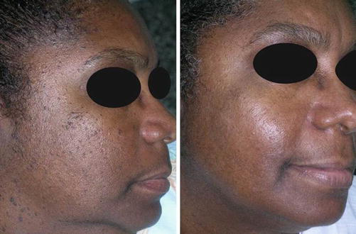
Just after procedure we apply steroid cream for the first 3 days associated with a restorative cream, which is maintained until complete healing. After treatment, patients are advised to wear sunscreens with broad spectrum, and it is fundamental to avoid sun exposure (Figs. 1, 2, 3, 4, 5, 6, and 7).
Conclusion
Nowadays, ethnic skin patients are looking for dermatologic treatments, and according to advances in lasers’ technology, the best results can be achieved. It is fundamental to photograph the patients before and after each procedure, to use the appropriate parameters according to the indications and to follow the patients during the post-procedure period until complete skin recovery.
Take Home Messages
- Ethnic skin is a term used to define the darker skin corresponding to Fitzpatrick’s IV, V, and VI skin types.
- Patients with ethnic skin have an increased risk of problems related to pigmentation, such as post-inflammatory hyperpigmentation or hypopigmentation.
- The risk of pigmentary changes is correlated with laser injury and thermal damage. It depends on laser wavelength, fluence, and density.
- Over time, lasers devices have evolved, and recent techniques make its use safer, with less downtime and, consequently, with less damage and risk of post-inflammatory pigmentation.
- When performed by skilled, well-trained dermatologists, fractional ablative and nonablative lasers become safe and viable for many treatments.
References
Aldahan AS, Shah VV, Mlacker S, Samarkandy S, Alsaidan M, Nouri K. Laser and light treatments for striae distensae: a comprehensive review of the literature. Am J Clin Dermatol. 2016;17(3):239–56.
Battle Jr EF, Hobbs LM. Laser therapy on darker ethnic skin. Dermatol Clin. 2003;21:713–23.
Bhatt N, Alster TS. Laser surgery in dark skin. Dermatol Surg. 2008;34(2):184–94.
Ciocon DH, Engelman DE, Hussain M, Goldberg DJ. A split-face comparison of two ablative fractional carbon dioxide lasers for the treatment of photodamaged facial skin. Dermatol Surg. 2011;37(6):784–90.
Cole PD, Hatef DA, Kaufman Y, Pozner JN. Laser therapy in ethnic populations. Semin Plast Surg. 2009;23(3):173–7.
IBGE – Brazilian Institute of Geography and Statistics, Brazil 500 Years. Rio de Janeiro; 2000.
Jackson BA. Lasers in ethnic skin: a review. J Am Acad Dermatol. 2003;48(Suppl 6):S134–8.
Kadunc B, Palermo E, Addor F, Hetsavaht L, Rabello L, Hattos R, Martin S. Textbook of dermatologic surgery, cosmiatry and laser. In: Osório N, Macéa J, editors. Ablative fractional lasers for photoaging. 1st ed. Rio de Janeiro; 2012. p. 772. Elsevier.
Kalil C, Campos V. Practical laser handbook and other sources of electromagnetic energy. In: Boechat A, editor. Laser and interaction with tissues. 1st ed. Rio de Janeiro; 2012. p. 19. Elsevier.
Kim BJ, Lee DH, Kim MN, Song KY, Cho WI, Lee CK, Kim JY, Kwon OS. Fractional photothermolysis for the treatment of striae distensae in Asian skin. Am J Clin Dermatol. 2008;9(1):33–7.
Kim JE, Won CH, Bak H, Kositratna G, Manstein D, Dotto GP, Chang SE. Gene profiling analysis of the early effects of ablative fractional carbon dioxide laser treatment on human skin. Dermatol Surg. 2013;39(7):1033–43.
Kono T, Chan HH, Groff WF, Manstein D, Sakurai H, Takeuchi M, Yamaki T, Soejima K, Nozaki M. Prospective direct comparison study of fractional resurfacing using different fluences and densities for skin rejuvenation in Asians. Lasers Surg Med. 2007;39(4):311–4.
Lee SE, Kim JH, Lee SJ, Lee JE, Kang JM, Kim YK, Bang D, Cho SB. Treatment of striae distensae using an ablative 10,600-nm carbon dioxide fractional laser: a retrospective review of 27 participants. Dermatol Surg. 2010;36(11):1683–90.
Macrene RA, Jeffrey SD, Kenneth AA. Fractional laser skin resurfacing. J Drugs Dermatol. 2012;11:1274–87.
Manstein D, Herron GS, Sink RK, Tanner H, Anderson RR. Fractional photothermolysis: a new concept for cutaneous remodeling using microscopic patterns of thermal injury. Lasers Surg Med. 2004;34(5):426–38.
Mateus A, Palermo E. Cosmiatria and laser practice in the doctor’s office. In: Almeida G, Golovaty R, Araújo R, editors. Dark skin. 1st ed. São Paulo; 2012. p. 504. Gen.
Munavalli GS, et al. Combining confluent and fractionally ablative modalities of a novel 2790nm YSGG laser for facial resurfacing. Lasers Surg Med. 2011;43(4):273–82.
Orringer JS, Kang S, Johnson TM, Karimipour DJ, Hamilton T, Hammerberg C, Voorhees JJ, Fisher GJ. Connective tissue remodeling induced by carbon dioxide laser resurfacing of photodamaged human skin. Arch Dermatol. 2004;140(11):1326–32.
Ortiz AE, Tremaine AM, Zachary CB. Long-term efficacy of a fractional resurfacing device. Lasers Surg Med. 2010;42(2):168–70.
Riggs K, Keller M, Humphreys TR. Ablative laser resurfacing: high-energy pulsed carbon dioxide and erbium:yttrium-aluminum-garnet. Clin Dermatol. 2007;25:462–73.
Shah S, Alster TS. Laser treatment of dark skin: an updated review. Am J Clin Dermatol. 2010;11(6):389–97.
Sriprachya-anunt S, Marchell NL, Fitzpatrick R, et al. Facial resurfacing in patients with Fitzpatrick Skin Type IV. Lasers Surg Med. 2002;30(2):86–92.
Taylor SC, Cook-Bolden F, Rahman Z, Strachan D. Acne vulgaris in skin of color. J Am Acad Dermatol. 2002;46(2 Suppl Understanding):S98–106.
Tierney EP, Eisen RF, Hanke CW. Fractionated CO2 laser skin rejuvenation. Dermatol Ther. 2011;24(1):41–53.
Wat H, Wu DC, Chan HH. Fractional resurfacing in the Asian patient: current state of the art. Lasers Surg Med. 2017;49(1):45–59.
Xu XG, Luo YJ, Wu Y, Chen JZ, Xu TH, Gao XH, He CD, Geng L, Xiao T, Zhang YQ, Chen HD, Li YH. Immunohistological evaluation of skin responses after treatment using a fractional ultrapulse carbon dioxide laser on back skin. Dermatol Surg. 2011;37(8):1141–9.
Yang YJ, Lee GY. Treatment of striae distensae with nonablative fractional laser versus ablative CO2 fractional laser: a randomized controlled trial. Ann Dermatol. 2011;23(4):481–9.







