
Erbium Laser for Scars and Striae Distensae
Erbium Laser for Scars and Striae Distensae
Paulo Notaroberto Serviço de Dermatologia, Hospital Naval Marcílio Dias, Rio de Janeiro, Brazil
Abstract
Scars are a very common complication of skin injuries such as burns, surgeries, and trauma (lacerations or abrasions) affecting millions of people every year. The appearance of scars can be very disturbing to patients both physically and psychologically, being aesthetically unacceptable and impacting negatively on the quality of life. Treatment of scarring may require many different kinds of treatments, depending on the kind of scarring present; however, skin vaporization and residual thermal damage can only be achieved by ablative lasers and explain the superiority of ablative laser treatment over chemical peels and dermabrasion. The present chapter addresses the issue of the ablative Erbium (Er:YAG) laser, which is highly absorbed by water and which, together with the possibility of being modulated by variations in pulse duration, makes it a precise, safe, and effective tool in managing scars. The aims of the ablative Erbium laser in treating atrophic scars are reducing the depth of the scar borders and stimulating neocollagenesis to fill depressions.
Keywords LaserErbiumAblativeAblationResurfacingScarStriae distensaeStretch mark
Introduction
Scars are a very common complication of skin injuries such as burns, surgeries and trauma (lacerations or abrasions) affecting millions of people every year. The appearance of scars can be very disturbing to patients both physically and psychologically (Harithy and Pon 2012), being aesthetically unacceptable and impacting negatively on the quality of life. Scars can also cause pruritus, tenderness, pain, sleep disturbance, anxiety, and depression in postsurgical patients (Oliaei et al. 2012).
Acne is a common disorder that affects up to 80% of people aged between 11 and 30 years (Oliaei et al. 2012; Fife 2011; Al-Saedi et al. 2014) and over 90% of adolescents (Fabbrocini et al. 2010). Several factors are involved in the pathogenesis of acne, but the severe inflammatory response involved in the process may result in permanent scars, an unfortunate complication of acne vulgaris (Fife 2011). The incidence of acne scarring is not well studied, but it may occur to some degree in 95% of patients with acne vulgaris. Studies report the incidence of acne scarring in the general population to be 1–11%. Having acne scars can be emotionally and psychologically distressing to patients. Acne scars may be linked to poor self-esteem, social ostracism, withdrawal from society, depression (Al-Saedi et al. 2014), anxiety, altered social interactions, body image alterations, lowered academic performance, and unemployment, and they are a risk factor for suicide (Fife 2011).
There is no general consensus in the literature as to what is the best treatment (Harithy and Pon 2012). In the last 15 years, laser resurfacing has emerged at the forefront of acne scar treatment. The first lasers to be used for acne scarring were the ablative CO2 and Er:YAG lasers, which emit radiation at wavelengths of 10,600 and 2,940 nm, respectively; having a high affinity for water, they ablate the epidermis and stimulate collagen synthesis (Hession and Grabber 2015) (see chapter “CO2 Laser for Scars,” this volume). Determining which laser system to use depends upon the type and severity of acne scarring, the amount of recovery a patient can tolerate, and the ultimate goals and expectations of each patient (Sobanko and Alster 2012). No treatment is 100% effective in “erasing” scars, and the best result is improvement, not perfection. Treatment of scarring may require many different kinds of treatments, depending on the kind of scarring present (Keyal et al. 2013); however, skin vaporization and residual thermal damage can only be achieved by ablative lasers and explain the superiority of ablative laser treatment over chemical peels and dermabrasion (Alster and Zaulyanov-Scanolon 2007).
The clinician who deals with scar treatment must understand the pathophysiology of scar formation. The process of wound healing is didactically separated into three stages: inflammation, proliferation, and maturation (Harithy and Pon 2012; Fabbrocini et al. 2010). By examining biopsy specimens of acne lesions from the backs of patients with severe scars and of patients without scars, Holland et al. found that the inflammatory stage was stronger and had a longer duration in patients with scars than in those without (Fabbrocini et al. 2010).
Erbium 2,940 nm Photothermal Ablation
Erbium (Er:YAG) laser is a flashlamp-excited system that emits light at an invisible infrared wavelength of 2,940 nm. The chromophore for ablative lasers is water. It is not an exaggeration to affirm that the laser target is the skin per se, since the skin is made up of approximately 80% water. Erbium 2,940 nm wavelength light is between 12 and 18 times better absorbed by tissue water when compared to the 10,600 nm wavelength emitted by the CO2 laser. The first generation of Erbium lasers was approved for cutaneous resurfacing by the FDA (Food and Drug Administration) in 1996 (Riggs et al. 2007), and it works by emitting a short pulse (SP) of 250–350 μs that is shorter than the thermal relaxation time of the skin, which is 1 ms (Al-Saedi et al. 2014). The ablation threshold of the first-generation Er:YAG laser for human skin has been calculated at 1.6 J/cm2, as compared with 5 J/cm2 calculated for high-energy, short-pulse CO2 laser systems. Because the Er:YAG laser is so exquisitely absorbed by water, the SP Erbium laser causes 10–40 μm of tissue ablation and as little as 5 μm of thermal damage to the surrounding tissue (Al-Saedi et al. 2014). The second generation of Erbium lasers has variable and longer pulses (500 μs–10 ms) and was FDA approved in 1999. Longer-pulsed Er:YAG lasers have been shown to increase the underlying thermal effect zone to approximately 120 μm (Lukac et al. 2010), leading to coagulation and skin tightening but increasing the risk of secondary side effects such as erythema and dyschromia (hypo- and hyperchromia) (Alster and Zaulyanov-Scanolon 2007). Side effects and complications after Er:YAG laser resurfacing are similar to those observed after CO2 laser skin resurfacing, but they tend to be less severe in duration, incidence, and intensity (Keyal et al. 2013; Alexiades-Armenakas et al. 2008).
Resurfacing lasers are high-energy pulsed lasers that generate photothermal ablation, which occurs with rapid heating when tissue absorbs enough laser energy, leading to tissue vaporization. The thermal effect also occurs in the area surrounding the ablated zone due to thermal diffusion (the zone of thermal damage). Modulated Erbium lasers with longer pulse durations result in larger areas of thermal coagulation when compared to the first-generation SP Er:YAG 2,940 nm laser devices (Carrol and Humphreys 2006). Pozner and Goldberg conducted a study on the histologic effect of a variable-pulsed Er:YAG laser and concluded that the thermal effect desired from the CO2 laser can be observed by using longer (50 ms pulse width) Er:YAG laser pulses (Pozner and Goldberg 2000; Khatri 2001). The ablative Erbium laser produces moderate immediate intraoperative contraction, but subsequent wound healing results in dermal shrinkage identical to that seen with CO2 ablative laser devices (Sapijaszko and Zachary 2002).
Response rates to the first-generation short-pulse Er:YAG lasers ranged from 25% to 90% (Fabbrocini et al. 2010). In order to address these shortcomings, longer-pulsed Er:YAG lasers were developed. In a prospective study of 35 patients with pitted acne scars, results were excellent (>75% improvement) in 36% of patients and good (50–75% improvement) in 57% (Hession and Grabber 2015). The combination of short pulses (for ablation) with longer pulses (for coagulation) is called dual mode Er:YAG, and systems working this way range in pulse duration from 500 μs to 10 ms. As a group, these lasers have been shown to produce deeper tissue vaporization, greater control of hemostasis, and collagen shrinkage leading to clinical skin tightening. This translates into greater clinical improvement in mild to moderate acne scars than achieved by their short-pulsed predecessors, and they thus represent a good compromise between the CO2 laser and the first-generation Er:YAG laser (Keyal et al. 2013).
The aims of the ablative laser in treating atrophic scars are reducing the depth of the scar borders and stimulating neocollagenesis to fill depressions. Focused vaporization can be used for treating isolated scars, but performing the treatment over an entire cosmetic unit (field treatment) is highly recommended for increasing the overall collagen tightening effect, which promotes improvement of distensible scars. Field treatment also decreases the chance of a sharp demarcation between treated and untreated sites (Alster and Zaulyanov-Scanolon 2007). A feather treatment using gentler energy should be performed on the periphery of the treated area with the goal of smoothing the transition between treated and untreated areas.
The concept of ablative fractional photothermolysis (AFP) was introduced in 2003 as an option for low-risk, short-downtime and effective resurfacing techniques. Fractionated lasers work by thermally altering a “fraction” of the skin, leaving up to 95% of the skin untouched, leading to faster healing, shorter downtime, and fewer adverse effects in comparison to non-fractional ablative laser devices (Loesch et al. 2014; Zgavec and Stopajnik 2014). AFP induces small three-dimensional zones of thermal damage known as microscopic treatment zones (MTZs) (Harithy and Pon 2012). In spite of not ablating a large surface, AFP generates MTZs which are real “columns of heat” capable of generating collagen contraction (skin tightening) and inflammation, which stimulates neocollagenesis. With AFP, the depth of penetration is directly proportional to the energy delivered in each MTZ, and the intensity of the treatment increases at the same rate that the density of spots is increased. Density can be reported as either the percentage of laser coverage in a treated area or the number of MTZs per square centimeter (Harithy and Pon 2012).
Protocols
Resurfacing
The treatment must be planned, executed, and followed up carefully in order to maximize results and minimize adverse effects.
The skin must be prepared with topical use of glycolic acid or tretinoin associated with vitamin C at least 1 month before performing the procedure. The topical use of hydroquinone on darker phototypes during the pretreatment period to minimize the risk of residual hyperchromia is not a consensus. All patients should be advised about the need to use sunscreens with very high UVA and UVB protection and to avoid sun exposure pre- (at least 1 month) and posttreatment (at least 2 months).
An oral prophylactic anti-herpetic therapy regimen should be started 2 days before the procedure and must be sustained for 5 days; it is mandatory whenever there is a previous history of herpes simplex or when an aggressive treatment will be performed (especially if the treatment affects the peri-oral area). Prophylactic antibiotic therapy is required when a non-fractional ablative treatment will be performed.
Topical anesthesia used to be enough for pain control, but oral pain relief (such as trometamol ketorolac), infiltrative anesthesia, or nerve blocks may be necessary. The use of corticosteroids (topically or orally) can reduce erythema and edema in the posttreatment period, but its use is quite controversial because many doctors believe it may prejudice the final outcome, since neocollagenesis is mainly due to inflammation.
Ointments such as petrolatum must be used during the period of reepithelialization, which takes 2 or 3 days depending on the treatment intensity.
Post-trauma Scars
Kim et al. conducted a prospective trial enrolling 12 patients of Fitzpatrick skin types III–V with 15 scars resulting from facial trauma and repair by suturing on the day of the trauma. Fractionated Er:YAG laser (LOTUSII, Laseroptek, Sungnam, Korea) treatment was initiated at least 4 weeks after primary repair of the wound, and each patient underwent four sessions at 1-month intervals. Two passes combining short (0.35 ms) and long (1 ms) pulses were performed in each session, and all patients were submitted to the same pulse and energy parameters. This Korean study demonstrated that ablative fractional Er:YAG laser treatment improved scars based on objective results and patient satisfaction rates (Kim et al. 2012).
A study carried out at the University of Verona (Italy) by Dr. Nocini et al. enrolled ten patients with scarring after unilateral and bilateral cleft lip surgery. All subjects underwent four passes combining different depths of ablation and coagulation (first pass, 100 μm ablation without coagulation; second pass, 80 μm ablation with 50 μm coagulation; third pass, 60 μm ablation with 25 μm coagulation; and fourth pass, 40 μm ablation to smooth the margins of the surgical area) per session. The authors reported clinical improvement of the scars treated with the non-fractional Er:YAG laser (Contour, Sciton, Palo Alto, California, USA) after the first treatment, with continued improvement after the second laser session (Nocini et al. 2003).
Acne Scars
Acne scarring can occur as a result of damage to the skin during the healing of active acne and can be classified into three different types depending on whether there is a net loss or gain of collagen: atrophic, hypertrophic, or keloid. Atrophic acne scars are by far the most common type, ranging between 80% and 90% of total acne scars, and are divided into ice pick, boxcar, and rolling scars. Regarding atrophic scars, the ice pick type represents 60–70% of total scars, the boxcar 20–30%, and rolling scars 15–25%. The ice pick scar is a narrow (less than 2 mm), punctiform and deep scar which does not undergo visible alteration when the skin is stretched, and typically the opening is wider than the deeper infundibulum, forming a “V” shape. The rolling scar is a result of dermal tethering of the dermis to the subcutaneous tissue, and these scars are usually wider than ice picks, ranging from 4–5 mm. These scars give a rolling or undulating appearance to the skin and tend to improve in clinical appearance when distended. The boxcar scar is round or oval with well-established vertical edges. These scars tend to be wider at the surface than an ice pick scar and do not have the tapering V shape. Instead, they can be visualized as a “U” shape with a wide base and can be shallow or deep (Fabbrocini et al. 2010). Often all three types of atrophic scars can be observed in the same patients, and it can be very difficult to differentiate between them. As the skin ages, the appearance of acne scars often worsens due to a relative weakness in the dermis rather than fading (Fife 2011; Weinstein 1999).
The pathogenesis of atrophic acne scarring is not completely understood but is most likely related to inflammatory mediators and enzymatic degradation of collagen fibers. It is not clear why some acne patients develop scars while others do not, as the degree of acne does not always correlate with the incidence or severity of scarring. Once scarring has occurred, it is usually permanent (Fife 2011). Histologically, post-acne scars are usually limited to the epidermis and upper papillary dermis and are thus amenable to treatment with a variety of techniques, including ablative and non-ablative lasers for skin resurfacing (Keyal et al. 2013).
After physical examination, understanding the patient’s concerns and expectations relating to his or her acne scars is the next step in the management of the acne scar and is determinant to success (Fife 2011).
A study conducted by Woo et al. at Korea University included 158 volunteers of Fitzpatrick skin phototypes III–V with atrophic acne scars who were separated into three groups and treated with short pulse (350 μs – group 1), long pulse (7 ms – group 2), and dual-mode (350 μs followed by 8 ms – group 3) non-fractional ablative Erbium (2,940 nm) laser. The patients treated with the short-pulsed Er:YAG showed better improvement of ice pick scars when compared with the long-pulsed treatment. On the other hand, longer pulses induced greater improvement of deep and shallow box scars and of rolling scar types when compared to the short-pulse results. The group submitted to dual mode treatment showed the best overall improvement and the best improvement for each type of scar (Woo et al. 2004). Jeong and Kye from the University of Korea treated 35 patients presenting atrophic scars with a long-pulsed (10 ms) non-fractional Er:YAG (2,940 nm) laser and observed an excellent outcome in 36%, a good outcome in 57%, and a fair outcome in 7% (Jeong and Kye 2001).
Deng et al. performed a prospective study at Shanghai Jiao Tong University, Shanghai (China), with 26 patients presenting moderate to severe atrophic acne scarring. Five treatment sessions with a fractional Erbium laser device (Pixel 2,940, Harmony, Alma Lasers, Ltd., Caesarea, Israel), with fluences ranging from 800 to 1,400 mJ/cm2 at 49 MTZ/cm2 and long-pulse duration (2 ms), were applied to the treated area using 8–10 passes, with minimal discomfort and insignificant collateral effects. The authors observed improvement of at least 50% in 100% of all subjects (Deng et al. 2009). Hu et al. from Taiwan (China) conducted a study enrolling 34 volunteers who were submitted to a single session of a fractional ablative Er:YAG laser (Profractional-XC, Sciton Inc., Palo Alto, California, USA) providing 150 μm of thermal damage. This trial revealed a satisfaction rate of 72.7% among patients, with minimal side effects (Hu et al. 2011).
The fractional ablative Erbium treatment combines the gentleness of the fractional technique with a more intense coagulation mode, promoting remarkable skin remodeling and dermal tightening. Nirmal et al. from India performed a clinical trial including 25 patients and noticed that rolling and superficial box scars showed significantly greater improvement when compared with ice pick and deep box scars after 2 ms pulse-duration fractional ablative Er:YAG treatments (Nirmal et al. 2013).
Figures 1a, b and 2a, b illustrate my clinical experience with the Erbium laser for acne scar treatment. These figures show improvement in severe acne scars after 3 treatment sessions with a 1-month interval between them. A fractional Erbium laser device (Pixel 2,940, Harmony, Alma Lasers, Ltd., Caesarea, Israel) was used with fluences of 1,400 mJ/cm2 at 49 MTZ/cm2, long-pulse duration (2 ms), and four overlapping “shots” (stacking).
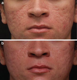
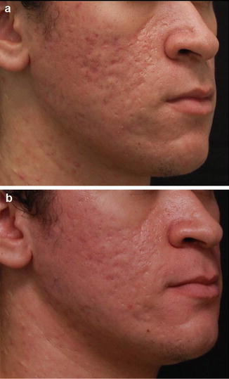
Often a combination of techniques (e.g., subcision or filler injections combined with fractional resurfacing) will ensure a better result compared to one procedure alone (Fife 2011). Yin et al. performed a prospective study enrolling 40 subjects presenting severe acne. Patients were treated with 15% 5-aminolevulinic acid (ALA) photodynamic therapy and subsequently received ablative fractional Er:YAG (2,940 nm) five times at 4-week intervals. After 6 months, the lesions showed overall improvement in all subjects (good to excellent improvement in acne inflammatory lesions), with 80% overall improvement in acne scars.
After 12 months, most subjects had improved hypertrophic and atrophic scars (good to excellent improvement in 85%), and no one had recurrent acne inflammatory lesions. Patient self-evaluation also revealed good to excellent improvements (on average) in acne lesions and scarring, with significant improvements in self-esteem 6 months after treatment. The authors suggested that the combination of ALA-PDT and fractional resurfacing with Er:YAG is a promising option for the management of severe acne, preventing scar formation (Yin et al. 2014).
Hypertrophic Scars and Keloids
Hypertrophic scars and keloids are not a hallmark among the indications for ablative lasers. The Er:YAG laser seems to be more suitable for treating hypertrophic scars and keloids because, as it is 12–18 times more selective for water than the CO2 laser due to its shorter wavelength (2,940 nm), it causes less residual thermal injury and less inflammation (Oliaei et al. 2012; Al-Saedi et al. 2014). Er:YAG at 5 J/cm2 vaporizes tissue to a depth of 20–25 μm with an additional 5–10 μm zone of thermal necrosis. Er:YAG lasers have shown moderate improvement of hypertrophic scars and keloids. These ablative lasers target water in the tissue, resulting in tissue vaporization (Harithy and Pon 2012).
Burn Scars
Treatment with ablative full-field CO2 and Er:YAG lasers has been used to treat burn scars but has been associated with prolonged recovery times and contradictory results. Burn scars, especially new ones, need to be treated gently. Ablative fractional lasers have the capability to act on a well-controlled percentage of skin while stimulating new collagen formation, remodeling the burn scar tissue and subsequently normalizing the texture, elasticity, and color of the scar. A few case reports have shown that ablative fractional resurfacing is safe and effective in the treatment of burn scars; however, further studies are needed to determine parameters (Harithy and Pon 2012).
Stretch Marks (Striae Distensae)
Stretch marks (also called striae distensae) are histologically characterized as scars; although there is no break in the continuity of the epidermis, they demonstrate microscopic evidence of thinning and flattening of the epidermis, a normal or decreased number of melanocytes, and thinning and retraction of dermal collagen and elastin (Godberg et al. 2005; Maia et al. 2010). In the early phase, inflammatory changes are remarkable, but later the epidermis is thin and flattened. Recent stretch marks show a deep and superficial perivascular lymphocytic infiltrate. Collagen bands in the upper third of the reticular dermis are stretched and aligned parallel to the surface of the skin. In the latter stages, there is thinning of the epidermis due to flattening of the epidermal ridges and loss of collagen and elastin (Elsaie et al. 2009). Clinically, stretch marks appear as erythematous (striae rubra), linear dermal scars in the early phase, or as hypopigmented (striae alba) linear dermal scars with epidermal atrophy in the late phase.
There is little good-quality research focused on the physiopathology of stretch marks (Cordeiro and Moraes 2009). Although the etiology of stretch marks is not well understood, it is accepted that the combination of mechanical stretching of the skin, genetics, endocrine disorders, and possibly secretion of relaxin during pregnancy, alone or in combination, plays a role in the physiopathology of striae distensae (Maia et al. 2010).
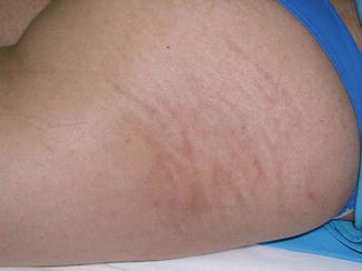
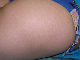
Stretch mark treatment is based on stimulating neocollagenesis and restoring epidermal architecture. The use of ablative technologies such as the Erbium laser induces clinical improvement on body areas, but almost all patients develop significant post-inflammatory hyperpigmentation, especially in darker skin tones. The sequence of Figs. 3, 4, 5, and 6 illustrates the course of treatment of stretch marks on the thigh area of a patient with a type II Fitzpatrick skin phototype. A non-fractional Erbium laser device (Fidelis, Fotona Lasers Ltd., Ljubljana, Slovenia) was used with the LP (long pulse) 600 μs pulse duration.
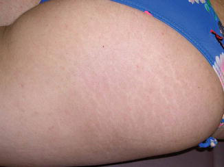
Longer pulse durations induce low ablation and intense coagulation and inflammation (Fig. 4).
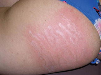
The residual post-inflammatory hyperchromia is a hallmark of ablation on body areas (Fig. 5), despite the significant final result (Fig. 6). This is the reason that sublative and non-ablative lasers are the first options among lasers for managing stretch marks.
Post-Procedure Care
The ablative laser procedure does not end when the surgical act is finalized. Post-procedure care is an important underlying part of guaranteeing the expected aesthetic result, accelerating the healing process with a smooth recovery and avoiding complications. Open wound care is performed with frequent application of ointments to the surface of the treated area, while the occlusive approach requires the use of occlusive bandages. Open and closed wound care helps to control pain and accelerates the healing process. Unlike open dressings, occlusive bandages increase the risk of infection (Costa et al. 2011) and do not allow visualization of the wound.
Pain can be controlled most of the time with cold compresses or cold-water sprays (Costa et al. 2011). Effective pain control can be achieved with the use of oral analgesics (paracetamol, codeine), combined or not with an anxiolytic (lorazepam) (Costa et al. 2011). Edema can be managed with the application of ice bags or cold-water compresses, but the use of an oral corticosteroid (40–60 mg prednisone daily for a variable period of 3–5 days) or an intramuscular corticosteroid can be useful in isolated cases (Oliaei et al. 2012; Costa et al. 2011). Ointments and antihistamines can be used to relieve intense pruritus.
Complications and Side Effects
Ablative lasers induce thermal destruction of the skin with an adjacent coagulation area. Therefore, some manifestations are expected and desirable in the post-procedure period. Recovery time depends on the amount of energy targeted to the skin, the pulse duration, and the delivery system (full ablation or fractional). The healing process after fractional treatment is significantly faster compared with full ablative (non-fractional) treatment (Zgavec and Stopajnik 2014).
Erythema and minimal crusting, which disappeared within 7 days, are the expected side effects after fractional treatment (Zgavec and Stopajnik 2014). The mean duration of erythema tends to be 2 days, and mean crusting is around 5 days (Nirmal et al. 2013). On the other hand, extensive crusting after treatment with the non-fractionated handpiece is observed even at a 14-day follow-up (Zgavec and Stopajnik 2014). Pain as evaluated by patients is milder when using fractionated handpieces in comparison with full ablative treatment (Zgavec and Stopajnik 2014) and disappears by the second day after treatment (Zgavec and Stopajnik 2014; Costa et al. 2011). Pain rarely occurs after the second day of the postoperative period and must be investigated if it occurs (dryness and infection are common causes) (Costa et al. 2011).
Edema usually varies from mild to moderate, peaks on the second and third days, and can last for up to 1 week (Costa et al. 2011). Edema is usually more intense in the peri-orbital areas and eyelids. Longer pulse durations with less ablation and more coagulation (deeper heating) usually lead to more pronounced edema. Pruritus affects more than 90% of patients undergoing ablative treatments in the first 2 weeks after the procedure and is due to the healing process (Costa et al. 2011). When pruritus is intense and persistent, a secondary infection must be investigated. Desquamation and post-fractional, non-ablative laser xerosis occur in 60% and 87% of cases, respectively (Costa et al. 2011). Purpura can occur and resolves spontaneously (Costa et al. 2011).
Viral, bacterial, and fungal infections are rare manifestations that occur during the first post-procedure week and require proper identification and treatment to avoid further complications (AlNomair et al. 2012), such as persistent erythema or scar formation. Infection must be considered when intense or persistent pain, erythema, and edema occur. The most common type of infection after fractional laser skin resurfacing is caused by HSV and has been reported in 0.3–2% of cases (Costa et al. 2011; AlNomair et al. 2012). Patients may not present with classic herpetiform vesicopustules but instead may demonstrate only superficial erosions that develop during the first week after treatment (AlNomair et al. 2012). Given that most patients present subclinical levels of HSV, prophylactic use of oral antivirals such as aciclovir, famciclovir, or valaciclovir is always recommended preventively in perioral or full-face ablative resurfacing. Prophylaxis with antivirals taken at regular doses for HSV infection must start 1 or 2 days before the laser procedure and continue until the skin is completely healed. Prophylaxis notwithstanding, herpetic infection sometimes does occur. In such cases, doses of oral antivirals equivalent to those used in treating herpes zoster virus must be used (Zhang and Obagi 2009). Rates of bacterial infection in traditional resurfacing tend to be low (0.5–4.5% of cases) and are even rarer when fractional, non-ablative lasers are used, occurring in only 0.1% of cases (Costa et al. 2011; AlNomair et al. 2012). Studies suggest that most bacterial infections related to laser ablation occur with the use of occlusive bandages in the post-procedure period (Costa et al. 2011). When infection is suspected, secretions must be cultured, an antibiogram test must be carried out, and a broad-spectrum systemic antibiotic (penicillin, a first-generation cephalosporin, or ciprofloxacin) is administered while waiting for the results of the bacterial culture and antibiogram (Costa et al. 2011).
Candida albicans is the most frequent agent related to fungal infections occurring after skin ablation, and the infection starts between the first and second weeks of the post-procedure period. Patients presenting pruritus, pain, and whitish erosions on a highly erythematous base, as well as satellite lesions outside the treated area, must be suspected of having a fungal infection. A direct mycological examination and fungal culture must be performed if infection is suspected (Costa et al. 2011).
Acneiform eruptions have been described as a frequent complication of fractional skin resurfacing, and they can be a result of aberrant follicular epithelialization during healing or secondary to the use of ointments during the recovery period (Costa et al. 2011; AlNomair et al. 2012). The development of milia cysts has been reported in as many as 19% of cases (AlNomair et al. 2012) and appears between 3 and 8 weeks after laser treatment as a consequence of the use of occlusive bandages, oils, or creams during the healing process (Costa et al. 2011).
Post-inflammatory hyperpigmentation (PIH) after skin resurfacing can be transient or long lasting and is one of the most common complications after ablative resurfacing (Costa et al. 2011). Hyperpigmentation is much less frequent with fractional laser skin resurfacing than with full-ablative resurfacing but is observed in 1–32% of patients, depending on the system used, the parameters applied, and the skin phototypes treated (AlNomair et al. 2012). Patients with darker skin phototypes (Fitzpatrick III–VI) or melasma have a higher likelihood of developing post-inflammatory hyperpigmentation (Costa et al. 2011; AlNomair et al. 2012). Some studies report up to 68% hyperpigmentation after skin ablation (Costa et al. 2011). Patients prone to developing PIH must be prepared during the 3 months prior to the procedure with a combination of hydroquinone and glycolic acid or tretinoin, or with hydroquinone cream used alone, in addition to the use of sunscreens (Costa et al. 2011). PIH must be treated as soon as possible, avoiding aggressive approaches before reepithelialization is complete, as they can worsen the condition (Costa et al. 2011). The regular use of broad-spectrum sunscreen and avoiding exposure to the sun for at least 6 to 8 weeks before and after the procedure are important in preventing the development of PIH (Costa et al. 2011). In addition to sunscreen, blemish agents such as hydroquinone, tretinoin and kojic, azelaic, and glycolic acids are also first-line treatments. Superficial chemical peels and microdermabrasion can be used to accelerate the lightening response (Costa et al. 2011).
Scarring is another known and rare complication of fractional ablative resurfacing (AlNomair et al. 2012), with serious and devastating consequences (Costa et al. 2011) for the final aesthetic result. It is a more frequent complication with the CO2 laser than with Erbium laser skin resurfacing. There are several potential explanations for hypertrophic scarring, including the use of excessively high-energy densities, postoperative infection of the skin, and lack of technical skill. The neck is a well-recognized site that is especially susceptible to the development of scarring and synechia because of the small number of pilosebaceous units and poor vasculature in this region, which are essential for wound healing. In addition, the thin skin of the neck renders it more susceptible to thermal injury. Other scar-prone anatomic locations that require more conservative treatment protocols include the periorbital and mandibular regions, the chest, and other areas over bony prominences (Fife 2011).
Conclusion
The ablative Erbium laser is highly absorbed by water, and this, together with the possibility of being modulated by variations in pulse duration, makes it a precise, safe, and effective tool in managing scars.
Take Home Messages
- Erbium (Er:YAG) laser is a flashlamp-excited system that emits light at an invisible infrared wavelength of 2,940 nm, and it is highly absorbed by water.
- Erbium (Er:YAG) can be modulated by variations in pulse duration, making it a precise, safe, and effective tool in managing scars.
- The aims of the ablative Erbium laser in treating atrophic scars are reducing the depth of the scar borders and stimulating neocollagenesis to fill depressions.
- No treatment is 100% effective in “erasing” scars, and the best result is improvement, not perfection.
- Treatment of scarring may require many different kinds of treatments, depending on the kind of scarring present; however, skin vaporization and residual thermal damage can only be achieved by ablative lasers and explain the superiority of ablative laser treatment over chemical peels and dermabrasion.
References
Alexiades-Armenakas M, Dover JS, Arndt KA. The spectrum of laser skin resurfacing: nonablative, fractional, and ablative laser resurfacing. J Am Acad Dermatol. 2008;58:719–37.
AlNomair N, Nazarian R, Marmur E. Complications in lasers, lights, and radiofrequency devices. Facial Plast Surg. 2012;28:340–6.
Al-Saedi S, Al-Hilo MM, Al-Shami SH. Treatment of acne scars using fractional Erbium:YAG laser. Am J Dermatol Venerol. 2014;3(2):43–9.
Alster T, Zaulyanov-Scanolon L. Laser scar revision: a review. Dermatol Surg. 2007;33:131–40.
Carrol L, Humphreys TR. Laser – tissue interactions. Clin Dermatol. 2006;24:2–7.
Cordeiro RCT, Moraes AM. Striae distensae: physiopathology. Surg Cosmet Dermatol. 2009;1(3):137–40.
Costa FB, El Ammar ABPC, Ampos VB, Kalil CLPV. Complications in laser dermatologic surgery. Part II: fractional and non-fractional ablative laser and fractional non-ablative laser. Surg Cosmet Dermatol. 2011;3(2):135–46.
Deng H, Yuan D, Yan C, Lin X, Ding X. A 2940 nm fractional photothermolysis laser in the treatment of acne scarring: a pilot study in China. J Drug Dermatol. 2009;8(11):978–80.
Elsaie ML, Baumann LS, Elsaaiee LT. Striae distensae (stretch marks) and different modalities of therapy: an update. Dermatol Surg. 2009;35:563–73.
Fabbrocini G, et al. Acne scars: pathogenesis, classification and treatment. Dermatol Res Pract. 2010;2010:893080.
Fife D. Practical evaluation and management of atrophic acne scars: tips for the general dermatologist. J Clin Aesthet Dermatol. 2011;4(8):50–7.
Godberg DJ, Marmur ES, Schmults C, Hussain M, Phelps R. Histologic and ultrastructural analysis of ultraviolet B laser and light source treatment of leukoderma in striae distensae. Dermatol Surg. 2005;31:385–7.
Harithy R, Pon K. Scar treatment with lasers: a review and update. Curr Dermatol Rep. 2012;1:69–75.
Hession MT, Grabber EM. Atrophic acne scarring: a review of treatment options. J Clin Aesthet Dermatol. 2015;8(1):50–8.
Hu S, et al. Ablative fractional erbium-doped yttrium aluminum garnet laser with coagulation mode for the treatment of atrophic acne scars in Asian skin. Dermatol Surg. 2011;37:939–44.
Jeong JT, Kye YC. Resurfacing of pitted facial acne scars with a long-pulsed Er:YAG laser. Dermatol Surg. 2001;27:107–10.
Keyal U, Huang X, Bhatta AK. Laser treatment for post-acne scars – a review. Nepal J Med Sci. 2013;2(2):165–70.
Khatri KA. The effects of variable pulse width of erbium laser on facial skin. Dermatol Surg. 2001;27(4):332–4.
Kim SG, et al. The efficacy and safety of ablative fractional resurfacing using a 2,940-nm Er:YAG laser for traumatic scars in the early posttraumatic period. Arch Plast Surg. 2012;39:232–7.
Loesch MM, Somani AK, Travers JB, Spandau DF. Skin resurfacing procedures: new and emerging options. Clin Cosmet Investig Dermatol. 2014;7:231–41.
Lukac M, Perhavec T, Nemes K, Ahcan U. Ablation and thermal depths in VSP Er:YAG laser skin resurfacing. J Laser Health Acad. 2010;1:56–71.
Maia M, Marçon CR, Rodrigues AB, Aoki T, Amaro AR. Stretch marks in pregnancy: a comparative study of risk factors among primiparae in private and public health system maternity hospitals. Surg Cosmet Dermatol. 2010;2(3):165–72.
Nirmal B, et al. Efficacy and safety of erbium-doped yttrium aluminium garnet fractional resurfacing laser for treatment of facial acne scars. Indian J Dermatol Venereol Leprol. 2013;79:193–8.
Nocini PF, D’Agostino A, Trevisiol L, Bertossi D. Treatment of scars with Er:YAG laser in patients with cleft lip: a preliminary report. Cleft Palate Craniofac J. 2003;40(5):518–22.
Oliaei S, Nelson JS, Fitzpatrick R. Laser treatment of scars. Facial Plast Surg. 2012;28:518–24.
Pozner JM, Goldberg DJ. Histologic effect of a variable pulsed Er:YAG laser. Dermatol Surg. 2000;26(8):733–6.
Riggs K, Keller M, Humphreys TR. Ablative laser resurfacing: high-energy pulsed carbon dioxide and erbium:yttrium-aluminum-garnet. Clin Dermatol. 2007;25:462–73.
Sapijaszko MJA, Zachary CB. Er:YAG laser skin resurfacing. Dermatol Clin. 2002;20(1):87–96.
Sobanko JF, Alster TS. Management of acne scarring, part I. Am J Clin Dermatol. 2012;13(5):319–30.
Weinstein C. Modulated dual mode erbium/CO2 lasers for the treatment of acne scars. J Cutan Laser Ther. 1999;1:203–8.
Woo SH, Park JH, Key YC. Resurfacing of different types of facial acne scar with short-pulsed, variable-pulsed, and dual-mode Er:YAG laser. Dermatol Surg. 2004;30:488–93.
Yin R, Lin L, Xiao Y, Hao F, Hamblim MR. Combination ALA-PDT and ablative fractional Er:YAG laser (2,940 nm) for the treatment of severe acne. Lasers Surg Med. 2014;46:165–72.
Zgavec B, Stopajnik N. Clinical and histological evaluation of Er:YAG ablative fractional skin resurfacing. J Laser Health Acad. 2014;1:1–6.
Zhang AY, Obagi S. Diagnosis and management of skin resurfacing-related complications. Oral Maxillofac Surg Clin North Am. 2009;21(1):1–12.







