
CO2 Laser for Stretch Marks
CO2 Laser for Stretch Marks
Guilherme Almeida, Elaine Marques and Rachel Golovaty Department of Dermatology, Hospital Sirio Libanes, Brazil – Private office: Clinica Dermatologica Dr Guilherme de Almeida, Rua Barata Ribeiro 414, Sao Paulo, CEP 01308000, Brazil Clinica Dermatologica Dr Guilherme de Almeida, Sao Paulo, Brazil
Abstract
Stretch marks or striae distensae (SD) are a well-recognized, common dermatologic entity, which affect patients of all ages, genders, and ethnicities and rarely cause any significant medical problems but can have a deep psychological impact on affected patients. Risk factors have been reported, but much remains to be understood about their epidemiology. Although there is no standard treatment for SD, many topical applications, peeling, light, and laser systems, have been tried. Considering the many modalities used to improve SD, lasers have recently become a popular therapeutic alternative. The aim of this chapter is to discuss the causes and possible treatments described in literature, to approach the clinical efficacy and safety of fractional CO2 laser in the treatment of SD, and to show 5 years of our experience using this device.
Keywords Stretch marksStriae distensaeStriae rubraStriae albaStriae atrophicansStriae gravidarumLaser therapyLight therapyAcid peel treatmentsCollagen injectionLaser lipolysisRadiofrequencyMicrodermabrasionNonablative lasersFractional laser resurfacing
Introduction
Roederer first described striae in 1773, and Troisier and Menetrier made the first histological descriptions in 1889 (Troisier and Ménétrier 1889). Striae distensae (SD), also denominated stretch marks (SM), striae rubra, striae alba, striae atrophicans, striae gravidarum (SG), are a well-recognized common dermatologic entity, which affect patients of all ages, genders, and ethnicities. SD is common in adolescence, pregnancy, and obesity. The most commonly affected sites are the breasts, upper arms, abdomen, buttocks, and thighs. Initially, SD present as edematous red or pink linear plaques called striae rubra. Over time, the color fades, and the lesions become hypopigmented, atrophic, and permanent (striae alba). It is rarely caused by systemic diseases but commonly represents a deep psychological impact on affected patients (Al-Himdani et al. 2014; Watson et al. 1998; Ud-Din et al. 2016).
SD is a known feature of several clinical conditions, both chronic and acute, with very distinct pathophysiology (e.g., pregnancy, adolescent growth spurts, obesity, large weight gain, Cushing syndrome, Marfan syndrome, diabetes mellitus, long-term systemic or topical steroid use), making it difficult to determine their true etiology. Most SD research has focused on pregnant women and adolescents. A positive family history is a risk factor in both of these groups alike. Among adolescents, BMI and childhood obesity both influence risk of developing SD (Troisier and Ménétrier 1889).
Epidemiology
The prevalence of SD reported in the literature varies a lot, ranging from 6% to 88% (Cho et al. 2006; Sisson 1954; Kelekci et al. 2011; Thomas and Liston 2014; Chang et al. 2014; Ghasemi et al. 2017; Osman et al. 2017; Davey 1972; Elton and Pinkus 1966; García-Hidalgo et al. 1999; García-Hidalgo 2002). The prevalence ranges from 6% to 86% in adolescents and from 43% to 88% in pregnant women (Cho et al. 2006; Chang et al. 2014; Ghasemi et al. 2017; Osman et al. 2017; Atwal et al. 2006; Canpolat et al. 2010; Maia et al. 2009; Jaramillo-Garcia et al. 2009; Cohen et al. 1997). Among obese individuals with a BMI of 27–51, the prevalence is reported to be 43% (García-Hidalgo et al. 1999). The prevalence among nonpregnant women and adult male varies a lot in literature (Kelekci et al. 2011; Elton and Pinkus 1966; Cohen et al. 1997; Murphy et al. 1992). Risk factors in pregnant women may be constitutional (maternal age and BMI) or pregnancy related (birth weight, gestational age, weight gain during pregnancy, and polyhydramnios) (Ghasemi et al. 2017; Osman et al. 2017; Atwal et al. 2006; Canpolat et al. 2010; Murphy et al. 1992).
Many risk factors have been suggested for the development of SG, such as pregnancy maternal weight (Ersoy et al. 2016; Liu 1974; Thailand J-Orh et al. 2008), weight gain during pregnancy (Osman et al. 2017), maternal age (Atwal et al. 2006), skin structure (Ghasemi et al. 2017), family history (Chang et al. 2014), race, and birth weight (Liu 1974). These have been investigated, but their effect has not been clearly proven (Davey 1972; Liu 1974; Thomas and Liston 2014; Muzaffar et al. 1998; Kartal Durmazlar and Eskioglu 2009). Surgical interventions and medications have also been associated with SD (Osman et al. 2017; Pinkus et al. 1966; Di Lernia et al. 2001; McKusick 1971; Rolleston and Goodall 1931; Shafir and Gur 1999; Tsuji and Sawabe 1993; Gupta 2000).
Ersoy et al. (2016) published a new study to determine individual risk factors related to SD and reported some preventive measures. This prospective observational study included 211 primiparous pregnant women who were hospitalized for birth and did not have systemic diseases or other risk factors (drugs use or polyhydramnios). The use of preventive oil or drugs, smoking status, skin type, water intake, and level of financial income did not significantly predict the appearance of SG.
According to the logistic regression analysis, including all variables found to be significant in one-by-one comparisons, i.e., age, pregnancy BMI, BMI at admission, abdominal circumference, birth weight, family history, sex of the infant, and maternal education level, it was established that each unit of decrease in maternal age increased the risk of SG by 1.15-fold (Ghasemi et al. 2017; Muzaffar et al. 1998; Kartal Durmazlar and Eskioglu 2009; Thomas and Liston 2014).
Histopathogenesis of Striae Distensae
Three main theories relating to SD formation are described: mechanical stretching of the skin, hormonal changes, and an innate structural disturbance of the tegument. Mechanical stretching of the skin is postulated due to the perpendicularity of SD to the direction of the skin. However, contradictory studies dispute this theory (McKusick 1971; Nigam 1989). Adrenocorticotrophic hormone (ACTH) and cortisol are thought to promote fibroblast activity, leading to increased protein catabolism, modifying collagen and elastin fibers (Klehr 1979). Pregnancy-related hormones are also believed to influence SD formation (Osman et al. 2017; Nigam et al. 1990; Cordeiro et al. 2010; Lurie et al. 2011). Disorder of extracellular matrix’s gene expression is also postulated as a possible mechanism involved in SD formation (Etoh et al. 2013; Friedman et al. 1993).
The exact pathogenesis of striae is still controversial. Early histological dermal alterations may be visualized on electron microscopy including mast cell degranulation and macrophage activation leading to elastolysis of the mid-dermis. Release of enzymes by mast cell, including elastases, is proposed as a key initiatory process in SD pathogenesis. The inflammatory process induces collagen, elastin, and fibrillin modifications. The reorganization of fibrillin and elastin are thought to play an important role in SD pathogenesis, and those who are predisposed to developing SD may have an underlying deficiency of fibrillin (Watson et al. 1998; Sheu et al. 1991).
A deep and superficial perivascular lymphocytic infiltrate with occasional eosinophils and dilated vessels with edema of the upper dermis are characteristic of newly acquired striae. SD in this stage is referred to as “striae rubra” (SR).
In late stage, elongated collagen bands are concentrated within the upper third of the reticular dermis and arranged parallel to the surface of the skin. In the “terminal” stages of SD, there is a thinning of the epidermis due to blunting of the rete ridges and a paucity of collagen and elastic fibers. SD in this stage are classified as “striae alba” (SA) and are considered permanent (Watson et al. 1998; Ackerman Ab et al. 1997; Arem and Kischer 1980).
Striae distensae can be considered a form of dermal scarring, and their clinical and histological features are similar to those of scar remodeling. For whatever reason, dermal collagen ruptures or separates, and the resulting gap is replaced with newly formed collagen that orients itself in the direction of local stress forces (Sisson 1954). Irrespective of the underlying pathology that may incite a cascade of uncertain events, a final common pathway results in the breakdown and tearing of the dermal matrix, which manifests clinically as SD.
A recent study investigates early molecular alterations that may promote laxity of mature striae gravidarum (SG). They investigated the dermal elastic fibers network, which provides elastic properties of the human skin. They obtained skin samples of newly developed, erythematous abdominal SG in healthy pregnant women. Elastic fibers were examined by Verhoeff stain and immunofluorescence. The normal elastic fiber network appeared markedly disrupted in SG, compared with perilesional abdominal skin or control (normal-appearing hip skin). This disruption was accompanied by the emergence of short, disorganized, thin, threadlike “fibrils,” which were observed prominently in the mid-to-deep dermis. These fibrils were rich in tropoelastin (the main component of normal elastic fibers) and persisted into the postpartum period without forming normal-appearing elastic fibers. The emergence of these fibrils was accompanied by increased gene expression of tropoelastin and fibrillin-1 but not other elastic fiber components such as fibrillin-2 and fibulin-1, fibulin-2, and fibulin-5. They concluded that in early SG, the elastic fiber network appears markedly disrupted and newly synthesized tropoelastin-rich fibrils emerge as an uncoordinated synthesis of elastic fiber. Because they are thin and disorganized, tropoelastin-rich fibrils do not function as normal elastic fibers. These findings help to elucidate the pathogenic mechanism by which laxity occur in SG (Wang et al. 2015).
Treatment
Striae distensae is a considerable challenge in terms of their treatment. They rarely resolve without intervention. Even with intervention, improvement rather than complete resolution is a more realistic goal. The best results are achieved when treating SD in the early phase. Once SD reaches a mature, static phase, they are significantly more resistant to treatment.
Various treatment modalities are reported to treat or prevent SD. Among them are laser therapy (Cho et al. 2006; Belo and Arceo-Cruz 2009; Alexiades-Armenakas et al. 2011), light therapy (Sadick et al. 2007), chemical peelings (Mazzarello and Farace 2012), percutaneous collagen induction (Aust et al. 2010), laser lipolysis (Freedman 2010), radiofrequency techniques (Suh et al. 2007), and microdermabrasion (Abdel-Latif and Albendary 2008). No single therapy has been advocated to completely eradicate these lesions (see also the following chapters: “Non-ablative Lasers for Stretch Marks,” this volume; “Transepidermal Drug Delivery with Ablative Methods (Lasers and Radiofrequency),” this volume).
Even when procedures are indicated, topical treatment is considered the most used treatment and can be used before or associated with procedures (Kelekci et al. 2011). A recent article assessed the evidence for the use of topical treatments for SD. They reviewed the published literature in English language, from 1980 onward (Kelekci et al. 2011). The products were categorized by their mechanisms of action, including those which act in stimulating collagen production, increasing elasticity, and improving cell proliferation and those with anti-inflammatory and rehydration properties. The results showed that there are few studies (n = 11) that investigate the efficacy of topical in management of SD. Trofolastin and Alphastria creams demonstrated level 2 evidence of positive results for their prophylactic use in SD. Additionally, tretinoin used therapeutically showed varied results, while cocoa butter and olive oil did not demonstrate any effect. Overall, there was a distinct lack of evidence for each topical formulation. The majority of topical products failed to mention their effect on early SD vs. later stages of SD (striae rubrae compared to striae albae) and their role in both prevention and treatment. In conclusion, there is no topical formulation that is shown to be most effective in eradicating or improving SD. A structured approach in identification and targeted management of symptoms and signs with the appropriate topical is required. Randomized controlled trials are necessary to assess the efficacy of topical products for treatment and prevention of different stages of SD (Ud-Din et al. 2016).
Lasers and Light Devices
The 585 nm flash lamp-pulsed dye laser (PDL) at low energy densities is commonly used to target the dilated blood vessels of striae rubra. An increase in the amount of collagen has been reported after a series of PDL treatment (McDaniel et al. 1996; Alster 1997). The PDL has a moderate, beneficial effect in reducing the degree of erythema in striae rubra but no apparent benefit in striae alba. Because of the potential for adverse effects, PDL should be performed with extreme caution in patients with Fitzpatrick V–VI skin type. McDaniel et al. undertook a controlled study of 39 patients with SD. Treatment sites included the abdomen, thighs, and breasts. Four treatment protocols were used with different spot distances and fluences. Untreated SD was the control. Outcomes were measured by subjective analysis, shadow profilometry, and histological analysis. A significant reduction in skin shadowing was reported in patients with SD in all protocols compared to controls. Additionally, elastin regained its normal appearance in SD treated with low-fluence PDL (McDaniel et al. 1996; Hernández-Pérez et al. 2002).
Intense pulsed light (IPL), characterized by a noncoherent filtered flash lamp with a broadband spectrum (515–1200 nm), has been shown to replace dermal elastosis with neocollagen, thus improving the appearance of mature SD after a series of treatments (Zelickson et al. 2004).
Radiofrequency (RF) devices produce heat which converts electrical current to thermal energy that is uniformly dispersed to different tissue depths. It increases collagen production by inducing collagen type I mRNA expression (Manuskiatti et al. 2009).
The long-pulse 1064 Nd:YAG is a nonablative treatment for facial wrinkles, and an increase in dermal collagen has been reported after treatment. It also has a strong affinity to vascular targets, making it a useful modality in the treatment of SR. The 1064 Nd:YAG laser can be safely used, even in patients with dark skin types (Goldman et al. 2008).
The 308-nm xenon-chloride excimer laser (XeCl) used in psoriasis, vitiligo, and post-inflammatory hypopigmentation has been used to repigment SD. Posttreatment biopsies showed increased melanin pigment, hypertrophy, and increased number of melanocytes; however, they failed to demonstrate any improvement in skin atrophy (Goldberg et al. 2003, 2005). Alexiades-Armenakas et al. conducted a randomized-controlled trial of 31 patients with SD. Lesions were randomized by alternate allocation to receive treatment or not. Treatments were performed at biweekly intervals until a maximum of ten treatments were undertaken; 75% increase in colorimetric measurements relative to baseline or 100% visual pigment correction was obtained. Outcome measures included visually assessed pigment correction relative to control assessed by three blinded observers and skin pigmentation levels measured on a colorimeter. A statistically significant improvement in pigmentation was identified in treated SD vs. site-matched controls. Improved visual pigmentation levels compared to controls were also reported, but this declined toward baseline after 6 months. Alternate allocations in this study were blinded to treatment. Attrition bias may be another concern as there is no report of how many patients were in the final analysis (Alexiades-Armenakas et al. 2004).
Ablative lasers, as short pulse 10,600 nm CO2 laser, trigger epidermal vaporization and coagulation of the underlying dermis. They present a risk of hyperpigmentation, particularly in those with dark skin (Alster and Lupton 2002; Lee et al. 2010). Fractional photothermolysis (FP) was developed to overcome adverse effects associated with traditional ablative laser resurfacing and low efficacy of nonablative lasers (Lee et al. 2010; Geronemus 2006).
Fractional laser resurfacing can be delivered in either an ablative or nonablative mode. These laser devices generate focused laser energy that is delivered in a microarray pattern, producing small columns of tissue destruction in the epidermis and dermis, termed microscopic treatment zones (MTZs), with intervening islands of healthy tissue. Within these cones of destruction, the induction of tissue remodeling and synthesis of new collagen and elastic fibers occurs. The surrounding unaffected, healthy tissue serves as structural scaffolding as well as provides nutritional support for the treated zones, offering the advantage of significantly reduced healing times (Fisher and Geronemus 2005). The difference between ablative and nonablative FP lies in the variable degree of vaporization of columns of tissue (ablative) versus thermal injury with residual epidermal necrotic debris (nonablative). The nonablative technique achieves only minimal efficacy and requires multiple treatment sessions over an extended period of time, while the fractional ablative technique boasts superior efficacy, however, with more discomfort, postoperative erythema, and recovery time (Suh et al. 2007).
Fractional laser resurfacing devices demonstrate superior efficacy over other modalities of treatment techniques for photorejuvenation and have proven particularly effective for acne scars, deep facial rhytides and atrophic scarring. Given the clinical and histologic similarity of striae to the dermal scarring characteristic of these conditions, comparable outcomes could theoretically be achieved in SD. The fractional ablative 10,600 nm carbon dioxide CO2 laser has been shown to be highly efficacious for skin resurfacing as well as for the treatment of atrophic scars due to its ability to stimulate collagen and elastin regeneration and remodeling. Additionally, it has been documented that the fractional CO2 laser induces neocollagenesis to a greater degree than the nonablative lasers (Rahman et al. 2009).
Due to the high risk of pigmentary alteration in ethnic skin, the use of the CO2 laser in patients with phototypes IV–VI has largely been discouraged; however, when used with appropriate caution, it appears that the fractionated CO2 systems are safe and efficacious for the treatment of SD with no appreciable increase in risk for PIH.
Combination therapy may be the future for treating SD. Multiple simultaneous approaches may afford the use of lower fluences, ultimately decreasing adverse effects. Strict adherence to laser parameters and standardization of photography will be essential to ensure valid results. While a variety of energy devices could theoretically be used in combination, only a handful of well-powered studies have been performed, so it is hard to say which combination will be at the forefront (Aldahan et al. 2016).
Author Experience
In our daily practice, both striae rubra and striae alba, located in different areas of the body, have been treated with CO2 fractional laser (UltraPulse, Deep FX) in the last 5 years. During this period, we have documented treatment of 500 Brazilian patients (Fitzpatrick skin types III to V) with SD who were followed up for 2 years.
For the treatment, topical lidocaine associated with tetracaine cream was applied on the skin 20–60 min before laser therapy. Treatment consists of applying two passes of laser. The first pass was performed over the SD using a linear pattern, energy of 5–20 mJ, density 5–15%, with single pulses. The second pass was performed using a square pattern, energy of 2.5–10 mJ, densities 5–15%, with single pulses. This second pass was performed not only over the SD but also around the SD. Patients were advised to kindly wash the area, to apply a healing cream twice a day for 2 weeks, and to avoid sun exposure for 3 weeks. After this period, they were oriented to wear chemical and physical topical sunscreen.
Clinical results were evaluated 3 months and 2 years after treatment, through image software, which quantifies SD volume before and after treatment through an overlap of before and after images.
Three months after treatment, 100% of patients had improved. Among them, 15% were considered excellent improvement, 65% good improvement, and 20% moderate improvement. These results were sustained during 2 years of follow-up (Figs. 1, 2, 3, 4, and 5).
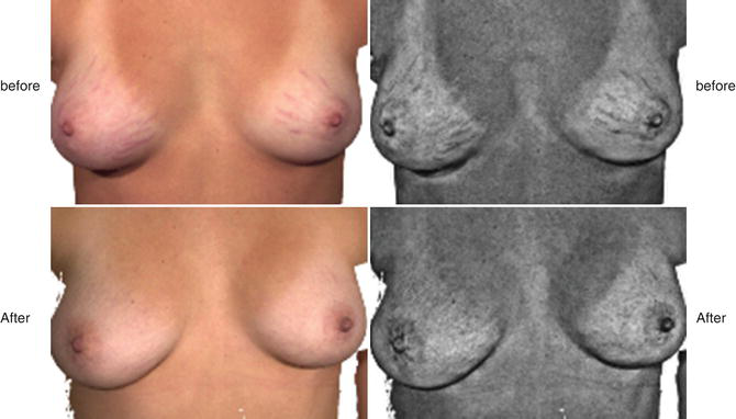
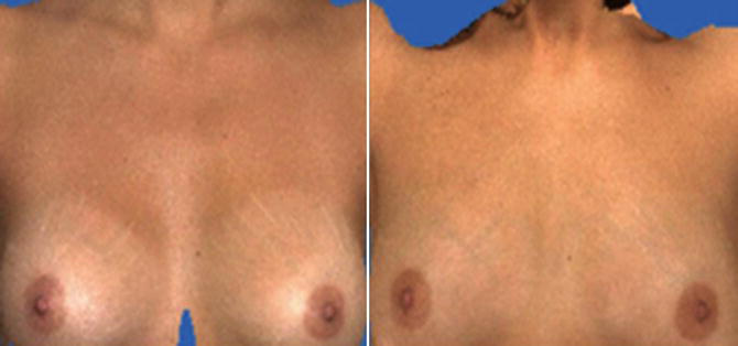
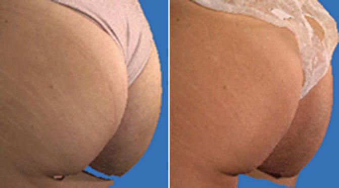
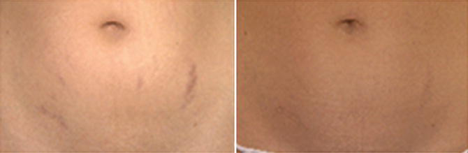
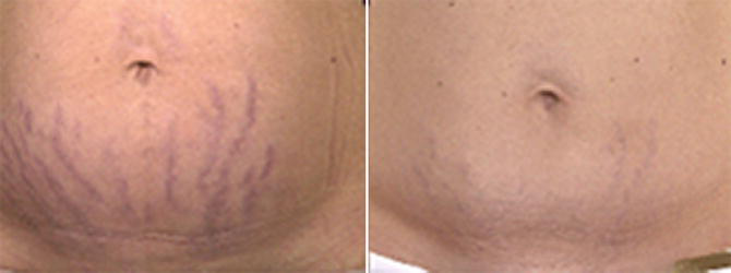
Best results were achieved for striae rubra, compared with striae alba, and for SD located on the breast. The second best anatomy region to have good results was the abdomen and then the thighs.
The degree of patient satisfaction was considered excellent in 10% and great in 90%.
Post-inflammatory hyperchromia was a transitory side effect. To avoid dyschromia, we advocate the use of low densities.
Conclusion
Striae distensae are a well-recognized, common dermatologic entity, which affect patients of all ages, genders, and ethnicities and rarely cause any significant medical problems but can have a deep psychological impact on affected patients. Many treatment modalities are available for treatment and prevention; however, striae distensae is still a challenge for dermatology. Laser treatment can be a good option when performed by experts. Fractional CO2 laser can bring very good results, but appropriate parameters must be adjusted according to the device. Low density is an important parameter to avoid side effects.
Take Home Messages
- Striae distensae commonly occur in pregnancy, puberty, and obesity.
- Proposed etiological mechanisms are hormones, physical stretch, and structural alterations to the tegument.
- Striae distensae does not have one standard treatment but many different treatment modalities.
- Treatments include topical agents, radiofrequency, percutaneous collagen induction, microneedling, IPL, and nonablative and ablative lasers.
- Expectations must be realistic, but the fractional CO2 laser has recently shown improvement of these aesthetically distressing lesions.
- Best results were achieved for striae rubra, compared with striae alba, and for SD located on the breast. The second best anatomy region to have better results was the abdomen.
- Parameters should be adjusted according to the device. Low density is advocated. Photoprotection and topical care are recommended to avoid post-inflammatory hyperchromia.
References
Abdel-Latif AM, Albendary AS. Treatment of striae distensae with microdermabrasion: a clinical and molecular study. JEWDS. 2008;5:24–30.
Ackerman Ab CN, Sanchez J, Guo Y, et al. Histologic diagnosis of inflammatory skin diseases: an algorithmic method based on pattern analysis. Baltimore: Williams & Wilkins; 1997. p. 734–6.
Aldahan AS, Shah VV, Mlacker S, Samarkandy S, Alsaidan M, Nouri K. Laser and light treatments for Striae Distensae: a comprehensive review of the literature. Am J Clin Dermatol. 2016;17(3):239–56.
Alexiades-Armenakas MR, Bernstein LJ, Friedman PM, et al. The safety and efficacy of the 308-nm excimer laser for pigment correction of hypopigmented scar and striae alba. Arch Dermatol. 2004;14:955–60.
Alexiades-Armenakas M, Sarnoff D, Gotkin R, Sadick N. Multi-center clinical study and review of fractional ablative CO2 laser resurfacing for the treatment of rhytides, photoaging, scars and striae. J Drugs Dermatol. 2011;10:352–62.
Al-Himdani S, Ud-Din S, Gilmore S, Bayat A. Striae distensae: a comprehensive review and evidence-based evaluation of prophylaxis and treatment. Br J Dermatol. 2014;170(3):527–47.
Alster TS. Laser treatment of hypertrophic scars, keloids and striae rubra. Dermatol Clin. 1997;15:419–29.
Alster TS, Lupton JR. Prevention and treatment of side effects and complications of cutaneous laser resurfacing. Plast Reconstr Surg. 2002;109:308–16.
Arem AJ, Kischer CW. Analysis of striae. Plast Reconstr Surg. 1980;65:22–9.
Atwal GS, Manku LK, Griffiths CE, et al. Striae gravidarum in primiparae. Br J Dermatol. 2006a;155:965–9.
Aust MC, Knobloch K, Vogt PM. Percutaneous collagen induction therapy as a novel therapeutic option for Striae distensae. Plast Reconstr Surg. 2010;126:219–20.
Belo V, Arceo-Cruz MC, Buse E, et al. The efficacy of fractional resurfacing using an erbium glass laser in the treatment of stretch marks on Asian skin. Lasers Surg Med. 2009;41:92.
Canpolat F, Akis HK, Cemil BC, et al. Investigation of risk factors associated with striae gravidarum in Turkish primipara pregnants. Turkderm Deri Hastaliklari ve Frengi Arsivi. 2010;44:28–31.
Chang ALS, Agredano YZ, Kimball AB. Risk factors associated with striae gravidarum. J Am Acad Dermatol. 2014;51:881–5.
Cho S, Park ES, Lee DH, et al. Clinical features and risk factors for striae distensae in Korean adolescents. J Eur Acad Dermatol Venereol. 2006;20:1108–13.
Cohen HA, Matalon A, Mezger A, et al. Striae in adolescents mistaken for physical abuse. J Fam Pract. 1997;45:84–5.
Cordeiro RC, Zecchin KG, de Moraes AM. Expression of estrogen, androgen, and glucocorticoid receptor in recent striae distensae. Int J Dermatol. 2010;49:30–2.
Davey CM. Factors associated with the occurrence of striae gravidarum. J Obstet Gynaecol British Commonwealth. 1972;79:1113–4.
Di Lernia V, Bonci A, Cattania M, et al. Striae distensia (rubrae) in monozygotic twins. Pediatr Dermatol. 2001;18:261–2.
Elton RF, Pinkus H. Striae in normal men. Arch Dermatol. 1966;94:33–4.
Ersoy E, et al. Is it possible to prevent Striae Gravidarum? J Chinese Med Assoc. 2016;79:272–5.
Etoh M, Jinnin M, Makiko K, et al. MicroRNA-7 down regulation mediates excessive collagen expression in localized scleroderma. Arch Dermatol Res. 2013;305:9–15.
Fisher GH, Geronemus RG. Short-term side effects of fractional photothermolysis. Dermatol Surg. 2005;31:1245–9. discussion 1249.
Freedman B. Striae reduction following laser lipolysis. Lasers Surg Med. 2010;42:96–7.
Friedman DW, Boyd CD, Mackenzie JW, et al. Regulation of Collagen gene expression in keloids and hypertrophic scars. J Surg Res. 1993;55:214–22.
García-Hidalgo L. Dermatological complications of obesity. Am J Clin Dermatol. 2002;3(7):497–506.
García-Hidalgo L, Orozco-Topete R, Gonzalez-Barranco J, et al. Dermatoses in 156 obese adults. Obes Res. 1999;7:299–302.
Geronemus RG. Fractional photothermolysis: current and future applications. Lasers Surg Med. 2006;38:169–76.
Ghasemi A, Gorouhi F, Rashighi-Firoozabadi M, et al. Striae gravidarum: associated factors. J Eur Acad Dermatol Venereol. 2017;21:743–6.
Goldberg DJ, Marmur ES, Hussain M. 308 nm excimer laser treatment of mature hypopigmented striae. Dermatol Surg. 2003;29:596–9.
Goldberg DJ, Marmur ES, Schmults C, et al. Histologic and ultrastructural analysis of ultraviolet B laser and light source treatment of leukoderma in striae distensae. Dermatol Surg. 2005;31:385–7.
Goldman A, Rossato F, Pratti C. Stretch marks: treatment using the 1,064 nm Nd:YAG laser. Dermatol Surg. 2008;34:1–7.
Gupta M. Medroxyprogesterone acetate injections. Development of striae. British J Family Plan. 2000;26:104–5.
Hernández-Pérez E, Colombo-Charrier E, Valencia-Ibiett E. Intense pulsed light in the treatment of striae distensae. Dermatol Surg. 2002;28:1124–30.
Jaramillo-Garcia CM, Lopera-Calderon MC, Zuluaga de Cadena A. Related factors with atrophic stretch marks in adolescent female students from two private educational establishments from the city of Medellin, 1997-1999. CES Med. 2009;23:s69–79.
Kartal Durmazlar SP, Eskioglu F. Striae gravidarum: associated factors in Turkish primiparae. J Turk Acad Dermatol. 2009;3:93401a.
Kelekci KH, Kelekci S, Destegul E, et al. Prematurity: is it a risk factor for striae distensae? Int J Dermatol. 2011;50:1240–5.
Klehr N. Striae cutis distensae: morphokinetic examinations in vitro. Acta Derm Venereol suppl (Stockh). 1979;59:105–8.
Lee SE, Kim JH, Lee SJ, et al. Treatment of striae distensae using an ablative 10,600-nm carbon dioxide fractional laser: a retrospective review of 27 participants. Dermatol Surg. 2010;36:1683–90.
Liu DT. Striae gravidarum. Lancet. 1974;1:625.
Lurie S, Matas Z, Fux A, et al. Association of serum relaxin with striae gravidarum in pregnant women. Arch Gynecol Obstet. 2011;283:219–22.
Maia M, Marcon CR, Rodrigues SB, et al. Striae distensae in pregnancy: risk factors in primiparous women. An Bras Dermatol. 2009;84:599–605.
Manuskiatti W, Boonthaweeyuwat E, Varothai S. Treatment of striae distensae with a tripolar radiofrequency device: a pilot study. J Dermatol Treat. 2009;20:359–64.
Mazzarello V, Farace F, Ena P, et al. A superficial texture analysis of 70% glycolic acid topical therapy and striae distensae. Plast Reconstr Surg. 2012;129:589–90.
McDaniel DH, Ash K, Zukowoski M. Treatment of stretch marks with the 585 nm flashlamp-pumped pulsed dye laser. Dermatol Surg. 1996;22:332–7.
McKusick VA. Transverse striae distensae in the lumbar areas in father and two sons. Birth Defects Orig Artic Ser. 1971;58:169–76.
Murphy KW, Dunphy B, O’Herlihy C. Increased maternal age protects against striae gravidarum. J Obstet Gynaecol. 1992;12:297–300.
Muzaffar F, Hussain I, Haroon TS. Physiologic skin changes during pregnancy: a study of 140 cases. Int J Dermatol. 1998;37:429–31.
Nigam PK. Striae cutis distensae. Int J Dermatol. 1989;28:426–7.
Nigam K, Mishra R, Ramesh V. 17-ketosteroid levels in striae cútis distensae. Indian J Dermatol Venereol Leprol. 1990;56:125–6.
Osman H, Rubeiz N, Tamim H, et al. Risk factors for the development of striae gravidarum. Am J Obstet Gynecol. 2017;196:62.e1–5.
Pinkus H, Keech MK, Mehregan AH. Histopathology of striae distensae, with special reference to striae and wound healing in the Marfan syndrome. J Investig Dermatol. 1966;46:283–92.
Rahman Z, Macfalls H, Jiang K, et al. Fractional deep dermal ablation induces tissue tightening. Lasers Surg Med. 2009;41:78–86.
Rolleston JD, Goodall EW. Two cases of striae atrophicae following typhoid fever. Proc R Soc Med. 1931;25:593–600.
Sadick NS, Magro C, Hoenig A. Prospective clinical and histological study to evaluate the efficacy and safety of a targeted high-intensity narrow band UVB/UVA1 therapy for striae alba. J Cosmet Laser Ther. 2007;9:79–83.
Shafir R, Gur E. Re: Striae distensae of augmented breasts after oral contraceptives. Ann Plast Surg. 1999;43:220.
Sheu HM, Yu HS, Chang CH. Mast cell degranulation and elastolysis in the early stage of striae distensae. J Cutan Pathol. 1991;18:410–6.
Sisson WR. Colored striae in adolescent children. J Pediatr. 1954;45:520–30.
Suh DH, Chang KY, Son HC, et al. Radiofrequency and 585-nm pulsed dye laser treatment of striae distensae: a report of 37 Asian patients. Dermatol Surg. 2007;33:29–34.
Thailand J-Orh R, Titapant V, Chuenwattana P, Tontisirin P. Prevalence and associated factors for striae gravidarum. J Med Assoc Thai. 2008;91:445–51.
Thomas RGR, Liston WA. Clinical association of striae gravidurum. J Obstet Gynecol. 2014;24:270–1.
Troisier E, Ménétrier P. Histologie des vergetures. Ann Gynecol. 1889;31:2016.
Tsuji T, Sawabe M. Hyperpigmentation in striae distensae after bleomycin treatment. J Am Acad Dermatol. 1993;28:503–5.
Ud-Din S, McGeorge D, Bayat A. Topical management of striae distensae (stretch marks): prevention and therapy of striae rubrae and albae. J Eur Acad Dermatol Venereol. 2016;30(2):211–22.
Wang F, Calderone K, Smith NR, Do TT, Helfrich YR, Johnson TR, Kang S, Voorhees JJ, Fisher GJ. Marked disruption and aberrant regulation of elastic fibres in early striae gravidarum. Br J Dermatol. 2015;173(6):1420–30.
Watson RE, Parry EJ, Humphries JD, Jones CJ, Polson DW, Kielty CM, et al. Fibrillin microfibrils are reduced in skin exhibiting striae distensae. Br J Dermatol. 1998;138(6):931–7.
Zelickson BD, Kist D, Bernstein E, et al. Histological and ultrastructural evaluation of the effects of radiofrequency-based nonablative dermal remodeling device: a pilot study. Arch Dermatol. 2004;140:204–9.







