
CO2 Laser for Scars
CO2 Laser for Scars
Thales Lage Bicalho Bretas, Aline Tanus, Marcia Linhares and Maria Claudia Almeida Issa Universidade Federal Fluminense, Rua Miguel de Frias, 77 Sala 1215, Icaraí, Niterói, RJ, Brazil Brazilian Society of Dermatology, Rua Visconde de Pirajá, 595/402, Ipanema, Rio de Janeiro, RJ, Brazil Department of Clinical Medicine – Dermatology, Fluminense Federal University, Rua Visconde de Pirajá, 595/402, Ipanema, Niterói, RJ, Brazil
Abstract
Scars result from the substitution of damaged skin by a new and abnormal tissue following an injury. Ablative devices, including the erbium and carbon dioxide lasers, have been shown to be effective in improving the appearance of scars, including mature burn scars. The CO2 laser promotes thermal fractionated ablation of the skin, and the resultant selective healing stimuli improve the altered tissue. The scars then become more homogeneous with the surrounding skin. The carbon dioxide laser can be combined with other interventions to achieve optimal results. It can also be used to allow drug penetration into the dermis homogeneously, a technique called drug delivery. The use of drugs such as corticoids to reduce hypertrophic scars and poly-L-lactic acid (PLLA) to increase collagenesis in atrophic scars has been reported. In post-procedure/post-treatment and burn scars, the early use of the CO2 laser is important to remodel the tissue before the maturation of the scar, therefore bringing better cosmetic results. In this chapter, we will expatiate on the multiple kinds of scars, the peculiarities of the ablative fractional carbon dioxide CO2 laser and the approach to different types of scars through the use of this technology. We will also discuss the precautions to be taken before the procedure (pre-treatment), the procedure itself, the post-procedure/post-treatment care, the most commonly seen side effects and how to deal with them.
Keywords CO2 LaserScarsAblativeFractionalCarbon dioxide
Introduction
The healing process involves the release of inflammatory mediators, cytokines, and cell chemotaxis, leading to reepithelialization and deposition of types III and I collagen within the dermis, respectively. Scars are a consequence of abnormal substitution of damaged tissues after trauma, with an imbalance of collagen types and their fiber thickness within the dermis, as well as histological disorganization. They represent unaesthetic marks capable of decreasing self-esteem and quality of life, therefore demanding the start of an efficient treatment as quickly as possible, in order to offer the patient satisfactory and aesthetic results. Treating scars has never been an easy task for dermatologists, and it usually involves a multitherapeutic approach, with a combination of drugs and technologies. There are unnumbered treatments described, such as ablative and nonablative lasers, corticosteroid injection, intralesional 5-fluorouracil or bleomycin, silicone patches, cryotherapy, chemical peels, dermabrasion, fillers, punch excisions, punch elevations, microneedling, subcision, and many others (Ozog et al. 2013; Wolfram et al. 2009; Reish and Eriksson 2008).
In the past decades, the appearance of fractional lasers (ablative or not) took scar management to another level, offering good results with minimally invasive procedures. The recent use of the carbon dioxide (CO2) ablative fractional laser (AFL) for the treatment of scars has delivered good results when correctly managed. This laser promotes thermal ablation of multiple small channels between nondamaged skin, called microthermal zones (MTZ), causing healing stimuli that result in neocollagenesis and remodeling of the altered tissue. The expression of some cellular markers of dermal wound healing and neocollagenesis, such as collagen III, heat shock protein 70, alpha-smooth muscle actin, and proliferating cell nuclear antigens has been reported in treated areas. Therefore, scars become shallower and narrower, and their surface tends to look more homogeneous with the surrounding skin (Manstein et al. 2004; Laubach et al. 2006; Walgrave et al. 2008; Alexiades-Armenakas et al. 2011; Rkein et al. 2014).
The CO2 laser itself, without being combined with other techniques, already offers visibly good improvement in the skin texture, shape, and aspect of the scar, as well as its similarity to the surrounding skin. But it has also been used concomitantly with the application of several medications; it produces small channels that allow the penetration of drugs into the dermis, homogeneously, a technique called drug delivery. In hypertrophic scars, for example, the CO2 laser itself does not represent a good option as monotherapy; it is often used to deliver drugs such as corticoids and bleomycin that act synergistically with the laser to reduce the hypertrophy and improve even more the aspect of the scar. This method demands special caution and knowledge from the operator, as the laser itself could cause new scars in a person who has some genetic tendency of developing hypertrophic scars, as well as overstimulate those scar fibroblasts and worsen the fibrosis already generated by an overreaction of that body to a previous damage stimulus. In atrophic and acne scars, the CO2 laser can also be used with the delivery of drugs like poly-L-lactic acid (PLLA) to increase collagenesis and fulfill the depressions of the scar tissue (Vrijman et al. 2011; Shamsaldeen et al. 2011; Fife et al. 2009; Avram et al. 2009).
Scars
Scars represent abnormal substitution of a damaged tissue after any trauma that reaches the dermis. Functionally, they are less malleable than normal tissue and they lack cutaneous annexes, such as hair follicles, sebaceous and sweat glands. They can be either hypertrophic or atrophic and are frequently generated by harms such as burns, surgery, tattoos, accidents, or some inflammatory diseases like acne. There is also a particular kind of hypertrophic scar, known as keloid, which is a result of a very exacerbated inflammatory response of the host to skin damage, and demands a different and more cautious approach.
Hypertrophic scars result from uncontrolled proliferation of fibroblasts and extracellular matrix. The reason why this abnormal proliferation occurs is primarily genetic, and afro-descendants are usually more prone to this alteration. Hypertrophic scars are higher than the surrounding skin, and they do not go beyond the originating scar borders. Typically, hypertrophic scars can be seen on the shoulders, superior trunk, and ears (Alster and Tanzi 2003; Verhaeghe et al. 2013; Kuo et al. 2004).
A keloid, by definition, goes over the boundaries of the original injury, has a higher recurrence index, and represents a challenging task to dermatologists. Besides its visual impairment, the keloid is usually painful, rigid, with thick fibrosis and low mobility. It does not regress spontaneously, and the affected patients have a high genetic predisposition (Reiken et al. 1997; Alster and Williams 1995; Sherling et al. 2010).
Atrophic scars result from inefficient collagen remodeling and abnormal regeneration of the fibrous tissue. They represent depressions on the skin surface and can originate from unaware care of a wound or also be influenced by genetic predisposition (Vrijman et al. 2011).
Acne scars originating from moderate to severe acne in teenagers and young adults represent frequent and important complaints in a dermatological consultation. Their etiology is related to an exaggerated inflammatory reaction that lasts too long, an inefficient immune response of the host to the tissue damage, and an anomalous healing capacity with low collagen synthesis (Jeremy et al. 2003; Taylor et al. 2011; Fabbrocini et al. 2010; Holland et al. 2004; Sobanko and Alster 2012). Acne scars are classified by the amount of collagen lost or gained by the tissue, by its thickness, its stretching capability, its depth, and its architecture.
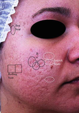
Atrophic scars are the most common kind of acne scars and occur because of the destruction of dermal structures by the deep inflammatory infiltrate. They can be divided into ice pick scars, rolling scars, and boxcar scars (Fig. 1). Ice pick scars are narrow, spotted, and deep scars, clinically shown as multiple small points of depression on the skin surface. They usually have less than 2 mm of width and grow vertically until the deep dermis or subcutaneous tissue, assuming a cone figure. Rolling scars are shallower and wider, with a width of 4–5 mm, with sloping edges, and they occur when the dermis becomes attached to the subcutaneous tissue, producing a superficial ripple. Boxcar scars are rounded or oval, with various widths, from shallow to deep, and a diameter between 1.5 and 4 mm (Levy and Zeichner 2012; Jacob et al. 2001; Lee et al. 2013).
Fractional CO2 Laser
Basic Principles
The CO2 laser is an ablative fractional laser (AFL), with a wavelength of 10,600 nm, and its target is water. The AFL produces microscopic columns of ablated tissue, which extend from the epidermis to the dermis (the so-called microthermal zones, MTZ), saving healthy tissue areas between these columns. Collagen degeneration and focal epidermal necrosis stimulate a process of fast reepithelialization promoted by the surrounding healthy skin cells. The AFL technology gave us a new perspective for facial resurfacing treatments, decreasing the side effects and complications of the standard ablative resurfacing so far feared, since it did not use to preserve any healthy skin area.
Other kinds of ablative fractional lasers are the Er:YAG (yttrium, aluminium, garnet) 2,940 nm, and the new Er:YSG (yttrium, sapphire, garnet) 2,790 nm. They only differ from each other in the wavelength and the water absorption coefficient, as the target is the same: the emanating energy is absorbed by the amount of water in the tissue (Manstein et al. 2004; Laubach et al. 2006; Walgrave et al. 2008; Alexiades-Armenakas et al. 2011; Alster and Nanni 1998).
The Er:YAG has the biggest water absorption coefficient, as it promotes immediate vaporization of cells without thermal damage. It is capable of absorbing 12–18 times more water than the CO2. The pulse duration of the Er:YAG is much smaller than that of the CO2 laser, resulting in less surrounding thermal diffusion. Nevertheless, the inability of the Er:YAG to cause thermal injury results in less collagen contraction, making it useless to promote perioperative hemostasis (Alster and Nanni 1998).
The CO2 laser device has a handpiece that projects small spots on the skin. Depending on the model used, it is possible to adjust the shot format (triangle, square, or round, for example), the spot density, and the diameter of each spot, which can range from 125 µm to 1.25 mm. It has the smallest water absorption coefficient, causing large collateral thermal damage and promoting excellent hemostasis when used in the standard, nonfractional mode (Rkein et al. 2014).
There are, basically, four or five parameters to be adjusted in the CO2 laser, bearing in mind the skin area to be treated, the depth, the diameter, and the ablation degree:
- Energy: the amount of power (in watts) delivered to the tissue in a given time (the laser pulse duration, in seconds). It is directly proportional to the depth of penetration and the thermal injury promoted, meaning that the higher the energy, the deeper the laser reaches and the more injury it promotes in the surrounding tissue.
- Fluence/Density: the amount of energy delivered to a certain area – the overall size of the application area or the “spot size” produced by the laser handpiece. Thus, the energy density or fluence is measured in J/cm2. The higher the fluence, the faster the temperature increases in the tissue and, consequently, the bigger the intensity of the desired effect. The effect of the treatment is achieved both by varying the laser output energy and the laser pulse duration at the tissue application area.
- Pulse duration: in milliseconds (ms), it is directly proportional to thermal injury of the tissue. The longer the pulse lasts, the bigger the damage made in the target tissue and surroundings.
- Distance between dots: determines the balance between the density of energy delivered and the preserved skin in the treated area. It is inversely proportional to density, which means that the smaller the distance between the dots, the more energy will be delivered into the target area, with denser thermal ablation. Higher densities mean dots overlapping, leaving a smaller healthy skin surface preserved and making the post-procedure period more uncomfortable.
Mechanism of Action of the Fractional CO2 Laser
Heat shock proteins (HSP) are upregulated and have their role in cutaneous remodeling after thermal injuries. They have anti-inflammatory and cell-protecting actions. Histologic studies have demonstrated enhanced levels of HSP, such as HSP70 and HSP47, that promote the process of collagen generation, leading to dermal thickening and improvement of scar appearance (Magnani and Schweiger 2014). HSP70 is a procollagen chaperone and plays a crucial role in wound healing, promoting neocollagenesis and the expression of other growth factors, like transforming growth factor β (TGF-β), essential to healing. HSP47 also plays a key role in promoting neocollagenesis. Its peak expression is at 1 month posttreatment, and it remains high at 3 and 6 months. Remodeling and new collagen formation were noted at 3 and 6 months postprocedure. The long-term expression of these two heat shock proteins supports the long-term efficacy of fractional CO2 laser resurfacing (Xu et al. 2011).
The CO2 laser also enhances the expression of tissue collagenases, such as matrix metalloproteinases (MMP), that act in collagen degradation. As MMPs are not normally expressed in the skin, the balance between collagen formation and degradation results in individual responses for each patient (Vrijman et al. 2011).
As already mentioned, the CO2 laser produces small dots in between preserved skin in the treated area, the MTZ, with vaporization of the corneal layer over these channels. An area of undamaged tissue surrounds each ablative zone. Spared, viable keratinocytes in these areas migrate to the MTZs and promote the healing process, with collagen formation and reepithelialization. Immediately after the laser pulse, hypochromic dotted macules may be seen on the skin surface, representing this corneal layer’s vaporization. These macules evolve to erythema, more visible within 3 days after the treatment. At that time, serosanguinous drainage and swelling occur in various degrees, according to the parameters used. Bleeding occurs between the stratum corneum and stratum granulosum. Regeneration and desquamation of the epithelium is observed, with preservation of the basal lamina. There is also disappearance of elastin in elastic fibers located in the superficial dermis of scar areas (Magnani and Schweiger 2014).
The healing process finishes with the formation of small brown crusts, corresponding to the extrusion of damaged keratinocytes. Necrotic debris are eliminated within 1–2 weeks (it can take even longer in certain patients), leaving the skin surface more “tanned” until its complete exfoliation (Rkein et al. 2014).
Three weeks after treatment, almost complete regeneration of the epithelium is observed. Microscopically, elastin can be seen in electron-dense deposits, and elastic fibers look fragmented and elaunin-like, which is indicative of the dermal remodeling process (Omi et al. 2011). A visible improvement of scars can be seen within 3 months, but it has already been described up to 12–18 months after treatment, with neocollagenesis and dermal remodeling evidenced by histological analyses.
The Use of Fractional CO2 Laser in Treating Different Kinds of Scars
Hypertrophic Scars
Although the CO2 laser has been used to treat hypertrophic scars, the pulsed dye laser (PDL) 585 nm has higher evidence of efficacy based on recent findings in the literature. The overexpression and abnormal activity of TGF-β1 and TGF-β2 are involved in the pathogenesis of hypertrophic scars and keloids. The mechanism of action of the PDL is not yet a consensus, but it is believed that it reduces the expression of TGF-β, fibroblast proliferation, and collagen III deposition in the scar. It is also described that, among the mechanisms of action of the PDL, are the selective photothermolysis of blood vessels, the release of histamine and interleukins by mast cells, and collagen degradation with consequent reestablishment of the dermis (Alster and Nanni 1998; Patel and Clement 2002; Reish and Eriksson 2008; Alster and Zaulyanov 2007).
Intralesional corticosteroid infiltration aiming to inhibit collagen synthesis immediately after a PDL session is technically easier due to tissue swelling, offering less resistance to needle penetration into the scar (Mustoe et al. 2002; Jalali and Bayat 2007; Roques and Téot 2008; Manuskiatti and Fitzpatrick 2002; Gupta and Sharma 2011; Chowdri et al. 1999).
The fractional CO2 laser is often used to improve thickness, stiffness, and abnormal texture of more mature scars by ablative destruction and resurfacing (Waibel et al. 2013). Treatment with the fractional CO2 ablative laser can be started from 12 months after injury or following the conclusion of PDL treatment, in case they are combined. Laser sessions are delivered with 4–6-week intervals until a plateau in improvement is observed. Generally, only one modality is used per session, but more than one platform can be used on different sites in the same session.
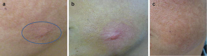
The microscopic thermal zones generated by the CO2 laser have also been used as channels to enhance the penetration of triamcinolone (Fig. 2a–c) and other topical drugs into the skin. This technique, called drug delivery, is less painful than drug injection and allows a more uniform distribution of the medication throughout the scar’s dermis, acting synergistically with the laser. The corneal stratum is normally impermeable to molecules weighing more than 500 Da; nevertheless, the channels created by the laser allow higher penetration and bioavailability of topical medications (Goodman 2006).
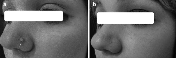
With all that information in mind, the CO2 laser is usually combined with the drug delivery technique to achieve better results in treating keloids and hypertrophic scars (Fig. 3a, b). Knowing that the laser itself can promote a strong healing response in people who are already genetically prone to scar formation, it is always extremely advised to start slowly at the first few laser sessions, with low potencies/fluencies and low pulse durations so as not to worsen scars by feeding their exacerbated inflammatory chain secondary to healing stimuli.
Atrophic Scars in General and Atrophic Acne Scars
Atrophic scars are better treated with AFL, like the CO2 laser, when compared to nonablative fractional lasers, because the energy reaches the deep dermis (1.5–1.6 mm of depth) and the remodeling process can last months. The AFL increases heat shock protein (HSP) expression, which in turn regulates the tissue response to thermal injury through activation of epidermal stem cells that will replace the just-damaged cells. IL-1, TNF-alfa, TGF, and MMP signalize the removal of the damaged cells and neocollagenesis.
The CO2 laser treatment reduces the scar’s width and depth and stimulates the synthesis and organization of collagen fibers, filling in atrophic areas. It is highly recommended to treat the whole aesthetic unit surface, and neither only the affected areas nor the scar itself, in order to avoid clear demarcation between treated and nontreated areas. As a resurfacing method, the CO2 laser promotes a higher collagenesis stimulus and a better aesthetic result. It improves the homogeneity of the skin, progressively making the atrophic areas seem flatter, shallower, more superficial, and reintegrating them to the surrounding normal skin.
We must never forget the possibility of the concomitant use of drugs that stimulate collagenesis and reverse the atrophy through the drug delivery system, with the homogeneous penetration of medications to improve the skin texture and regain its volume and vitality. In atrophic scars, poly-L-lactic acid (PLLA) is the substance currently used to improve collagenesis through the drug delivery technique, straight after the laser session. PLLA, when introduced, induces a local inflammatory response with activation and production of collagen, acting in a synergic way with the laser.
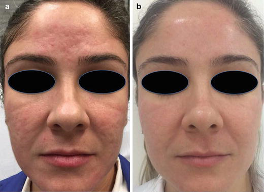
Acne scars are mostly atrophic, subdivided into different types as already mentioned (ice pick, boxcar, and rolling scars). The great majority of affected patients have multiple types simultaneously. With the resurfacing technique, there is good improvement in the general aspect of the treated aesthetic unit, but multiple laser sessions are usually needed to achieve impressive results. All subtypes of acne scars experience great amelioration, but boxcar scars are often more responsive. Patients shall be oriented about the need for subsequent sessions, from four to six, to obtain the best outcome (Fabbrocini et al. 2010; Magnani and Schweiger 2014; Omi et al. 2011) (Fig. 4a, b).
Postsurgical/Post-Traumatic Scars
Fast and early action on these scars is essential to improve their thickness and texture. Depigmentation is the hardest alteration to treat. Due to the greater depth they have in comparison to acne scars, postsurgical scars usually reach worse results. Fibroblasts and myofibroblasts start migration during the first week and begin to form scar tissue after the second week. Therefore, the correct approach shall be made readily within the first week after surgery.
Many studies use the Vancouver Scar Scale (VSS) for clinical evaluation of the scar, including pigmentation (0 = normal, 1 = hypopigmentation, 2 = hyperpigmentation), vascularity (0 = normal, 1 = pink, 2 = pink to red, 3 = red, 4 = red to purple, 5 = purple), pliability (0 = normal, 1 = supple, 2 = yielding, 3 = firm, 4 = banding, 5 = contracture), and height (0 = normal, 1 = <2 mm, 2 = 2–5 mm, 3 = >5 mm) (Chowdri et al. 1999).
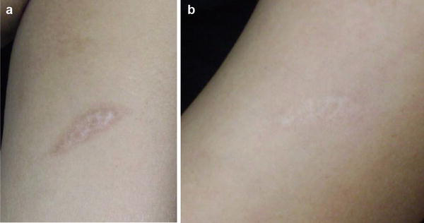
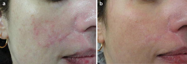
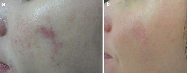
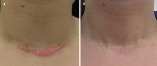
Apparently, the CO2 laser can be used to treat postsurgical and post-traumatic scars (Figs. 5a, b, 6a, b, 7a, b, and 8a, b), just as it can promote the formation of a new scar. The occurrence of hypertrophic scars secondary to treatment with the CO2 laser has been reported. Many suggested protocols for laser treatment are available, but the energy delivered and the area to be treated must be thoroughly chosen. The suggested densities to approach facial scars range from 30% to 50%, whereas in extrafacial areas they range from 20% to 30%, as the chances of hypertrophic scars are higher at those areas, especially on the neck, as previously mentioned. Moreover, lower energies also offer the best results and lower the risks of erythema and permanent depigmentation after the laser treatment (Roques and Téot 2008; Manuskiatti and Fitzpatrick 2002; Sobanko et al. 2015; Zachariae 1988).
Burn Scars
Burn scars have a complex formation mechanism that involves trauma responses, and their treatment must focus on aesthetic and functional improvements. Apart from all adequate surgical and curative care, it is common to see patients complaining about local pain, burning, or itching. In managing burn scars, we shall focus on addressing the aberrant collagen deposition typically shown in mature scars. Mature scars, like adult skin, have a predominance of type I collagen over type III, and this proportion is inverted in fetal skin.
The use of the fractional CO2 laser in burn scars has been proven to decrease collagen I and increase collagen III in the treated tissue, rearranging the scar tissue to reestablish the normal collagen deposition seen in young and nondamaged skin. This finding may be responsible for the related gain in mobility of the scar region after treatment.
Besides the improvement of the skin architecture, there is no way to regain the lost adnexal structures, like hair follicles, sebaceous and sweat glands. Therefore, the aesthetic improvement might be limited in cases of extensive skin areas affected (Ozog et al. 2013).
Preprocedure
The procedure itself depends on each manufacturer’s protocols. But there are some general precautions and instructions to be followed, especially before and after the procedure. Starting from a well-done anamnesis and a minimalist physical exam, dermatologists must pay special attention to the conditions mentioned below:
- Active infections: infections of any kind (fungal, bacterial, or viral) are relative contraindications to the laser and should be treated properly before the laser session.
- Inflammatory skin disorders: disorders on the area to be treated, like eczema or psoriasis, are not to be submitted to the procedure until their complete resolution. Patients with inflammatory skin diseases that have the Köbner phenomenon (lichen planus, psoriasis, perforating dermatosis, vitiligo, etc.), even outside the area to be treated, shall be well informed of this possibility.
- Active acne: patients with acne scars and active disease should be treated properly with topical or oral treatment prior to proceeding to scar management, in order to control the inflammatory lesions before the laser sessions. There is no point in treating acne scars when new ones are still appearing, since it would prolong the treatment and make it inefficient.
- Recent oral isotretinoin use: concerning the possibility of healing impairment in patients taking isotretinoin, as well as the increased risk of keloid formation, it is recommended to wait at least 6 months after the end of treatment to start the laser sessions. There is still a lack of studies evaluating this wound-healing impairment, but a consensual interval of 6 months has been established (Zachary and Rofagha 2012).
- Herpes simplex personal history: patients with a previous history of herpes simplex infection must undergo prophylaxis with antiviral agents. The duration is not yet a consensus, but oral aciclovir (400 mg every 8 h), valacyclovir (1 g once daily), or famciclovir (250 mg every 12 h) should be started at least 48 h prior to the procedure and kept until reepithelialization, usually 5 days following the laser session (Zachary and Rofagha 2012).
- Higher skin phototypes (Fitzpatrick IV to VI): darker skin tones have a higher risk of postinflammatory hyperpigmentation related to the procedure. Therefore, the procedure shall be discouraged, since the risks would exceed the benefits. If extremely necessary, lower fluencies and densities can be used with caution.
Procedure
Proceeding to the laser session, a topical anesthetic should be applied in order to gently cover the area to be treated. After its action time, the skin must be well cleaned and a small area might receive the first laser shot to test skin ablation and the patient’s tolerance to the procedure.
The laser session is to be based on the manufacturer’s orientations concerning the specific laser device and its personal parameters for the patient’s situation. Both the patient and the applicant must wear safety goggles. The skin then receives the laser shots, and the operator must avoid overlapping, since this practice can increase laser potency and cause harm to the skin. When a certain area is treated with multiple overlapping, there is higher thermal damage, less healthy skin is left behind, hence elevating the risk of scar formation.
Immediately after the laser application, the doctor has a chance to perform drug delivery, spreading some active ingredients over the ablated area depending on the patient’s indications, like triamcinolone or bleomycin for hypertrophic scars and PLLA for atrophic ones.
Post Procedure
Immediately after the laser session, the skin presents erythema and swelling due to tissue vaporization and an exuberant serous discharge. A refreshing and calming mask can be placed over the patient’s face, since one can be experiencing a burning sensation. Meanwhile, orientations for the post-procedure period are to be given to the patient, like hygiene routines and the application of topical healing creams on the treated area twice a day until complete reepithelialization. The doctor must be sure that patients know about the procedure’s downtime, recovery, precautions that need to be taken at home, and how to manage expectations. It is very important to tell patients that, during the first few days, the skin is going to look worse than before treatment, followed by the appearance of crusts that get better in 7–10 days, when the healing and reepithelialization process is going to be completed.
The results are really promising and tend to improve after subsequent sessions, with 2–6 weeks of interval between them. Higher potencies can be progressively used depending on the patient’s tolerance and response to the treatment.
Side Effects
Aside from the fact that the fractional CO2 laser offers many fewer side effects than the standard ablative CO2 laser, they can still occur. Some precautions are to be taken in order to avoid them, such as using lower energies and not overlapping more than twice at the same place. Side effects occur more often in patients submitted to either higher energies or densities (Vrijman et al. 2011; Shamsaldeen et al. 2011; Fife et al. 2009; Avram et al. 2009; Alster and Tanzi 2003).
Secondary infection is the most frequently reported side effect, and herpes simplex prophylaxis is usually needed to avoid it. Erythema, swelling, and pain can be signs of infection, and patients have to be well oriented to readily contact their doctor in case of any of these changes.
Acneiform eruptions can either be a response to thermal injury or a reaction to the greasy healing creams prescribed after the procedure, and the latter must be replaced by more fluid options. The introduction of some mild topical medicines for acne also optimizes the resolution of acneiform eruptions.
Occlusive dressings and antibiotics can cause contact dermatitis, better treated with topical corticosteroids. Late complications occur weeks after the procedure and include hyperpigmentation, persistent erythema, a spotted appearance of the skin, ectropion, hypertrophic scars, and hypopigmentation.
Hyperpigmentation and hypertrophic scars are rare, becoming more usual in extrafacial areas (particularly the neck) and when there have been multiple overlaps or excessively aggressive parameters used.
Hypopigmentation is even rarer. In cases of dyschromia, topical agents like hydroquinone, retinoic acid, thioglycolic acid, and others can be used. Lasers and light devices can also play an important role in cases of persistent erythema (intense pulsed light, ruby, alexandrite) and hyperchromia (intense pulsed light, Q-switched Nd:YAG).
Conclusions
The CO2 laser offers a new and safe treatment for unaesthetic scars, with low incidence of side effects, good tolerance, and high efficacy, making it a promising technique for this common complaint in dermatology offices. When well indicated, the results are truly satisfying. Nevertheless, there are some precautions to be taken before application of the laser: evaluate skin type and the patient’s expectations, antiviral prophylaxis when needed, and good orientation of patients. They need to know about the downtime of this ablative laser, the possible side effects, how to deal with recovery, and how to make it easier and faster, keeping the good results.
Concerning the laser device, each one carries its manufacturer’s manual, as well as the correct parameters taking the patient’s indication and skin characteristics into account. However, concepts of fluency, potency, energy, spot size, and pulse duration are values that allow us to understand it as a patterned device: the higher the parameters, the stronger and deeper the laser will reach the skin.
Take Home Messages
- Scars are a consequence of abnormal substitution of damaged tissues after trauma.
- The recent use of the carbon dioxide (CO2) ablative fractional laser (AFXL) has delivered good results when correctly managed.
- Treating scars is never an easy task for dermatologists, and involves a multitherapeutic approach, with a combination of drugs and technologies.
- The CO2 laser creates microthermal zones (MTZ) that stimulate a healing process involving heat shock proteins, metalloproteinases, cytokines, and chemotaxis, with neocollagenesis and reorganization of the scar tissue and its surroundings.
- The CO2 laser can be used to treat scars as monotherapy or combined with other techniques, such as pulsed dye laser, corticosteroid/bleomycin injections, topical drugs, and others to achieve better results, especially in hypertrophic scars.
- The CO2 laser can also be used with the concomitant application of topical drugs that will penetrate into the MTZ and reach the dermis, acting synergistically in a technique called drug delivery.
- In hypertrophic scars and keloids, the use of the CO2 laser must be even more careful, since it may stimulate the overreacting healing inflammatory reaction found in genetically predisposed people.
- In acne scars, there is usually a concomitant presence of multiple kinds of atrophic scars, with general improvement after the CO2 laser session. Patients often need more than one session, usually three to six, with 1–3 months of interval. Ice pick scars are the worst responders.
- In postsurgical and burn scars, early use is the key for success.
- Patients must be very well oriented about the procedure, its downtime, its pre- and post-procedure precautions, as well as the possible side effects and signs of infection/complications.
- If the patient has a personal history of herpes simplex infection, he/she must undergo prophylaxis schemes before the laser application.
- The presence of infections or active inflammatory diseases in the skin area to be treated must be completely solved before the laser session, as well as inflammatory skin diseases with the Köbner phenomenon shall lead to discouraging the procedure.
- Before the laser application, topical anesthesia with lidocaine, tetracaine, and others must be applied onto the skin area to be treated and then removed with correct asepsis of the skin.
- The laser session should be based on the manufacturer’s protocol, considering the patient’s Fitzpatrick skin type, the indication, and the parameters that match all this information.
- The post-procedure period involves the use of restorative creams and general precautions like sun protection. If the patient is currently under herpes simplex prophylaxis, it must be continued until complete skin reepithelialization, around 5–10 days after the laser session.
- The most common side effects are infections, dyschromia, persistent erythema, hypertrophic scarring, and hypopigmentation, the latter being rare. The doctor must feel capable of readily handling these intercurrences.
Cross-References
- CO2 Laser for Photorejuvenation
- Erbium Laser for Scars and Striae Distensae
- Light-Emitting Diode for Acne, Scars, and Photodamaged Skin
- Non-ablative Fractional Lasers for Scars
References
Alexiades-Armenakas M, Sarnoff D, Gotkin R, Sadick N. Multi-center clinical study and review of fractional ablative CO2 laser resurfacing for the treatment of rhytides, photoaging, scars and striae. J Drugs Dermatol. 2011;10(4):352–62.
Alster TS, Nanni CA. Pulsed dye laser treatment of hypertrophic burn scars. Plast Reconstr Surg. 1998;102(6):2190–5.
Alster TS, Tanzi EL. Hypertrophic scars and keloids: etiology and management. Am J Clin Dermatol. 2003;4(4):235–43.
Alster TS, Williams CM. Treatment of keloid sternotomy scars with 585 nm flashlamp-pumped pulsed-dye laser. Lancet. 1995;345(8959):1198–200.
Alster T, Zaulyanov L. Laser scar revision: a review. Dermatol Surg. 2007;33(2):131–40.
Avram MM, Tope WD, Yu T, Szachowicz E, Nelson JS. Hypertrophic scarring of the neck following ablative fractional carbon dioxide laser resurfacing. Lasers Surg Med. 2009;41(3):185–8.
Chowdri NA, Masarat M, Mattoo A, Darzi MA. Keloids and hypertrophic scars: results with intraoperative and serial postoperative corticosteroid injection therapy. Aust N Z J Surg. 1999;69(9):655–9.
Fabbrocini G, Annunziata MC, D’Arco V, De Vita V, Lodi G, Mauriello MC, Pastore F, Monfrecola G. Acne scars: pathogenesis, classification and treatment. Dermatol Res Pract. 2010;2010:893080.
Fife DJ, Fitzpatrick RE, Zachary CB. Complications of fractional CO2 laser resurfacing: four cases. Lasers Surg Med. 2009;41(3):179–84.
Goodman G. Acne and acne scarring: the case for active and early intervention. Aust Fam Physician. 2006;35(7):503–4.
Gupta S, Sharma VK. Standard guidelines of care: keloids and hypertrophic scars. Indian J Dermatol Venereol Leprol. 2011;77(1):94–100.
Holland DB, Jeremy AH, Roberts SG, Seukeran DC, Layton AM, Cunliffe WJ. Inflammation in acne scarring: a comparison of the responses in lesions from patients prone and not prone to scar. Br J Dermatol. 2004;150(1):72–81.
Jacob CI, Dover JS, Kaminer MS. Acne scarring: a classification system and review of treatment options. J Am Acad Dermatol. 2001;45(1):109–17.
Jalali M, Bayat A. Current use of steroids in management of abnormal raised skin scars. Surgeon. 2007;5(3):175–80.
Jeremy AH, Holland DB, Roberts SG, Thomson KF, Cunliffe WJ. Inflammatory events are involved in acne lesion initiation. J Invest Dermatol. 2003;121(1):20–7.
Kuo YR, Jeng SF, Wang FS, Chen TH, Huang HC, Chang PR, Yang KD. Flashlamp pulsed dye laser (PDL) suppression of keloid proliferation through down-regulation of TGF-beta1 expression and extracellular matrix expression. Lasers Surg Med. 2004;34(2):104–8.
Laubach HJ, Tannous Z, Anderson RR, Manstein D. Skin responses to fractional photothermolysis. Lasers Surg Med. 2006;38(2):142–9.
Lee SH, Zheng Z, Roh MR. Early postoperative treatment of surgical scars using a fractional carbon dioxide laser: a split-scar, evaluator-blinded study. Dermatol Surg. 2013;39(8):1190–6.
Levy LL, Zeichner JA. Management of acne scarring part II – a comparative review of non-laser based, minimally invasive approaches. Am J Clin Dermatol. 2012;13(5):331–40.
Magnani LR, Schweiger ES. Fractional CO2 lasers for the treatment of atrophic acne scars: a review of the literature. J Cosmet Laser Ther. 2014;16(2):48–56.
Manstein D, Herron GS, Sink RK, Tanner H, Anderson RR. Fractional photothermolysis: a new concept for cutaneous remodeling using microscopic patterns of thermal injury. Lasers Surg Med. 2004;34(5):426–38.
Manuskiatti W, Fitzpatrick RE. Treatment response of keloidal and hypertrophic sternotomy scars: comparison among intralesional corticosteroid, 5-fluorouracil, and 585 nm flashlamp-pumped pulsed dye laser treatments. Arch Dermatol. 2002;138(9):1149–55.
Mustoe TA, Cooter RD, Gold MH, Hobbs FD, Ramelet AA, Shakespeare PG, Stella M, Téot L, Wood FM, Ziegler UE, International Advisory Panel on Scar Management. International clinical recommendations on scar management. Plast Reconstr Surg. 2002;110(2):560–71.
Omi T, Kawana S, Sato S, Bonan P, Naito Z. Fractional CO2 laser for the treatment of acne scars. J Cosmet Dermatol. 2011;10:294–300.
Ozog DM, Liu A, Chaffins ML, Ormsby AH, Fincher EF, Chipps LK, Mi QS, Grossman PH, Pui JC, Moy RL. Evaluation of clinical results, histological architecture, and collagen expression following treatment of mature burn scars with a fractional carbon dioxide laser. JAMA Dermatol. 2013;149(1):50–7.
Patel N, Clement M. Selective nonablative treatment of acne scarring with 585 nm flashlamp pulsed dye laser. Dermatol Surg. 2002;28(10):942–5.
Reiken SR, Wolfort SF, Berthiaume F, Compton C, Tompkins RG, Yarmush ML. Control of hypertrophic scar growth using selective photothermolysis. Lasers Surg Med. 1997;21(1):7–12.
Reish RG, Eriksson E. Scars: a review of emerging and currently available therapies. Plast Reconstr Surg. 2008;122(4):1068–78.
Rkein A, Ozog D, Waibel JS. Treatment of atrophic scars with fractionated CO2 laser facilitating delivery of topically applied poly-L-lactic acid. Dermatol Surg. 2014;40(6):624–31.
Roques C, Téot L. The use of corticosteroids to treat keloids: a review. Int J Low Extrem Wounds. 2008;7(3):137–45.
Shamsaldeen O, Peterson JD, Goldman MP. The adverse events of deep fractional CO(2): a retrospective study of 490 treatments in 374 patients. Lasers Surg Med. 2011;43(6):453–6.
Sherling M, Friedman PM, Adrian R, Burns AJ, Conn H, Fitzpatrick R, Gregory R, Kilmer S, Lask G, Narurkar V, Katz TM, Avram M. Consensus recommendations on the use of an erbium-doped 1,550-nm fractionated laser and its applications in dermatologic laser surgery. Dermatol Surg. 2010;36(4):461–9.
Sobanko JF, Alster TS. Management of acne scarring, part I – a comparative review of laser surgical approaches. Am J Clin Dermatol. 2012;13(5):319–30.
Sobanko JF, Vachiramon V, Rattanaumpawan P, Miller CJ. Early postoperative single treatment ablative fractional lasing of Mohs Micrographic Surgery facial scars: a split-scar, evaluator-blinded study. Lasers Surg Med. 2015 Jan;47(1):1–5.
Taylor M, Gonzalez M, Porter R. Pathways to inflammation: acne pathophysiology. Eur J Dermatol. 2011;21(3):323–33.
Verhaeghe E, Ongenae K, Bostoen J, Lambert J. Nonablative fractional laser resurfacing for the treatment of hypertrophic scars: a randomized controlled trial. Dermatol Surg. 2013;39(3 Pt 1):426–34.
Vrijman C, van Drooge AM, Limpens J, Bos JD, van der Veen JP, Spuls PI, Wolkerstorfer A. Laser and intense pulsed light therapy for the treatment of hypertrophic scars: a systematic review. Br J Dermatol. 2011;165(5):934–42.
Waibel JS, Wulkan AJ, Shumaker PR. Treatment of hypertrophic scars using laser and laser assisted corticosteroid delivery. Lasers Surg Med. 2013;45(3):135–40.
Walgrave S, Zelickson B, Childs J, Altshuler G, Erofeev A, Yaroslavsky I, Kist D, Counters J. Pilot investigation of the correlation between histological and clinical effects of infrared fractional resurfacing lasers. Dermatol Surg. 2008;34(11):1443–53.
Wolfram D, Tzankov A, Pülzl P, Piza-Katzer H. Hypertrophic scars and keloids – a review of their pathophysiology, risk factors, and therapeutic management. Dermatol Surg. 2009;35(2):171–81.
Xu XG, Luo YJ, Wu Y, Chen JZ, Xu TH, Gao XH, et al. Immunohistological evaluation of skin responses after treatment using a fractional ultrapulse carbon dioxide laser on back skin. Dermatol Surg. 2011;37:1141–9.
Zachariae H. Delayed wound healing and keloid formation following argon laser treatment or dermabrasion during isotretinoin treatment. Br J Dermatol. 1988;118:703.
Zachary C, Rofagha R. Laser therapy. In: Bologna J, Jorizzo J, Schaffer J, editors. Dermatology. 3rd ed. London: Elsevier; 2012. p. 2260–72.







