
CO₂ Laser for Other Indications
CO₂ Laser for Other Indications
Emmanuel Rodrigues de França, Alzinira S. Herênio Neta and Gustavo S. M. de Carvalho Department of Dermatology, University of Pernambuco – UPE, Recife, PE, Brazil Department of Dermatology, University of São Paulo, São Paulo, SP, Brazil
Abstract
The CO2 laser is an ablative laser and has a strong affinity for water. It has been widely used with the objective to promote the rejuvenation or improve the appearance of scars by stimulating new collagen. As well, its increasingly widespread use has allowed the safe and effective treatment of various dermatoses, from benign epithelial tumors to melanocytic lesions to premalignant lesions (actinic cheilitis). Patient education on pre- and post-laser care is essential to maintaining good result. We discuss below some CO2 laser indications in routine dermatology.
Keywords CO2 lasersLaser therapyLasersGasAblative laser
Introduction
Dermatologists and plastics surgeons often encounter in their clinical practice injuries initially considered as small, which frequently have a difficult resolution. Pathologies as syringoma, sebaceous hyperplasia, verrucous epidermal nevus, acrochordon, and viral warts can be solved by different methods, but the laser, particularly the CO2, can be very useful, with surprising results. We will cover the most common dermatologic conditions that can be benefited by this method.
Dermatosis Papulosa Nigra
Dermatosis papulosa nigra (DPN) is a common condition in the black population, particularly in women, with prevalence of 35% in the African-American population (Kundu and Patterson 2013). It begins in adolescence, women being the most affected. The number and size of the lesions increase with age. Clinically, it is represented by multiple hyperchromic, asymptomatic papules, typically affecting the head and neck. It histologically resembles the seborrheic keratosis, showing hyperkeratosis, irregular acanthosis, horn cysts, and marked hyperpigmentation of the basal layer. This is a benign lesion of genetically determined character – positive family history in around 40–54% of cases (Hairston et al. 1964).
No treatment is usually indicated for DPN. The condition, though indolent, can sometimes be symptomatic or aesthetically undesirable. In such cases, treatment options include shaving, curettage, cryotherapy, electrodessication, microdermabrasion, and laser. More aggressive approaches can be complicated by postoperative hyperpigmentation, hypopigmentation, or scars. Keloid formation is a potential complication (Fig. 1).
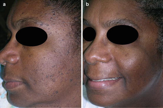
Lasers have been cited in the literature as useful therapeutic modalities for DPN, including CO2 (Bruscino et al. 2014), Nd:YAG, diode, pulsed dye, and erbium lasers. Among them, the CO2 laser has been shown to be safe, with low rates of recurrence or complications (scarring, hypo-/hyperpigmentation), and also a high degree of satisfaction by patients, even at the highest phototypes (Ali et al. 2016). Topical anesthesia is sufficient in most cases, and white petrolatum ointment once a day is indicated until reepithelialization of the lesions. There must be an interval of 3–4 months between the sessions.
Xanthelasma
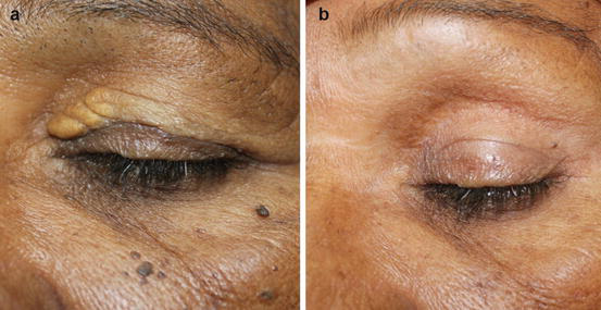
This is a benign disorder characterized by yellowish plaques typically located in the periorbital region, especially in the inner corner of the eyes and upper eyelids; it is also the most common form of skin xanthoma. The lesions have a tendency to progress and coalesce, with permanent character.
They are due to accumulation of fat within the histiocytes, known as foamy histiocytes, located mainly in the upper reticular dermis. The main component is accumulated cholesterol, which for the most part is esterified. In 50% of patients, normal serum levels of cholesterol are found. The main association is with hypertriglyceridemia, found in 50% of cases. A reduced HDL level can be found in some patients. In such cases, it may be considered a predictor of cardiovascular risk, severe ischemic heart disease, and atherosclerosis, especially if combined with hypertension, diabetes, obesity, and smoking. It is a rare disease in the general population and has a slight predominance in females. It has its peak incidence between the fourth and fifth decade of life.
The diagnosis is clinical, but it should be remembered that about half of patients have abnormal lipid levels; therefore, they should have their values measured frequently. Some drugs such as nilotinib, used to treat chronic myelogenous leukemia, may lead to the development of xanthelasma (Sayin et al. 2016).
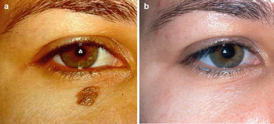
Treatment is based primarily on dietary restriction and lipid-lowering drugs if necessary. The aesthetics of xanthelasma is not addressed by isolated treatment of dyslipidemia. Numerous therapeutic options for the aesthetic treatment of xanthelasma are available, such as surgical removal, electrosurgery, chemical cauterization with trichloroacetic acid, and cryosurgery. Pingyangmycin, of the bleomycin family of antibiotics, can be injected into the lesions with good results (Wang et al. 2016). Electrocautery and cryosurgery can destroy superficial lesions, but require repeated treatments. Cryosurgery may cause scarring and hypopigmentation and should be discouraged. The use of ablative lasers such as ultrapulsed CO2, erbium:YAG (Güngör et al. 2014), Q-switched Nd:YAG, diode, pulsed dye laser, and KTP laser has become popular in the treatment of these lesions. CO2 ablative lasers are excellent options for localized xanthelasmas without involvement of muscles (Mourad et al. 2015; Pathania et al. 2015) (Fig. 2).
Surgical removal is best indicated for cases of diffuse xanthelasma with deep involvement of the dermis and/or muscle. Recurrence is common, with a rate of about 40%.
Sebaceous Hyperplasia
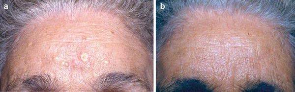
It is a common benign condition of the sebaceous glands of middle-aged adults or older. Lesions may be single or multiple and manifest themselves as small yellowish or skin-colored papules of 2–9 mm, normally with a central umbilication, located on the face (particularly the nose, cheek, and forehead). Occasionally they are seen on the breast, areola, mouth, scrotum, prepuce, and vulva. Rarely reported variants include a giant form, a linear or zosteriform arrangement, a diffuse form, and a familial form. Some consider rhinophyma a special form of sebaceous hyperplasia. Its frequency is about 1% in healthy elderly adults, but it can be as high as 10–16% in patients receiving long-term immunosuppression with cyclosporin A. Around 43.7% of neonates may present sebaceous hyperplasia. It has been reported in association with internal malignancy in Muir-Torre syndrome. It must be distinguished from basal cell carcinoma (BCC) – some papules have telangiectasia – and from molluscum contagiosum.
Histopathology shows a multilobulated sebaceous gland increased in size. The lobes have one or more layers of basal cells at their periphery, with undifferentiated sebocytes containing large nuclei and scant cytoplasmic lipid, in contrast to normal sebocytes, which are filled with lipids. The decrease in levels of circulating androgens, associated with aging, appears to be the cause of sebaceous hyperplasia. Ultraviolet radiation and immunosuppression have been postulated as cofactors (Fig. 3).
Treatment
Therapeutic options include photodynamic therapy, cryotherapy, cauterization or electrocoagulation, chemical topical treatment with trichloroacetic acid (TCA), and treatment with argon laser, carbon dioxide laser, and 1,450 nm and 1,720 nm diodes (No et al. 2004; Aghassi et al. 2000; Winstanley et al. 2012; Simmons et al. 2015a). The complications of these destructive, nonspecific therapies include depigmentation and atrophic scarring. Oral isotretinoin has been shown to be effective in removing some lesions after 2–6 weeks of treatment, but recurrence of the lesions is common after cessation of therapy.
Viral Wart
It is a frequent viral skin infection, with a limited course, caused by the human papillomavirus (HPV), able to produce epidermal proliferation characterized by acanthosis accompanied by papillomatosis, and it can be found in up to 10% of young adults and children. It is caused by papillomaviruses of the papovavirus group, double-stranded DNA viruses capable of eliciting a cytolytic effect on the infected cells, causing their death.
Clinically, it is characterized by exophytic papules or nodules with a roughened surface, sometimes with small darkened spots, which represent thrombosed capillaries. They are commonly located on the back of the hands and fingers, in the nail bed or periungual region, and on the knee folds. About 65% of common warts disappear spontaneously within 2 years. New warts may develop at sites of trauma, constituting the isomorphic Koebner phenomenon, which is usually less pronounced than for flat warts.
Infection is acquired by direct contact with patients with clinical and subclinical lesions, or through objects or contaminated surfaces (pools, gyms). It is believed that each new injury results from autoinoculation. Minor trauma predisposes to infection. Nail biting is associated with periungual warts. Trauma while shaving can spread the filiform warts of the beard area. Hyperhidrosis and flatfoot predispose to plantar warts. The average incubation period is 3 months, but it can range between 1 and 20 months. The papillomas caused by HPV are initially benign.
The incidence of warts, their malignant potential, and regression appear to be directly related to immune disorders mediated by host cells. Warts occur more frequently, last longer, and appear in large numbers in patients with AIDS and lymphomas and in those who take immunosuppressant drugs.
Treatment
Lesions in patients with a cell-mediated immunity deficit are generally resistant to treatment. Moreover, treatment of one lesion can lead to regression of many or all warts in immunocompetent individuals (Fig. 4).
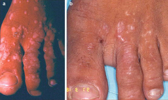
The objective is to destroy the infected cells using substances such as fuming nitric acid, salicylic acid, lactic acid, TCA, cantharidin, podophyllin, 5-fluorouracil, or intralesional bleomycin. Cryosurgery, photodynamic therapy, and even surgical procedures such as curettage and electrodessication may be used. Surgery with suture and radiotherapy are contraindicated. Among the lasers, the CO2 laser has been described, as well as hyperthermia by Nd:YAG laser (Oni and Mahaffey 2011). HPV is more vulnerable to hyperthermia than to cryotherapy. In resistant warts, the flashlamp-pumped pulsed dye laser (585 nm) has been used with 80% efficiency.
Recurrence of viral lesions (condylomas and warts) after treatment with the CO2 laser has not been more frequent than with isolated techniques. One study showed no recurrence of lesions in 12 months of follow-up after removal of warts with the CO2 laser and imiquimod 5% cream after epithelialization, applied once a day, five times a week for 2 weeks (Zeng et al. 2014).
Plantar Wart
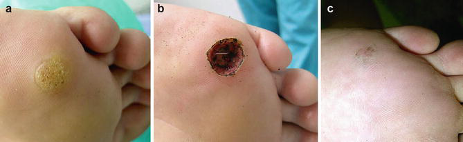
Plantar warts are notoriously more difficult to treat and eradicate. The use of artificial dermis (as a dressing) after CO2 laser ablation, and the use of salicylic acid on residual lesions, appear to be effective in these situations. Mitsuishi demonstrated the absence of HPV DNA in the upper epidermis of the treated sites after this technique, and the absence of significant scarring or severe pain (Mitsuishi et al. 2010) (Fig. 5).
Genital Warts
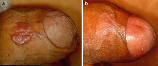
The use of the CO2 laser in genital warts is safe and effective (Padilla-Ailhaud 2006). The cure rate in a single session reaches 70%. Relapses are associated with multiple partners and involvement of the cervix in women. Combination therapy with the CO2 laser and photodynamic therapy with ALA (5-aminolevulinic acid) exhibits a lower recurrence rate than isolated CO2 laser therapy for refractory lesions (Huang et al. 2014). The treatment can be performed during pregnancy (Savoca et al. 2001). In a study of 18 pregnant women treated between 15 and 38 weeks of gestation, there were no abortions, premature births, or complications (infection, bleeding) from the procedure (Gay et al. 2003). Bowenoid papulosis corresponds to grade III intraepithelial neoplasia of the penis or vulva and is strongly associated with HPV 16 (Fig. 6).
Considerations
During use of the CO2 laser, the emitted smoke consists of gases and/or toxic vapors such as benzene, formaldehyde, and hydrogen cyanide, bioaerosols, steam, and live or dead cell remnants (including blood debris and viruses). The use of smoke-filter vacuum units, venting of smoke outdoors, gloves, and laser masks is advisable. The hose can be held by a helper 2 cm from the operative field or be coupled to the handpiece. Several studies have shown that the smoke resulting from vaporization of viral lesions by the CO2 laser is an aerosol containing viral particles that disperse over a diameter greater than 2 m, even under vacuum, contaminating the equipment and the people involved (skin and nasal/breast exposure) during surgery. For this reason, the CO2 laser is not a first-choice treatment for viral lesions such as common warts and genital warts.
Studies analyzing the smoke resulting from vaporization of human viral warts with the Er:YAG laser did not detect the presence of viral DNA; this laser is apparently safer than the CO2 laser. However, the case was described of a doctor who used the Nd:YAG laser to treat perianal warts and developed laryngeal papillomatosis. Viral particles of HIV and hepatitis C virus, in addition to HPV, were also found in the smoke caused by CO2 laser vaporization; therefore, treatment of patients with these infections by this method is not recommended (Hallmo and Naess 1991).
Melanocytic Nevus
Melanocytic nevus (MN) is a benign lesion of nevus cells that arises as a result of the proliferation of melanocytes. There are two fundamental types: congenital melanocytic nevus and acquired melanocytic nevus.
Congenital melanocytic nevi are present from birth. They usually present as small blemishes or brown papules, smooth or warty and sometimes hairy; even larger lesions can occupy entire limbs. When they are larger than 20 cm, they are called giant melanocytic nevi (0.002% of newborns). Common and giant MN often have a hairy surface (95%) and a roughened surface with color ranging from brown to black. Neurological disorders may be associated with giant MN according to the most affected area, such as spina bifida and meningocele, due to infiltration of melanocytes into the nerve structures, constituting neurocutaneous melanosis. Surgical excision is recommended because of the high risk of malignant transformation, but it is often a difficult treatment to carry out due to the extent of the lesion, making it necessary to resort to expanders, patchwork rotation, and placement of grafts. Other therapeutic options currently available for congenital MN include dermabrasion, chemical peels, and laser ablation. These methods improve the aesthetic appearance, but they are not effective at completely removing the deep nevus cells, since they are surface treatments. Only complete excision of the nevus with clear deep surgical margins can effectively reduce or eliminate the potential for malignant transformation in the future.
Acquired MN are common and usually occur between 12 and 30 years of age, although they can appear in childhood. They tend to slowly decline after the age of 35 and may increase in size during puberty, pregnancy, corticosteroid use, and sun exposure. Clinically they may present as flat, warty, domed, or pedunculated lesions. Histologically they can be junctional, compound, or intradermal. Usually no treatment is necessary. In cases of removal for various cosmetic reasons, methods such as surgical excision, cryosurgery, electrodissection, and more recently laser have been used. The nevus lesion should be excised with a margin of 1–2 mm and subjected to histopathological study when malignancy is suspected.
There are few data in the literature supporting the use of laser in melanocytic lesions. Nonablative methods produce selective photothermolysis of melanin pigment, with secondary destruction of the nevus cells, as performed with the Q-switched ruby, Nd:YAG, and alexandrite lasers. These produce a surface whitening effect; despite cosmetic improvement, there are recurrences in many cases, they may mimic a melanoma (pseudomelanoma), and they may alter the potential for malignant transformation, which requires long-term follow-up studies. Once the possibility of malignancy has been excluded by clinical evaluation and dermoscopy, intradermal and compound nevi may be removed by ablative lasers such as the CO2 or Er:YAG laser with satisfactory cosmetic results (Hague and Lanigan 2008; Bukvić et al. 2010; Baba and Bal 2006). The CO2 laser is currently preferred because it causes less scarring and less bleeding and because of the simplicity of the procedure. For nevi larger than 5 mm, some authors suggest serial ablation of the lesion at intervals of 2–4 weeks between sessions, varying the number of sessions according to the size of the lesion (Ozaki et al. 2014) (Fig. 7).
Verrucous Epidermal Nevus
These are circumscribed hamartomatous lesions formed almost exclusively by keratinocytes. They may arise at birth and during childhood or only become apparent in adulthood. Lesions are typically seen on the trunk, tend not to cross the midline, and follow Blaschko’s lines. Lesions on the limbs tend to be linear and verticalized. Initially, they appear as streaks or pigmented plates that darken with time and show an increasingly keratotic surface. When they extend over one-half of the body, they are called nevus unius lateris, and when widespread, ichthyosis hystrix.
A variant of verrucous nevus is ILVEN (inflammatory linear verrucous epidermal nevus), with constant itching and the appearance of a chronic eczematous dermatitis or psoriasis; women are more affected. Clinically it is characterized by the appearance, from birth, of recurrent chronic inflammatory phenomena, usually unilateral, with intense itching and refractoriness to treatment (Lee et al. 2001). Another variant is nevus comedonicus, corresponding to a set of papules with central keratin plugs.
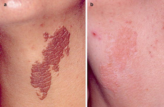
There is no ideal treatment, and it can often be disappointing because of relapses and unaesthetic scars. Therapy includes topical agents, dermabrasion, cryosurgery, photodynamic therapy, and laser cutting. The most commonly used are the ablative lasers, such as the CO2 or Er:YAG laser (Thual et al. 2006). The use of lasers allows satisfactory aesthetic results (Boyce and Alster 2002). The Er:YAG laser should be used for less warty lesions (Pearson and Harland 2004) (Fig. 8).
Syringoma
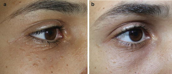
It is a fairly common benign tumor, usually multiple, represented by small rosy-yellowish papules smaller than 3 mm, symmetrical, located on the lower eyelids and in the periorbital region, mainly in adult women. Sometimes it can be translucent or cystic. They are largely of cosmetic significance.
Syringomas usually appear first in puberty; additional lesions may develop later. There is a form of sudden onset in adolescence that affects the neck, chest, abdomen, and penis: the eruptive hidradenoma. It can also be found on the vulva, armpits, and back of the hands. It is characterized histologically by cystic ducts and comma-shaped and solid epithelial cords surrounded by fibrous stroma. The histogenesis of syringomas is probably related to eccrine elements or pluripotent stem cells.
Friedman and Butler classify syringomas into four variants: (1) the localized form, (2) the form associated with Down’s syndrome, (3) the generalized form that encompasses multiple eruptive syringomas, and (4) a familial form. Rarely, syringomas may be associated with Brooke-Spiegler syndrome, an autosomal dominant disease characterized by the development of multiple cylindromas, trichoepitheliomas, and occasional spiradenomas. Syringomas occur with increased frequency in patients with Down’s syndrome (6–36% of cases), usually in women over 10 years old (Fig. 9).
Surgical Care
The main reason for treatment is cosmetic. Complete removal is often unsuccessful and recurrence is common, as syringomas are generally located in the dermis. Possible treatments include surgical excision with primary suture, electrocautery, cryosurgery, dermabrasion, TCA, carbon dioxide laser, or Er:YAG laser (Cho et al. 2011; Sajben and Ross 1999; Kitano 2016; Seo et al. 2016; Lee et al. 2015). Regarding use of the CO2 laser, recurrence of the tumor is associated with superficial ablation, and complications such as hypopigmentation and atrophy are associated with deeper ablation (Fig. 10).
Fordyce Spots
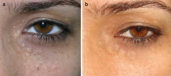
Fordyce granules are asymptomatic sebaceous glands commonly found on the oral mucosa, upper lip, and retromolar region. They are characterized by multiple whitish or yellowish papules with a diameter of 0.1 to 1 mm which occasionally may coalesce and form plaques. Only sebaceous glands visible through the epithelium should be considered Fordyce granules. In children they are usually not noticed until puberty, but they are histologically present. Their incidence increases with age, especially after the hormonal stimulation of puberty. Prevalence in adults ranges from 70% to 85%, with a slight predominance in males. Histopathologically, the lesions are indistinguishable from sebaceous glands, but they are not associated with a hair follicle and their duct opens directly onto the surface.
It is an entity of easy clinical diagnosis, and additional tests are not needed. The condition must be distinguished from other lesions of the oral cavity: small colonies of Candida albicans, miniature lipomas, Koplik’s spots, warts, papular mucosal lesions of Cowden syndrome, lichen planus, and leukoplakia. Despite its asymptomatic nature and its status as a normal variant, some patients seek treatment for cosmetic reasons. There are reports of cases in which dichloroacetic acid, the CO2 laser (Ocampo-Candiani et al. 2003), photodynamic therapy using 5-aminolevulinic acid, oral isotretinoin, and curettage with electrocoagulation were used (Chuang et al. 2004; Baeder et al. 2010) (Fig. 11).
Seborrheic Keratoses
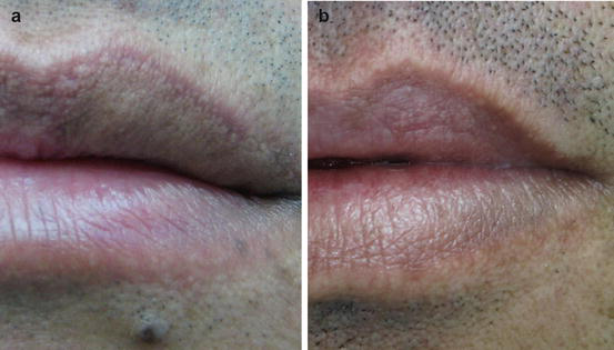
Seborrheic keratoses are limited keratotic papules or plaques arising from epidermal proliferation of keratinocytes. The lesions usually appear after age 40 and are more common in Caucasians. Their surface is rough and greasy, does not reflect light, and can show horn cysts or have a cerebriform appearance. They have very variable pigmentation, ranging from light brown to black, and may be confused with actinic keratosis, melanocytic nevus, or lentigo maligna (Fig. 12).
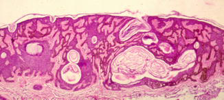
They are located mostly on the trunk and face. They may be single or number in the tens, and can be removed by curettage, cryosurgery, electrocoagulation, or CO2 laser (Fig. 13).
Rhinophyma
Rhinophyma is a benign dermatologic disease of the nose that primarily affects Caucasian men from the fifth to the seventh decades of life. There is hyperplasia of the sebaceous glands leading to a peau d’orange appearance. It is characterized by slowly progressive enlargement of the nose with irregular thickening of the nasal skin and nodular deformation. It is one clinical type of rosacea.
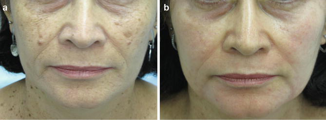
Rhinophyma shows prominence of the sebaceous glands, with the development of thickened, disfigured noses in extreme cases. The condition usually does not produce scars. Rhinophyma can occur as an isolated entity, without other symptoms or signs of rosacea. It can be disfiguring and distressing to patients. Some authors consider rhinophyma a distinct disease. The main reasons that lead patients to seek help are aesthetic and functional impairments, such as nasal obstruction and sleep apnea. However, 46 cases of malignancies such as BCC and SCC have been found associated with rhinophyma, which leads us to examine all excised tissue (Fig. 14).
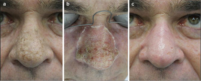
Various methods have been used to correct the malformations this disease produces in the nose, such as dermabrasion, electrocautery, and laser therapy. The treatment of choice for rhinophyma is surgical removal of the hyperplastic tissue. Use of the CO2 laser for the treatment of rhinophyma is an appropriate therapy with excellent aesthetic results, minimal surgical morbidity, and little risk. The pulsed dye laser can be used after the CO2 laser to improve the vascular component of rhinophyma (Moreira et al. 2010). The Er:YAG may also be used as an ablative laser (Orenstein et al. 2001). Electrocoagulation and cold scalpel cutting provide similar long-term results, but hemostasis is less efficient and operating time is longer. The postoperative healing period is faster and scars occur less often with the CO2 laser (Meesters et al. 2015; Baró et al. 2015; Serowka et al. 2014).
Actinic Cheilitis
Actinic cheilitis (AC) is considered a premalignant lesion or an incipient, superficial form of squamous cell carcinoma (SCC) of the lip. Genetically predisposed keratinocytes probably undergo molecular changes induced by ultraviolet B light, yielding neoplastic keratinocytes. Therefore, AC is in fact the result of clonal expansion of transformed keratinocytes, considered from the outset an SCC in situ. It is commonly found in individuals whose professional activities involve chronic sun exposure, particularly redheads with light skin and eversion of the lower lip. The lower lip is more vulnerable to sunlight because it has a thin epithelium, a thin keratin layer, and a lower melanin content. Smoking and lip infection with human papillomavirus can cause cytogenetic changes and increase the risk of progression of actinic cheilitis to SCC (Wood et al. 2011).
Clinical signs include atrophic, diffuse, poorly demarcated plaques or erosive keratotic lesions that may affect all or parts of the vermilion. The definitive diagnosis is obtained by biopsy. Histopathological changes range from atrophy to hyperplasia of the squamous epithelium at the vermilion border, with varying degrees of keratinization, disorderly maturation, increased mitotic activity, and cytological atypia. Apoptotic cells are often present, but the basement membrane is intact. The underlying connective tissue shows basophilic degeneration (solar elastosis). Actinic cheilitis should be considered an intraepithelial or in situ SCC, based on the abovementioned microscopic changes (Fig. 15).
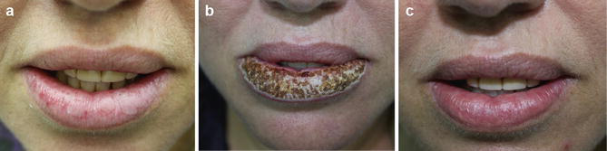
The risk of progression of AC to SCC varies from less than 1% to 20%. Clinically, pain, induration, large size, marked hyperkeratosis, ulceration, bleeding, rapid growth, and recurrence or persistence may be markers of progression of AC to SCC. The metastatic risk of SCC varies between 0.5% and 3%. However, lip SCC resulting from actinic cheilitis is more likely to metastasize than skin SCC, with rates ranging from 3% to 20% (Kwon et al. 2011). Treatment is of crucial importance due to the potential for malignant transformation. Surgical excision of the entire vermilion (vermilionectomy) with histological examination of serial sections is the preferred treatment. Other possible treatments include electrodissection, cryosurgery, photodynamic therapy, topical treatment with the antineoplastic agent 5-fluorouracil or the immunomodulator imiquimod, and lasers such as the CO2 and Er:YAG (Cohen 2013; Laws et al. 2000). However, with these approaches the tissue is not available for histological examination (Dinani et al. 2015). Prevention of AC can be achieved through reduction of cumulative exposure to UVB radiation. Protective clothing, reducing outdoor activities, and the use of sunscreens should be introduced very early in childhood and continue throughout life.
Exogenous Ochronosis
Ochronosis is a grayish-brown pigmentation of connective tissues that can be classified as endogenous or exogenous. The endogenous variety, also known as alkaptonuria, is a rare, congenital, autosomal recessive disorder that results from the absence of the enzyme that converts homogentisic acid into acetoacetic and fumaric acids. Affected individuals develop accumulation of homogentisic acid, an insoluble pigment that is deposited in various tissues such as cartilage, skin, and heart valves (Albers et al. 1992).
Exogenous ochronosis is clinically and histologically similar to the endogenous form, but it has no systemic involvement. It is characterized by blue-black or grayish, asymptomatic hyperpigmentation typically located on the face, neck, back, and extensor surfaces of the extremities. It most commonly follows the use of hydroquinone, but resorcinol, phenol, mercury, picric acid, and oral antimalarials may also be involved. It was initially considered to be caused only by the use of high concentrations of hydroquinone for an extended period, but recent case reports demonstrate development of this pathology with use of 2% hydroquinone for a period no longer than 3 months. The mechanism of hydroquinone-induced hyperpigmentation remains uncertain (Charlín et al. 2008). Activation of tyrosinase by high concentrations of hydroquinone has been reported, leading to stimulation of melanin synthesis. Other authors suggest that hydroquinone oxidase inhibits the activity of homogentisic acid in the skin, leading to accumulation of homogentisic acid, which then polymerizes and forms ochronotic pigment. Melanocytes could be involved; many cases are related to sun exposure, and there is a reported case of ochronosis that spared an area of vitiligo (Simmons et al. 2015b).
Clinical presentation: exogenous ochronosis can be identified in three stages. In stage I it presents only as erythema and mild pigmentation of the face and neck. With progression there are hyperpigmentation, “caviar-like” papules, and atrophy, which correspond to stage II. The last stage includes papulonodular lesions, surrounded or not by inflammation.
Histopathological examination of exogenous ochronosis lesions reveals yellow-brown or green, banana-shaped filaments in the papillary dermis. These filaments undergo degeneration, forming colloid milium, with progression to the papulonodular stage. In stage III there are inflammatory mediators, including giant cells, epithelioid cells, and histiocytes. Some biopsies show sarcoid-like granuloma formation surrounded by filaments. In severe cases, transepidermal elimination of pigment and pseudoepitheliomatous hyperplasia may also be described (Figs. 16, 17, and 18).
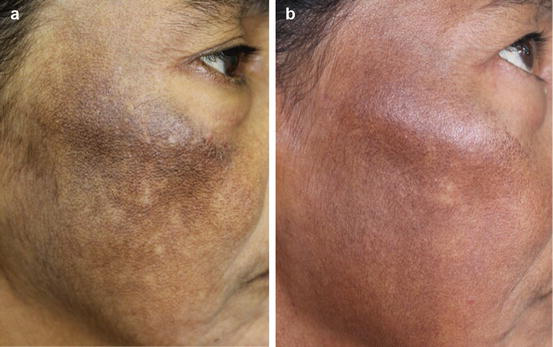
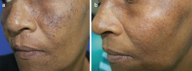
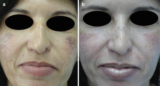
Therapy of exogenous ochronosis is difficult. Various treatments have been used, often with disappointing results. Avoiding the use of the causative substances is beneficial, but it can take several years for any result. Retinoic acid was effective in some patients but caused transient hyperpigmentation in others. The results of treatments with sunscreens and low-potency corticosteroids have been variable. There are reports of clinical improvement after use of oral tetracycline, dermabrasion, and the CO2 laser; however, the results are not uniform. Regarding dermabrasion, there is a case report in which hyperpigmentation was removed in a white patient. A combination of dermabrasion and CO2 laser with satisfactory results in the periorbital and nasal regions in a black woman was reported (Diven et al. 1990).
The use of Q-switched (Q-S) lasers for the treatment of pigmentary lesions and tattoos is well documented in the literature. The Q-S ruby laser (694 nm) and the 755 nm Q-S alexandrite laser were used to treat exogenous ochronosis with good results, based on the fact that the pigment of exogenous ochronosis is deposited in the dermis in a manner similar to tattoo pigment (Bellew and Alster 2004; Kanechorn-Na-Ayuthaya et al. 2013; Tan 2013).
There are reports on the effectiveness of intense pulsed light (IPL), like lasers, for the treatment of pigmented lesions. The mechanism of action of both is based on selective photothermolysis of pigmented cells. IPL has the advantage of pulse-width and wavelength adjustment according to skin type and the depth of pigment deposition in the skin. TCA peeling in different concentrations has been used for many years for the treatment of photoaging, acne scars, and pigmentation disorders. The use of TCA in hyperpigmentation is related to coagulative necrosis of epidermal cell proteins, followed by cell death. The depth of the process depends on the concentration used. A TCA solution between 15% and 25% produces only coagulative necrosis of the epidermis, resulting in superficial peeling. In our recently published work, TCA peeling was used as adjuvant therapy, applied immediately after the intense pulsed light sessions; this combination was observed to be effective for regression of the lesions.
Exogenous ochronosis is a disease that is difficult to treat, requiring a combination of several methods to obtain a satisfactory result (França et al. 2010).
Conclusion
The CO2 laser has great versatility in its use. It is indicated in various scenarios involving skin excision, vaporization, and coagulation; this laser has several modes of action, such as stimulating collagen and rejuvenation, removal of tumors, and removal of warts, xanthelasmas, and keratoses, among others. The CO2 laser is safe when used by trained dermatologists, allowing a dry surgical field with limited blood loss and induction of collagen and cicatrization. In the case of viral lesions (warts and condylomas), the laser can be used together with a smoke-evacuator filter.
Take Home Messages
- The CO2 laser is ablative with a high affinity for water, considered safe when used by properly trained physicians.
- The CO2 laser may be used in many scenarios, such as removal of benign epithelial tumors, keratoses, nevi, warts, and xanthelasma, among others.
- The CO2 laser has been shown to be safe in removing DPN, with low rates of recurrence or complications, and it also has a high degree of patient satisfaction even at the highest phototypes.
- The use of topical anesthetic for benign epithelial lesions is sufficient in most cases, and petrolatum ointment once a day is indicated until reepithelialization of the lesions.
- In cases of viral lesions, treatment of one lesion can lead to regression of many or all warts in immunocompetent individuals.
- The CO2 laser is not the first choice in the ablation of viral lesions, and the use of smoke-filter vacuum units, as well as gloves and goggles, is mandatory.
- Superficial ablation is associated with recurrence in the treatment of syringomas or nevi with the CO2 laser, while complications such as hypopigmentation and atrophy are associated with deeper ablation.
- The postoperative cicatrization period is faster in cases of rhinophyma treated with the CO2 laser compared with electrocoagulation.
- The CO2 laser permits removal of actinic keratoses, but there is no evaluation of the lesion margins.
- Exogenous ochronosis is difficult to treat, and the results with the CO2 laser are not uniform.
References
Aghassi D, González E, Anderson RR, Rajadhyaksha M, González S. Elucidating the pulsed-dye laser treatment of sebaceous hyperplasia in vivo with real-time confocal scanning laser microscopy. J Am Acad Dermatol. 2000;43(1 Pt 1):49–53.
Albers SE, Brozena SJ, Glass LF, Fenske NA. Alkaptonuria and ochronosis: case report and review. J Am Acad Dermatol. 1992;27(4):609–14.
Ali FR, Bakkour W, Ferguson JE, Madan V. Carbon dioxide laser ablation of dermatosis papulosa nigra: high satisfaction and few complications in patients with pigmented skin. Lasers Med Sci [Internet]. 2016;31(3):593–5.
Baba M, Bal N. Efficacy and safety of short-pulse erbium:YAG laser in the treatment of acquired melanocytic nevi. Dermatol Surg. 2006;32(2):256–60.
Baeder FM, Pelino JE, de Almeida ER, Duarte DA, Santos MT. High-power diode laser use on Fordyce granule excision: a case report. J Cosmet Dermatol. 2010;9(4):321–4.
Baró CJ, Gómez R, Serrat A. CO2 laser for the treatment of rhinophyma. Acta Otorrinolaringol Esp. 2015;66(1):61–2.
Bellew SG, Alster TS. Treatment of exogenous ochronosis with a Q-switched alexandrite (755 nm) laser. Dermatol Surg. 2004;30(4 Pt 1):555–8.
Boyce S, Alster TS. CO2 laser treatment of epidermal nevi: long-term success. Dermatol Surg. 2002;28(7):611–4.
Bruscino N, Conti R, Campolmi P, Bonan P, Cannarozzo G, Lazzeri L, Moretti S. Dermatosis papulosa nigra and 10,600-nm CO2 laser, a good choice. J Cosmet Laser Ther. 2014;16(3):114–6.
Bukvić Mokos Z, Lipozenčić J, Ceović R, Stulhofer Buzina D, Kostović K. Laser therapy of pigmented lesions: pro and contra. Acta Dermatovenerol Croat. 2010;18(3):185–9.
Charlín R, Barcaui CB, Kawahac B, Soares DB, Rabello-Fonseca R, Azulay-Abulafia L. Hydroquinone-induced exogenous ochronosis: a report of four cases and usefulness of dermoscopy. Int J Dermatol. 2008;47:19–23.
Cho SB, Kim HJ, Noh S, Lee SJ, Kim YK, Lee JH. Treatment of syringoma using an ablative 10,600-nm carbon dioxide fractional laser: a prospective analysis of 35 patients. Dermatol Surg. 2011;37(4):433–8.
Chuang YH, Hong HS, Kuo TT. Multiple pigmented follicular cysts of the vulva successfully treated with CO2 laser: case report and literature review. Dermatol Surg. 2004;30:1261–4.
Cohen JL. Erbium laser resurfacing for actinic cheilitis. J Drugs Dermatol. 2013;12(11):1290–2.
Dinani N, Topham E, Derrick E, Atkinson L. Ablative fractional laser-assisted photodynamic therapy for the treatment of actinic cheilitis. Br J Dermatol. 2015;173(1):15.
Diven DG, Smith EB, Pupo RA, Lee M. Hydroquinone-induced localized exogenous ochronosis treated with dermabrasion and CO2 laser. J Dermatol Surg Oncol. 1990;16(11):1018–22.
França E, Paiva V, Toscano L, Nunes G, Rodrigues T. Ocronose exógena: relato de caso. Surg Cosmet Dermatol [Internet]. 2010;2(4):319–21.
Gay C, Terzibachian JJ, Gabelle C, Reviron S, Ramanah R, Mougin C. Carbon dioxide laser vaporization of genital condyloma in pregnancy. Gynecol Obstet Fertil. 2003;31:214–9.
Güngör S, Canat D, Gökdemir G. Erbium:YAG laser ablation versus 70% trichloroacetic acid application in the treatment of xanthelasma palpebrarum. J Dermatolog Treat. 2014;25(4):290–3.
Hague JS, Lanigan SW. Laser treatment of pigmented lesions in clinical practice: a retrospective case series and patient satisfaction survey. Clin Exp Dermatol. 2008;33(2):139–41.
Hairston MA Jr, Reed RJ, Derbes VJ. Dermatosis papulosa nigra. Arch Dermatol. 1964;89:655.
Hallmo P, Naess O. Laryngeal papillomatosis with human papillomavirus DNA contracted by a laser surgeon. Eur Arch Otorhinolaryngol. 1991;248:425–7.
Huang J, Zeng Q, Zuo C, Yang S, Xiang Y, Lu J, Kang J, Tan L, Yu X, Xi C, Huang J, Kang L, Fan F, Chen J. The combination of CO2 laser vaporization and photodynamic therapy in treatment of condylomata acuminata. Photodiagnosis Photodyn Ther. 2014;11(2):130–3.
Kanechorn-Na-Ayuthaya P, Niumphradit N, Aunhachoke K, Nakakes A, Sittiwangkul R, Srisuttiyakorn C. Effect of combination of 1064 nm Q-switched Nd:YAG and fractional carbon dioxide lasers for treating exogenous ochronosis. J Cosmet Laser Ther. 2013;15(1):42–5.
Kitano Y. Erbium:YAG laser treatment of periorbital syringomas using the multiple ovoid-shape ablation method. J Cosmet Laser Ther. 2016;10:1–23.
Kundu RV, Patterson S. Dermatologic conditions in skin of color: part II. Disorders occurring predominantly in skin of color. Am Fam Physician. 2013;87(12):859–65.
Kwon NH, Kim SY, Kim GM. A case of metastatic squamous cell carcinoma arising from actinic cheilitis. Ann Dermatol. 2011;23(1):101–3.
Laws RA, Wilde JL, Grabski WJ. Comparison of electrodessication with CO2 laser for the treatment of actinic cheilitis. Dermatol Surg. 2000;26(4):349–53.
Lee BJ, Mancini AJ, Renucci J, Paller AS, Bauer BS. Full-thickness surgical excision for the treatment of inflammatory linear verrucous epidermal nevus. Ann Plast Surg. 2001;47(3):285–92.
Lee SJ, Goo B, Choi MJ, Oh SH, Chung WS, Cho SB. Treatment of periorbital syringoma by the pinhole method using a carbon dioxide laser in 29 Asian patients. J Cosmet Laser Ther. 2015;17(5):273–6.
Meesters AA, van der Linden MM, De Rie MA, Wolkerstorfer A. Fractionated carbon dioxide laser therapy as treatment of mild rhinophyma: report of three cases. Dermatol Ther. 2015;28(3):147–50.
Mitsuishi T, Sasagawa T, Kato T, Iida K, Ueno T, Ikeda M, Ninomiya R, Wakabayashi T, Kawasaki H, Motoki T, Kawana S. Combination of carbon dioxide laser therapy and artificial dermis application in plantar warts: human papillomavirus DNA analysis after treatment. Dermatol Surg. 2010;36(9):1401–5.
Moreira A, Leite I, Guedes R, Baptista A, Mota G. Surgical treatment of rhinophyma using carbon dioxide (CO2) laser and pulsed dye laser (PDL). J Cosmet Laser Ther. 2010;12(2):73–6.
Mourad B, Elgarhy LH, Ellakkawy HA, Elmahdy N. Assessment of efficacy and tolerability of different concentrations of trichloroacetic acid vs. carbon dioxide laser in treatment of xanthelasma palpebrarum. J Cosmet Dermatol. 2015;14(3):209–15.
No D, McClaren M, Chotzen V, Kilmer SL. Sebaceous hyperplasia treated with a 1450-nm diode laser. Dermatol Surg. 2004;30(3):382–4.
Ocampo-Candiani J, Villarreal-Rodriguez A, Quinones-Fernandez AG, Herz-Ruelas ME, Ruiz-Esparza J. Treatment of Fordyce spots with CO2 laser. Dermatol Surg. 2003;29(8):869–71.
Oni G, Mahaffey PJ. Treatment of recalcitrant warts with the carbon dioxide laser using an excision technique. J Cosmet Laser Ther. 2011;13(5):231–6.
Orenstein A, Haik J, Tamir J, Winkler E, Frand J, Zilinsky I, Kaplan H. Treatment of rhinophyma with Er:YAG laser. Lasers Surg Med. 2001;29(3):230–5.
Ozaki M, Suga H, Eto H, Kobayashi Y, Watanabe R, Takushima A, et al. Efficacy of serial excisions of melanocytic nevi on the face using a carbon dioxide laser: a cosmetic point of view. Aesthet Plast Surg [Internet]. 2014;38(2):316–21.
Padilla-Ailhaud A. Carbon dioxide laser vaporization of condyloma acuminata. J Low Genit Tract Dis. 2006;10(4):238–41.
Pathania V, Chatterjee M. Ultrapulse carbon dioxide laser ablation of xanthelasma palpebrarum: a case series. J Cutan Aesthet Surg. 2015;8(1):46–9.
Pearson IC, Harland CC. Epidermal nevi treated with pulsed erbium:YAG laser. Clin Exp Dermatol. 2004;29(5):494–6.
Sajben FP, Ross EV. The use of the 1.0 mm handpiece in high-energy, pulsed CO2 laser destruction of facial adnexal tumors. Dermatol Surg. 1999;25(1):41–4.
Savoca S, Nardo LG, Rosano TF, D’Agosta S, Nardo F. CO2 laser vaporization as primary therapy for human papillomavirus lesions: a prospective observational study. Acta Obstet Gynecol Scand. 2001;80(12):1121–4.
Sayin I, Ayli M, Oğuz AK, Cengiz Seval G. Xanthelasma palpebrarum: a new side effect of nilotinib. BMJ Case Rep. 2016;12:2016.
Seo HM, Choi JY, Min J, Kim WS. Carbon dioxide laser combined with botulinum toxin A for patients with periorbital syringomas. J Cosmet Laser Ther. 2016;31:1–5.
Serowka KL, Saedi N, Dover JS, Zachary CB. Fractionated ablative carbon dioxide laser for the treatment of rhinophyma. Lasers Surg Med. 2014;46(1):8–12.
Simmons BJ, Griffith RD, Falto-Aizpurua LA, Bray FN, Nouri K. Light and laser therapies for the treatment of sebaceous gland hyperplasia: a review of the literature. J Eur Acad Dermatol Venereol. 2015a;29(11):2080–7.
Simmons BJ, Griffith RD, Bray FN, Falto-Aizpurua LA, Nouri K. Exogenous ochronosis: a comprehensive review of the diagnosis, epidemiology, causes, and treatments. Am J Clin Dermatol. 2015b;16(3):205–12.
Tan SK. Exogenous ochronosis – successful outcome after treatment with a Q-switched Nd:YAG laser. J Cosmet Laser Ther. 2013;15(5):274–8.
Thual N, Chevallier JM, Vuillamie M, Tack B, Leroy D, Dompmartin A. CO2 laser therapy of verrucous epidermal nevus. Ann Dermatol Venereol. 2006;133(2):131–8.
Wang H, Shi Y, Guan H, Liu C, Zhang W, Zhang Y, Liu A, Qian Y, Zhao Y, Jiang H. Treatment of xanthelasma palpebrarum with intralesional pingyangmycin. Dermatol Surg. 2016;42(3):368–76.
Winstanley D, Blalock T, Houghton N, Ross EV. Treatment of sebaceous hyperplasia with a novel 1,720-nm laser. J Drugs Dermatol. 2012;11(11):1323–6.
Wood NH, Khammissa R, Meyerov R, Lemmer J, Feller L. Actinic cheilitis: a case report and a review of the literature. Eur J Dent. 2011;5(1):101–6.
Zeng Y, Zheng YQ, Wang L. Successful treatment of recalcitrant warts in combination with CO2 laser and imiquimod 5% cream. J Cosmet Laser Ther. 2014;16(6):311–3.







