
Actinic Cheilitis: Efficacy and Cosmetic Results
Actinic Cheilitis: Efficacy and Cosmetic Results
Marco Antônio de Oliveira A. C. Camargo Cancer Center, São Paulo, Brazil
Abstract
Actinic cheilitis (AC) is a common disease caused by long-term solar exposure and is considered a premalignant lesion of the lips. AC has the potential to develop into squamous cell carcinoma (SCC) and metastasis. SCC of the lip arising from AC is more prone to metastasis than the cutaneous form, with rates of the former varying between 3% and 20%, and it is the most common malignancy of the oral cavity. AC affects especially pale-skinned individuals with poorly pigmented lower lips. The overall 5-year survival rate is less than 75%. Treatments are difficult because surgical treatment can have significant adverse effects, whereas less invasive procedures may not be as efficient. A high degree of clinical suspicion should be maintained, given the malignant nature of the condition. We emphasize the need for regular follow-up, a rigorous clinical exam, and accurate pathologic analysis. The chapter will discuss the efficacy of photodynamic therapy (PDT) using methyl aminolevulinate and 5-aminolevulinic acid, application times, skin preparation techniques, pain, cosmetic outcome, and patient satisfaction.
Keywords Actinic cheilitisSolar cheilitisPhotodynamic therapy5-Aminolevulinic acidMethyl aminolevulinateCosmetic outcomeLip cancerSquamous cell carcinoma
Introduction
An understanding of the nature of actinic cheilitis can be achieved by studying and reflecting on comments about it by three dermatologists, namely Dubreuilh in 1896, Freudenthal in 1926, and Sutton Jr. in 1896 (Heaphy and Ackerman 2000). Actinic cheilitis is a very frequent disease of the lips (Dufresne and Curlin 1997). This ominous precursor of cancer occurs especially on the lower lip due to chronic sunlight exposure and the outdoor activities of people such as fishermen, farmers, beach workers, golfers, and surfers (Dufresne and Curlin 1997; Martins-Filho et al. 2011; Lucena et al. 2012; Ribeiro et al. 2014).
Basic Concepts
Actinic cheilitis, or solar cheilosis, attracts a number of clinical definitions, from malignant lesions to relatively mild descriptions of sun-damaged lips. This occurrence is sunlight-dose dependent (Table 1) and is cumulative over a 5–20-year exposure to ultraviolet radiation, immune status (organ transplantation), age, genetic predisposition (xeroderma pigmentosum, porphyria cutanea tarda, oculocutaneous albinism), drugs (voriconazole), geographic latitude (countries close to the equator), and use of lip protection (lipstick, lip sunscreen) (Rogers and Bekic 1997; Savage et al. 2010; Lucena et al. 2012; Jadotte and Schwartz 2012a). Other factors like tobacco, alcohol, poor oral hygiene, and chronic scarring in discoid lupus erythematosus are involved (Table 1) (Jadotte and Schwartz 2012a).
| Major risk factors | Minor risk factors |
|---|---|
| Genetic predisposition | Smoking |
| Skin pigmentation | Alcoholism |
| UV radiation history | Organ transplantation |
| Lupus discoid | |
| Chronic scars | |
| Poor oral hygiene |
As with actinic keratosis, the exact transition rate to the most common lip cancer, squamous cell carcinoma (SCC), is unknown, but the relative risk is felt to be 2.5 times higher. SCC is, however, the most frequent oral cavity cancer and head and neck malignant tumor, with an incidence in the United States of 1.8 per 100,000 (Ulrich et al. 2007). SCC is considered a disease of low aggression and favorable prognosis because it tends to progress slowly. The estimated occurrence of SCC is 89% on the lower lip, 3% on the upper lip, and 8% at the oral commissures (Picascia and Robinson 1987).
When diagnosed early it has a cure rate of 80–90% and a mortality rate between 10% and 15%. Metastases tend to occur in 3–20% of cases; in these cases the mean 5-year survival decreases to 25% (Kwon et al. 2011; Lim et al. 2014).
As already noted, AC occurs more often on the lower lip. Two forms exist. The acute form is less common and occurs after prolonged sunlight exposure. Edema and redness are characteristic of the mildest forms. Severe congestion, fissuring, and ulceration typify the most severe form. At times vesicles may appear on the vermilion edge. These vesicles rupture, causing superficial erosions, and take from days to weeks to heal (Nico et al. 2007).
The chronic form of AC usually manifests itself as slight scaling involving the whole area of the lower lip up to the commissures and is present in all seasons of the year. The scaling is not always uniform, and some areas may display more intensive hyperkeratosis than others (Picascia and Robinson 1987; Nico et al. 2007). Leukoplakia, a scaly plaque with a sandpapery feel on palpation with a gloved finger, is a common presentation (Jadotte and Schwartz 2012a).
White-gray or brown changes may appear. The vermilion edges lose their usual plasticity, which can be assessed by the appearance of marked folds. These wrinkles are parallel to one another and are perpendicular to the long axis of the lip (Picascia and Robinson 1987; Nico et al. 2007).
The differential diagnosis (Table 2) of AC covers a spectrum of neoplastic, inflammatory, eczematoid, and photosensitive disorders, along with a few rare but important diseases (Picascia and Robinson 1987; Jadotte and Schwartz 2012b).
| Condition |
|---|
| Squamous cell carcinoma |
| Amelanotic melanoma |
| Discoid lupus erythematosus |
| Oral lichen planus |
| Metastatic cancer |
| Plasma cell cheilitis |
| Cheilitis glandularis |
| Angular cheilitis |
| Granulomatous cheilitis |
| Eczematous cheilitis |
| Nutritional cheilosis |
| Actinic prurigo cheilitis |
| Factitious cheilitis |
| Necrotizing sialometaplasia |
| Sweet syndrome |
Evaluation of ultraviolet-ray-induced lesions of the lower lip can be difficult, as the clinical aspects often do not correspond to the severity of the microscopic epithelial alterations. When clinical signs are present in a well-demarcated area of the lip, the most severe histological changes are presumed to occur at this point. However, AC frequently presents as diffuse, poorly demarcated alterations along the vermilion, and choosing an area for incisional biopsy in these cases may be challenging (Nico et al. 2007). Carcinomatous transformation is generally characterized by induration, infiltration, or ulcers in the actinic area (Samimi 2016). Sometimes two or more biopsies are required. Currently, reflectance confocal microscopy seems to be a promising diagnostic tool for actinic cheilitis, as it allows noninvasive evaluation in real time and in vivo (Ulrich et al. 2011).
Treatment of actinic cheilitis provides relief of symptoms and improves the cosmetic appearance but, more importantly, prevents the development of squamous cell carcinoma (Picascia and Robinson 1987).
Surgical scalpel vermilionectomy offers the most definitive treatment to date for actinic cheilitis, followed by carbon dioxide laser (Nelson et al. 2007). There are various options to treat actinic cheilitis; however, these options very often have disabling side effects (Sotiriou et al. 2008).
The treatments that have been used for actinic cheilitis are shown in Table 3 (Robinson 1989; Dufresne et al. 2008; Lima Gda et al. 2010; Shah et al. 2010; Ulrich et al. 2011; Jadotte and Schwartz 2012b; Barrado Solís et al. 2015).
| Treatment |
|---|
| 3% diclofenac gel |
| 5% 5-fluoracil cream |
| 5% imiquimod cream |
| Carbon dioxide laser |
| Cryosurgery |
| Dermabrasion |
| Electrodesiccation and curettage |
| Er:YAG laser |
| Ingenol mebutate gel |
| Surgery (scalpel excision, vermilionectomy, Mohs Micrographic Surgery) |
| Trichloroacetic acid peel |
The treatment options for actinic cheilitis are diverse, and each modality has both distinct advantages and disadvantages. Each treatment can be safely performed in an outpatient setting, although each therapy option comes with different potential adverse effects. Some of them require operator expertise to perform. Apart from the therapeutic option, actinic cheilitis can potentially recur. Effective, well-established methods exist for actinic cheilitis treatment. Each modality has its own inherent risks, benefits, and anticipated sequelae. To determine the treatment of choice, age, comorbidities, mental health, previous carcinomas, and immunosuppression should be considered. Regarding the area, single or multiple lesions, localization, extension, induration, as well as ulcerated lesions should be considered. Isolated and well-defined lesions can be treated with destructive methods such as cryosurgery, dermabrasion, electrodesiccation and curettage, and lasers, while extensive areas can be fully treated with field treatments such as diclofenac, 5-fluoracil, imiquimod, lasers, dermabrasion, ingenol mebutate, photodynamic therapy, surgery, or trichloroacetic acid peel, as presented in Table 3.
Photodynamic therapy can be used for localized lesions or for the entire lip.
Photodynamic Therapy
PDT Mechanism of Action
Photodynamic therapy has been introduced as a therapeutic modality for epithelial skin tumors, revealing a high efficacy rate and satisfactory outcomes in cutaneous actinic keratosis, superficial basal cell carcinoma, and Bowen’s disease. Regarding actinic cheilitis, photodynamic therapy is considered a relatively new off-label therapeutic option (Rossi et al. 2008).
The advantages of PDT are its high efficacy and tolerability coupled with excellent cosmetic outcomes (Morton et al. 2008; Sotiriou et al. 2011). As the lip is a cosmetically sensitive site, treatments for actinic cheilitis must consider cosmetic outcomes (Morton et al. 2008; Sotiriou et al. 2011; Calzavara-Pinton et al. 2013).
Despite these facts, PDT for actinic cheilitis is not very common: there are only a few case series and studies that report the applicability and efficacy of this treatment modality for actinic cheilitis. In most of them, treatment outcome was assessed only by clinical evaluation (Sotiriou et al. 2010).
PDT Indications and Contraindications
Off-label clinical indications for PDT in actinic cheilitis are generally poor and based on individual case reports or small case series, and the few randomized clinical trials performed enrolled small numbers of patients with short follow-up. All authors agreed that the cosmetic outcome was excellent and tolerability was acceptable in the majority of patients (Calzavara-Pinton et al. 2013). In one study the cosmetic results were rated by the investigators as excellent in 81.8% of cases (Sotiriou et al. 2010).
If cosmetic outcome alone is considered, PDT offers positive cosmetic outcomes as observed by the investigators when compared with conventional treatments.
Ulceration, nodularity, atrophy, bleeding, blurred demarcation, and friability (especially when prolonged) indicate malignancy (Jadotte and Schwartz 2012a; Yazdani Abyaneh et al. 2015). In these cases, biopsy is mandatory and PDT should be avoided until histopathological elucidation.
All of the studies are descriptive series without control arms. A bias toward reporting and publishing series with positive findings should also be considered. Furthermore, various treatment parameters were used and lengths of follow-up ranged widely. Clinical responses were assessed subjectively by nonblinded investigators; histological confirmation of cure was established in only half of the subjects (Yazdani Abyaneh et al. 2015).
Furthermore, results are generally based on visual assessments without controlled parameters, such as histopathology and/or noninvasive imaging procedures (Calzavara-Pinton et al. 2013).
These factors have led to a wide range in clinical response and histological cure rates. There is a need for randomized controlled trials with long-term follow-up to evaluate the clinical and histological response of actinic cheilitis to PDT. More studies are also needed to determine the optimal number of treatment sessions and techniques (Yazdani Abyaneh et al. 2015).
PDT: Use and Doses
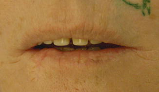
PDT is a noninvasive and precisely directed treatment. Patients are prepared for PDT first with gentle removal of scale and crust from the lips (Fig. 1).
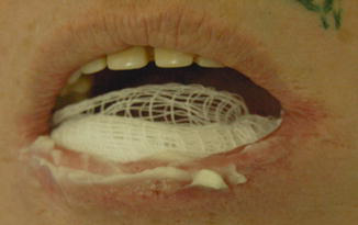
The procedure involves the topical application of photosensitizing agents, most often 20% 5-aminolevulinic acid (ALA) or 16% methyl 5-aminolevulinate cream (MAL), on the lip (Fig. 2).
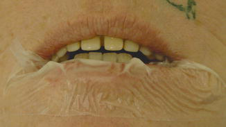
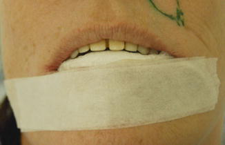
The area is occluded (Figs. 3 and 4) for a preestablished time of 3 h, after which visible-light irradiation (usually red or blue) is applied, with subsequent promotion of reactive oxygen species which in turn produce local tissue destruction (Barrado Solís et al. 2015; Choi et al. 2015).
Daylight PDT (DA-PDT) is a novel modality in which activation of the topical photosensitizer is induced by exposure to natural daylight without requiring preliminary occlusion. In two studies enrolling 12 patients, the use of DA-PDT showed benefits, decreasing the pain level (Levi et al. 2013; Fai et al. 2015).
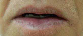
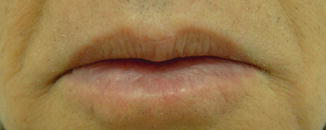
There are no specific data explaining exactly the cosmetic outcome of the lip treated with photodynamic therapy. In our experience we observed overall lip rejuvenation with a younger appearance (Figs. 5 and 6), uniform color, a better-demarcated vermilion transition, a soft and thinner epithelium, and less flaking.
PDT: Side Effects and Their Management
Pain and a burning sensation during irradiation are always present at different levels. Edema, erythema, blistering, hemorrhagic crusting, itchiness, erosions, paresthesias, scaling, and mild dryness can occur. These reactions resolve 5–14 days after the treatment, and cold, dry air helps in pain control (Sotiriou et al. 2010; Yazdani Abyaneh et al. 2015). After PDT, selective destruction of unhealthy tissue occurs, the local immune response increases, and the short-term treatment offers the advantages of minimal erythema, reduced scarring risk, and renewal of the skin epithelium (Alexiades-Armenakas 2007; Kodama et al. 2007; Sotiriou et al. 2008; Rossi et al. 2008; Castaño et al. 2009).
Conventional PDT for actinic cheilitis has been reported in a number of publications, with clinical and histological cure rates ranging between 47% and 100%. This wide variability in efficacy can partly be explained by different study designs, including differences in biological endpoints, light sources, and follow-up periods (Levi et al. 2013; Kim et al. 2013). Despite the observational clinical results, it is important to keep in mind the significantly higher histological recurrence rates (Sotiriou et al. 2010). Inadequate uptake of the photosensitizer, owing to dilution by saliva and rapid regeneration of the mucosal epithelium compared with the skin, may result in lower efficacy of the treatment. PDT offers a promising treatment option supported by excellent post-treatment cosmetic outcome, with the need for further study (Kim et al. 2013; Yazdani Abyaneh et al. 2015).
Authors’ Experience
The author always performs a lip biopsy before any actinic cheilitis treatment. It is possible to treat an isolated lesion as well as the entire lip. When photodynamic therapy is the treatment of choice, there is careful curettage of crusts and scales from the lip; a 1 mm thick layer of 20% methyl 5-aminolevulinate cream is applied over the entire lip. A cotton roll is placed at the gingival transition and the lip is covered by an occlusive bandage for 3 h. After removal of the cream, lesions are irradiated with 37 J/cm2 of red light (635 nm). Two treatment sessions are conducted 1 week apart.
Conclusion
Actinic cheilitis is a chronic premalignant condition affecting the lip. Untreated lesions may become squamous cell carcinoma, and treatment options include ablative, surgical, and topical modalities. Photodynamic therapy is focal and also a field-directed treatment for actinic cheilitis. The cosmetic outcome and results suggest that photodynamic therapy may offer a comparable response rate. Pain and high cost sometimes make the method unfeasible. However, increasing the efficacy of PDT through optimization of treatment parameters is still necessary.
Take Home Messages
- As with actinic keratosis, the exact transition rate to squamous cell carcinoma is unknown; the relative risk is felt to be 2.5 times higher.
- Treatment of actinic cheilitis should provide relief of symptoms and improve the cosmetic appearance but, more importantly, prevent the development of SCC.
- As the lip is a cosmetically sensitive site, treatments such as PDT for actinic cheilitis must consider cosmetic outcomes.
- PDT is advantageous in that it has transient, mild to moderate cosmetic adverse effects and minimizes patient discomfort.
Cross-References
- Daylight Photodynamic Therapy and Its Relation to Photodamaged Skin
- Photodynamic Therapy for Photodamaged Skin
- Transepidermal Drug Delivery and Photodynamic Therapy
References
Alexiades-Armenakas M. Aminolevulinic acid photodynamic therapy for actinic keratoses/actinic cheilitis/acne: vascular lasers. Dermatol Clin. 2007;25:25–33.
Barrado Solís N, Molés Poveda P, Lloret Ruiz C, Pont Sanjuan V, Velasco Pastor M, Quecedo Estébanez E, et al. Ingenol mebutate gel treatment for actinic cheilitis: report of four cases. Dermatol Ther. 2015;28:79–82.
Calzavara-Pinton PG, Rossi MT, Sala R. Italian Group For Photodynamic Therapy. A retrospective analysis of real-life practice of off-label photodynamic therapy using methyl aminolevulinate (MAL-PDT) in 20 Italian dermatology departments. Part 2: oncologic and infectious indications. Photochem Photobiol Sci. 2013;12:158–65.
Castaño E, Comunión A, Arias D, Miñano R, Romero A, Borbujo J. Tratamiento de queilitis actínicas con terapia fotodinâmica. Actas Dermosifiliogr. 2009;100:895–8.
Choi SH, Kim KH, Song KH. Efficacy of ablative fractional laser-assisted photodynamic therapy for the treatment of actinic cheilitis: 12-month follow-up results of a prospective, randomized, comparative trial. Br J Dermatol. 2015;173:184–91.
Dufresne Jr RG, Curlin MU. Actinic cheilitis. A treatment review. Dermatol Surg. 1997;23:15–21.
Dufresne Jr RG, Cruz AP, Zeikus P, Perlis C, Jellinek NJ. Dermabrasion for actinic cheilitis. Dermatol Surg. 2008;34:848–50.
Fai D, Romanello E, Brumana MB, Fai C, Vena GA, Cassano N, et al. Daylight photodynamic therapy with methyl-aminolevulinate for the treatment of actinic cheilitis. Dermatol Ther. 2015;28:355–68.
Heaphy Jr MR, Ackerman AB. The nature of solar keratosis: a critical review in historical perspective. J Am Acad Dermatol. 2000;43:138–50.
Jadotte YT, Schwartz RA. Solar cheilosis: an ominous precursor: part I. Diagnostic insights. J Am Acad Dermatol. 2012a;66:173–84. quiz 185–6.
Jadotte YT, Schwartz RA. Solar cheilosis: an ominous precursor part II. Therapeutic perspectives. J Am Acad Dermatol. 2012b;66:187–98. quiz 199–200.
Kim SK, Song HS, Kim YC. Topical photodynamic therapy may not be effective for actinic cheilitis despite repeated treatments. Eur J Dermatol. 2013;23:917–8.
Kodama M, Watanabe D, Akita Y, Tamada Y, Matsumoto Y. Photodynamic therapy for the treatment of actinic cheilitis. Photodermatol Photoimmunol Photomed. 2007;23:209–10.
Kwon NH, Kim SY, Kim GM. A case of metastatic squamous cell carcinoma arising from actinic cheilitis. Ann Dermatol. 2011;23:101–3.
Levi A, Wulf HC, Enk CD. Two cases of actinic cheilitis responsive to daylight-activated photodynamic therapy (DA-PDT). Photodermatol Photoimmunol Photomed. 2013;29:268–71.
Lim GF, Cusack CA, Kist JM. Perioral lesions and dermatoses. Dent Clin N Am. 2014;58:401–35.
Lima Gda S, Silva GF, Gomes AP, de Araújo LM, Salum FG. Diclofenac in hyaluronic acid gel: an alternative treatment for actinic cheilitis. J Appl Oral Sci. 2010;18:533–7.
Lucena EES, Costa DCB, Silveira EJD, Lima KC. Prevalence and factors associated to actinic cheilitis in beach workers. Oral Dis. 2012;18:575–9.
Martins-Filho PRS, Silva LCP, Piva MR. The prevalence of actinic cheilitis in farmers in a semi-arid northeastern region of Brazil. Int J Dermatol. 2011;50:1109–14.
Morton CA, McKenna KE, Rhodes LE. British Association of Dermatologists Therapy Guidelines and Audit Subcommittee and the British Photodermatology Group. Guidelines for topical photodynamic therapy: update. Br J Dermatol. 2008;159:1245–66.
Nelson CG, Spencer J, Nelson Jr CG. A single-arm, open-label efficacy and tolerability study of diclofenac sodium 3% gel for the treatment of actinic keratosis of the upper and lower lip. J Drugs Dermatol. 2007;6(7):712–7.
Nico MM, Rivitti EA, Lourenço SV. Actinic cheilitis: histologic study of the entire vermilion and comparison with previous biopsy. J Cutan Pathol. 2007;34:309–14.
Picascia DD, Robinson JK. Actinic cheilitis: a review of the etiology, differential diagnosis and treatment. J Am Acad Dermatol. 1987;17:255–64.
Ribeiro AO, Silva LCF, Martins-Filho PRS. Prevalence of the risk factors for actinic cheilitis in Brazilian fishermen and women. Int J Dermatol. 2014;53:1370–6.
Robinson JK. Actinic cheilitis. A prospective study comparing four treatment methods. Arch Otolaryngol Head Neck Surg. 1989;115:848–52.
Rogers 3rd RS, Bekic M. Diseases of the lip. Semin Cutan Med Surg. 1997;16:328–36.
Rossi R, Assad GB, Buggiani G, Lotti T. Photodynamic therapy: treatment of choice for actinic cheilitis? Dermatol Ther. 2008;21:412–5.
Samimi M. Chéilites: orientation diagnostique et traitement. Presse Med. 2016;45:240–50.
Savage NW, McKay C, Faulkner C. Actinic cheilitis in dental practice. Aust Dent J. 2010;55(Suppl 1):78–84.
Shah AY, Doherty SD, Rosen T. Actinic cheilitis: a treatment review. Int J Dermatol. 2010;49:1225–34.
Sotiriou E, Apalla Z, Koussidou-Erremonti T, Ioannides D. Actinic cheilitis treated with one cycle of 5-aminolaevulinic acid-based photodynamic therapy: report of 10 cases. Br J Dermatol. 2008;159:261–2.
Sotiriou E, Apalla Z, Chovarda E, Panagiotidou D, Ioannides D. Photodynamic therapy with 5-aminolevulinic acid in actinic cheilitis: an 18-month clinical and histological follow-up. J Eur Acad Dermatol Venereol. 2010;24:916–20.
Sotiriou E, Lallas A, Goussi C, Apalla Z, Trigoni A, Chovarda E, et al. Sequential use of photodynamic therapy and imiquimod 5% cream for the treatment of actinic cheilitis: a 12-month follow-up study. Br J Dermatol. 2011;165:888–92.
Ulrich C, Forschner T, Ulrich M, Stockfleth E, Sterry W, Termeer C. Management of actinic cheilitis using diclofenac 3% gel: a report of six cases. Br J Dermatol. 2007;156(Suppl 3):43–6.
Ulrich M, González S, Lange-Asschenfeldt B, Roewert-Huber J, Sterry W, Stockfleth E, et al. Non-invasive diagnosis and monitoring of actinic cheilitis with reflectance confocal microscopy. J Eur Acad Dermatol Venereol. 2011;25:276–84.
Yazdani Abyaneh MA, Falto-Aizpurua L, Griffith RD, Nouri K. Photodynamic therapy for actinic cheilitis: a systematic review. Dermatol Surg. 2015;41:189–98.







